
Abstract
Purpose:
In Kienböck’s disease, the wrist displays certain characteristic radiological parameters, which have been reported to differ among countries. In the present study, we aimed to identify specific radiological parameters of the unaffected wrists in patients with unilateral Kienböck’s disease and to determine the extent of the association of each parameter with the disease in Korea.
Methods:
This retrospective case–control study assessed the radiological parameters of patients with Kienböck’s disease (n = 53) and controls (n = 53), who visited our institution between January 2000 and May 2013. Ulnar variance (UV), radial inclination, lunate fossa inclination, lunate diameter, lunate height, lunate tilting angle (LTA), lunate covering index (LCI), and Ståhl index (SI) were measured and analyzed using a binary logistic regression model.
Results:
We observed that wrists with a high LTA and LCI, and low UV and SI had a tendency to develop Kienböck’s disease.
Conclusion:
In the Korean population, a high LTA and LCI, and low UV and SI of the unaffected wrists on plain radiography might be associated with Kienböck’s disease. The radiographic characteristics of the unaffected wrists can differ between patients with unilateral Kienböck’s disease and normal individuals.
Introduction
Kienböck was the first to describe the avascular necrosis of the lunate as lunatomalacia in 1910 1 ; however, the etiology of this disease remains unclear. A negative ulnar variance (UV), primary arterial ischemia, trauma, and hand-arm vibration are considered possible risk factors for Kienböck’s disease, and among these risk factors, negative UV appears to be the most commonly reported risk factor. 2 In fact, since Hulten first reported the relationship between negative UV and Kienböck’s disease, 3 several studies have confirmed this relationship. 4 –7 However, some studies have reported contrasting results. 8 –10 In particular, Afshar et al. suggested that the presence of negative UV as a risk factor for Kienböck’s disease may vary depending on the country of origin of the patient. 7
Other radiological parameters have been shown to be associated with Kienböck’s disease. Mirabello et al. indicated that the carpal index, lunate deformation, and radial slope are clinically related parameters in patients with Kienböck’s disease. 5 However, the parameters of the affected wrists are difficult to measure owing to the presence of lunate deformity and secondary arthritis. Two recent studies that measured the unaffected wrists reported that the lunate diameter (LD), lunate height (LH), and lunate tilting angle (LTA) were significantly associated with Kienböck’s disease, and Thienpont et al. indicated that wrists with certain characteristic radiological features may have a high risk of developing Kienböck’s disease.11,12
Although many radiological parameters have been studied, the influence of each parameter in Kienböck’s disease has not been estimated quantitatively. These parameters, including UV, should be analyzed on the basis of nationality/race. Therefore, in the present study, we aimed to identify specific radiological parameters of the unaffected wrists in patients with unilateral Kienböck’s disease and to determine the extent of the association of each parameter with the disease in Korea.
Materials and methods
This retrospective case–control study enrolled 91 consecutive patients diagnosed with Kienböck’s disease based on radiography and magnetic resonance imaging findings at the radiology department of our institution between January 2000 and May 2013. The study was approved by the institutional review board of our institution (IRB number: H-1404-009-569). The exclusion criteria were bilateral Kienböck’s disease (n = 7), concomitant inflammatory arthritis diagnosed in the rheumatologic department (n = 6), wrist or hand tumor (n = 3), previous wrist infection (n = 3), history of carpal bone fracture (n = 4), history of distal radius fracture (n = 8), history of forearm fracture (n = 2), and severe deformities of the unaffected wrist and forearm (n = 5). Therefore, 53 patients were finally included in the Kienböck’s disease group. The control group included patients who visited our institution with distal radius fracture during the study period, because we obtained plain radiographs of both the wrists as part of the routine diagnostic protocol for both Kienböck’s disease and distal radius fracture. We confirmed the absence of previous trauma, inflammatory arthritis, infection, tumors, and severe deformities in the unaffected wrist and forearm. As UV has been reported to differ according to age and sex, 13 we believed that other radiological parameters may also be influenced by age and sex. Therefore, we matched each patient from the Kienböck’s disease group to a subject from the control group according to age and sex (1:1 ratio).
Previous studies have shown that radiological parameters of a normal population do not significantly differ between the right and left wrists.11,14 Therefore, the unaffected wrists of the subjects in the control group were assessed in the present study.
Projection technique of the wrist
Standard posteroanterior wrist radiographs were obtained with the shoulder abducted to 90°, elbow flexed to 90°, forearm in the neutral position, and hand flat on a tabletop. Moreover, lateral radiographs were obtained with the elbow flexed to 90° and hand rotated to 90° in the sitting position. 15 All the images were obtained in a digital format using a Picture Archiving and Communication System.
Radiological parameters
For measuring the UV, the long axis of the radius was identified by bisecting its medullary width at 2 cm and 5 cm proximal to the distal radial cortex (Figure 1). 16 The UV was measured using the method of perpendiculars. A line was drawn perpendicular to the radial axis through the ulnarmost point of the articular surface of the radius. The distance between this line and the distal cortex of the ulna was measured.

Measurement of the UV. UV: ulnar variance.
For measuring the radial inclination (RI), a line was drawn from the ulnar aspect of the carpal surface of the radius to the radial styloid process (Figure 2). The angle between this line and the line perpendicular to the radial axis was measured. 16

Measurement of the RI and LFI. RI: radial inclination; LFI: lunate fossa inclination.
For measuring the lunate fossa inclination (LFI), a line was drawn from the ulnar aspect of the carpal surface of the radius to the radial prominence of the lunate fossa (Figure 2). The angle between this line and the line perpendicular to the radial axis was measured. 17
For measuring the LD, a line was drawn from the ulnar tip of the distal facet to the radial tip (Figure 3). Using this line as a base (the baseline of the lunate), the distance between the ulnar border of the lunate and the radial border was measured as the LD. 11

Measurement of the LD and LH. LD: lunate diameter; LH: lunate height.
For determining the LH, from the baseline of the lunate, the distance between the distal border of the lunate and the radial border was measured (Figure 3). 11
To determine the LTA, the angle between the baseline of the lunate and the line perpendicular to the radial axis was measured (Figure 4). 12
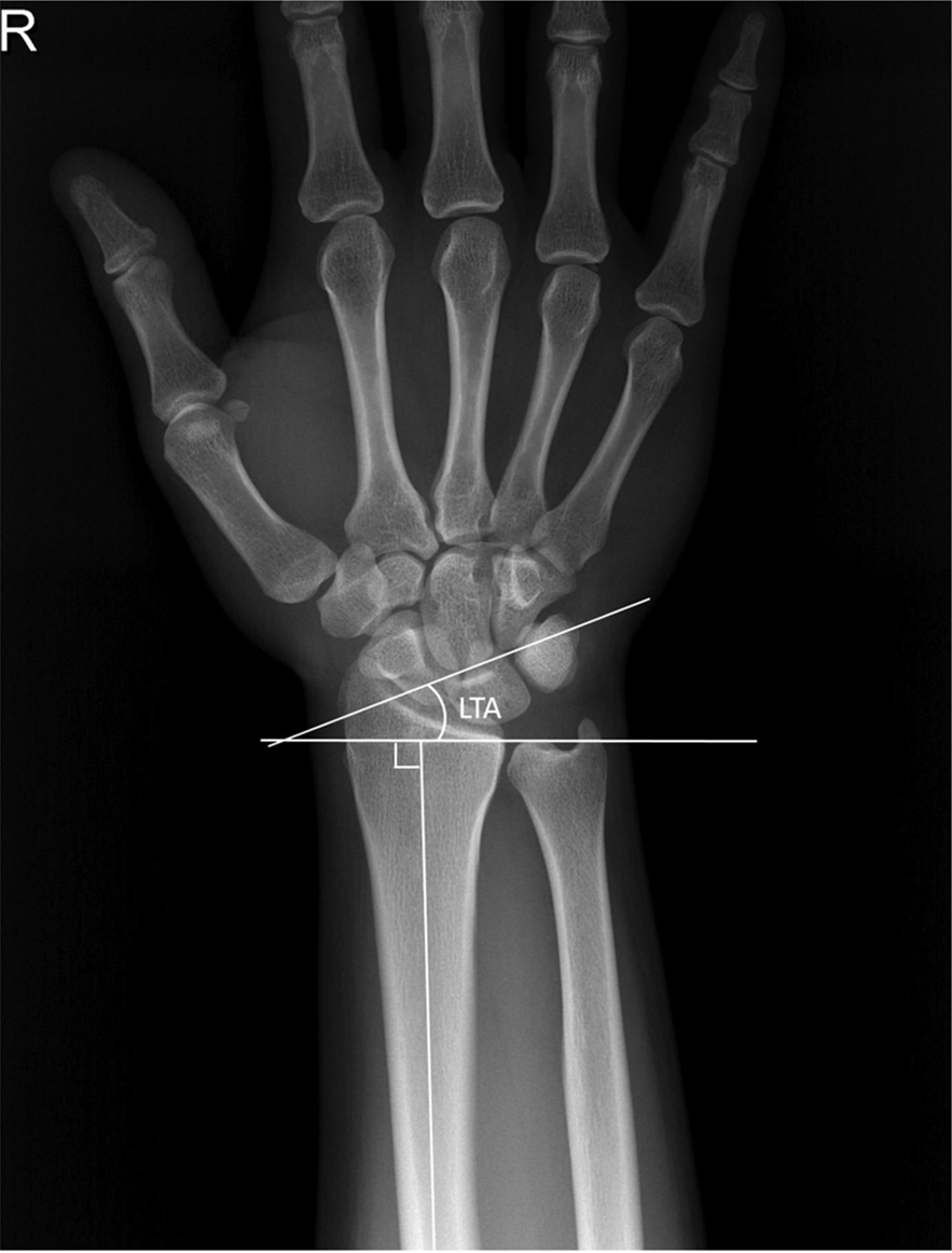
Measurement of the LTA. LTA: lunate tilting angle.
To determine the lunate covering index (LCI), the lunate was divided with a longitudinal line from the radial side of the distal radioulnar joint, which was parallel to the radial axis (Figure 5). The ratio of the lunate on the radius to the entire lunate was defined as the LCI, 18 and the LCI was represented as a percentage value for convenience.

Measurement of the LCI. LCI: lunate covering index.
Ståhl’s index (SI) was defined as the ratio of the longitudinal height to the dorsopalmar width of the lunate in lateral radiographs (Figure 6), 11 and the SI was represented as a percentage value for convenience.

Measurement of the SI. SI: Ståhl’s index.
Data measurements
All the parameters were measured up to the first decimal place. The interobserver reliability of the measurements was assessed by three orthopedic specialists. The intra-observer reliability was investigated by the first author, and was reassessed twice, at intervals of 3 weeks.
Statistical analysis
We evaluated the intra- and interobserver reliability for each parameter using the intra-class correlation coefficient (ICC) with 95% confidence intervals (CIs). An ICC of 1 indicates perfect reliability, and an ICC of 0 indicates no reliability. The multicollinearity between the parameters was determined using the variance inflation factor (VIF). A VIF of >10 indicates high multicollinearity, and a VIF of 1 indicates no multicollinearity. A radiological risk factor model was established using a binary logistic regression model with the backward elimination method. The level of significance was set at p < 0.05, and the CIs of odds containing 1.00 were excluded.
This study was approved by the IRB of our institution (IRB number: H-1404-009-569).
Results
The demographic data of the study subjects are presented in Table 1. The intra- and interobserver reliability and multicollinearity between the parameters are presented in Table 2. All the ICCs (>0.8) indicated a considerable degree of reliability between the investigators. All the VIFs (<10) indicated that the degree of multicollinearity among the parameters was low, suggesting that the interactions were considerably low.
Demographic information of the study subjects.

SD: standard deviation.
Results of the radiological parameters and reliability of the measurements.

ICC: intra-class correlation coefficient; VIF: variance inflation factor; CI: confidence interval; SD: standard deviation; LD: lunate diameter; LH: lunate height; LTA: lunate tilting angle; UV: ulnar variance; LFI: lunate covering index; RI: radial inclination; LCI: lunate covering index; SI: Ståhl’s index.
LD, LH, and LFU were excluded during logistic transformation, and RI was excluded as the CIs of odds contained 1.00. Additionally, the constant was omitted owing to the presence of a p value (0.139). The final logistic regression model was as follows (Table 3):
Results of binary logistic regression analysis assessing the influence of each parameter on Kienböck’s disease.

LTA: lunate tilting angle; UV: ulnar variance; LCI: lunate covering index; SI: Ståhl’s index; CI: confidence interval.

where Kt is the radiological tendency of Kienböck’s disease.
The Kt of the wrist with a mean radiological value was 0.66 in the Kienböck’s disease group and was 0.32 in the control group. Therefore, the radiological tendency for the development of Kienböck’s disease was two times higher in the Kienböck’s disease group than in the control group.
Discussion
Several mechanical factors have been reported to be involved in the development of Kienböck’s disease. Lluch et al. classified these into extrinsic and intrinsic factors. 19 The radiological parameters assessed in the present study were extrinsic factors, which can be corrected with surgical treatment. For instance, a joint-leveling procedure can be performed to correct negative UV, and radial closed wedge osteotomy can be performed to correct RI. However, only minimal information is available to determine the extent to which each parameter should be modified. The identified model indicates the extent to which each anatomical parameter contributes to the odds of developing Kienböck’s disease. The four parameters had low multicollinearity (VIFs < 10); therefore, the effect of each parameter should be discussed independently. Based on our model, we believe that a high LTA and LCI, and low UV and SI of the unaffected wrists might be associated with unilateral Kienböck’s disease.
Negative UV was first proposed as a risk factor of Kienböck’s disease by Hulten, and it has been investigated by many researchers (Table 4). 3,5 –14,17,20 –23 The number of studies that performed a direct comparison of previous data was low for the following reasons. First, although the UV was measured as a continuous value, some studies divided this value into categories such as negative, neutral, and positive UV. Second, the UV is thought to differ according to race, age, and sex; therefore, the results obtained from a study are only applicable to a population with similar demographic characteristics as those of the study population. Third, the studies used three different measuring methods. Many comparative studies have consistently reported that the UV is lower in the Kienböck disease group than in the control group.3,6,7,10,12,20,22 However, in three studies in the Caucasian population, the mean UV in the control group was negative.10,12,14 Moreover, in a previous study in a Korean population, the UV was positive in both the control and patient groups. 24 In the present study, the UV of the unaffected wrist was lower in the Kienböck disease group than in the control group, although the value was positive. Another previous study that used the same measurement technique also indicated that the mean UV value of the Korean population was positive. 25 Therefore, we believe that this relatively low UV compared with the reference UV may be characteristic in unaffected wrists. 24,25
Variable results according to different measurement methods, country, race, age, and gender in studies of ulnar variance in Kienböck’s disease.

UV: ulnar variance; SD: standard deviation.
The roles of lunate morphology (LD, LH, and SI) and the tilting angle (LTA) have been described previously. As part of the central column theory, Taleisnik stated that the lunate had an important mechanical role in the proximal carpal row. 26 A low LD and LH have been shown to be related with Kienböck’s disease, which suggests that a small lunate in the coronal plane may be associated with a high risk of the disease.11,12 In the present study, LD and LH from posteroanterior radiographs were not associated with a significant decrease in the risk of Kienböck’s disease and only an increase in the SI was associated with a significant decrease in the risk of the disease. As the SI represents lunate thickness from lateral radiographs, a thin lunate in the sagittal plane may indicate a high tendency for the development of Kienböck’s disease. Moreover, the LTA was significantly associated with the disease. The risk of Kienböck’s disease increased with an increase in the LTA, which has been reported in previous studies.11,12 This suggests that the vertical inclination of the lunate increased, and we believe that this may have led to a change in the lunate contact area or mechanical load shearing.
The area of the lunate covered by the radius has been reported to be associated with Kienböck’s disease. Razemon reported that, in cases with a high LCI, the pressure on the lunate may be more widely distributed, and hence, the risk of Kienböck’s disease is minimal. 18 In contrast, Thienpont et al. indicated that the LCI was not an important factor in Kienböck’s disease. 12 In these two studies, a line extending from the distal radioulnar joint was used to bisect the lunate. However, the distal radioulnar joint has three basic configurations, and the LCI value would change depending on the configuration. 27 Therefore, we chose a line from the distal radioulnar joint that was parallel to the radial axis. In the present study, a large coverage of the lunate by the radius was highly associated with Kienböck’s disease.
The coronal articular slopes of the radius (RI and LFI) have been examined previously. Mirabello et al. reported that patients with a low RI showed early onset of Kienböck’s disease. 5 Tsuge et al. and Thienpont et al. reported a significant relationship between the flattened radius and Kienböck’s disease; however, Thienpont et al. did not report any relationship between the LFI and Kienböck’s disease.11,12 Contrary to the anatomical findings of the RI, the clinical results of radial closed wedge osteotomy were good. 28 On biomechanical analysis, Watanabe et al. and Garcia-Elias et al. reported that the lunate was appropriately unloaded using lateral closed wedge osteotomy 28,29 ; however, Werner et al. and Kam et al. reported contrasting results in a cadaver model. 30,31 We believe that the use of a finite element model was a limitation of the former studies, and hence, the results of the latter studies are more reliable. The inconsistencies between the radiological findings and the clinical results, as well as the controversy regarding the biomechanical studies, indicate that other factors, besides simple lunate unloading, influence Kienböck’s disease.
The present study has some limitations. First, the parameters of the contralateral wrist might not reflect the parameters of the affected wrist in the patient group. Although two previous reports found no significant differences between some parameters of the right and left wrists in a normal population,11,14 the characteristics of the intact contralateral wrist might differ from those of the affected wrist. Second, the size of the control group was too small to represent the normal Korean population. As the difference in age between the groups was large, only a 1:1 ratio could be used during matching. Third, disease onset could not be estimated, and most patients visited the clinic only when they experienced symptoms. As the findings of wrist radiography can change with an increase in age, the time of disease onset may be crucial. We believe that the delay from onset to diagnosis may be due to time-related bias. 13 Fourth, we primarily focused on the osseous structures and not on the surrounding soft tissues. Additionally, a plain radiograph does not provide three-dimensional information. Finally, the subjects were matched for sex and age. Hence, the results may have limited value as a diagnostic tool. However, there were differences in the unaffected wrists between the patients and the normal subjects in our Korean study. The four radiological parameters evaluated using simple radiographic images were associated with Kienböck’s disease, according to our results.
In the Korean population, a high LTA and LCI, and low UV and SI of the unaffected wrists on plain radiography might be associated with Kienböck’s disease. Our results suggest that the radiographic characteristics of the unaffected wrists can differ between patients with unilateral Kienböck’s disease and normal individuals.
Footnotes
Declaration of conflicting interests
The author(s) declared no potential conflicts of interest with respect to the research, authorship, and/or publication of this article.
Funding
The author(s) received no financial support for the research, authorship, and/or publication of this article.