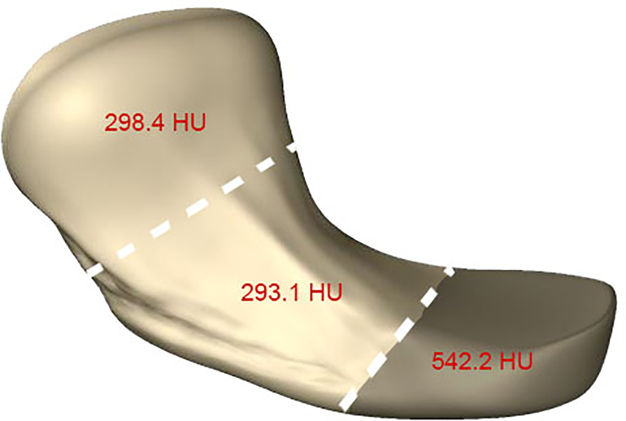
Introduction: The scaphoid bone is essential for wrist stability and movement. While fractures commonly occur at the scaphoid's waist, those at the proximal pole, though rarer, tend to result in nonunion, potentially linked to variations in bone mineral density (BMD). Previous studies have shown an increase in BMD at the proximal pole in fractured scaphoids, but BMD distribution in intact scaphoids has not been well studied. This study aims to map the BMD distribution of the intact scaphoid to better understand the etiology of fractures and optimize treatment approaches. Methods: Conducted under ethical approval, this study included 100 individuals, using computed tomography to assess the BMD in distal, middle, and proximal thirds of the scaphoid. Measurements were performed in the defined regions and analyzed using SPSS software, with significance accepted at P < .05. Results: The study comprised 34 females and 66 males, with no significant BMD difference between the right and left wrists. The proximal third exhibited significantly higher BMD (542.2 HU) compared to the middle (293.1 HU) and distal thirds (298.4 HU). A statistically significant higher BMD was observed in males, particularly in the proximal and distal thirds. A weak negative correlation between age and BMD was noted across all sections. Conclusion: The proximal scaphoid shows significantly higher BMD, potentially explaining its lower fracture incidence but higher nonunion rate. This insight into the BMD distribution within an intact scaphoid may guide the clinical management of scaphoid fractures, highlighting the need for targeted treatment strategies based on BMD variations.
GrewalRLutzKMacDermidJC, et al.Proximal pole scaphoid fractures: a computed tomographic assessment of outcomes. J Hand Surg Am. 2016;41(1):54-58.
2.
GholsonJJBaeDSZurakowskiD, et al.Scaphoid fractures in children and adolescents: contemporary injury patterns and factors influencing time to union. J Bone Joint Surg Am. 2011;93(13):1210-1219.
3.
MadeleyNJStephenABDowningND, et al.Changes in scaphoid bone density after acute fracture. J Hand Surg Br. 2006;31(4):368-370.
4.
SchreiberJJAndersonPARosasHG, et al.Hounsfield units for assessing bone mineral density and strength: a tool for osteoporosis management. J Bone Joint Surg Am. 2011;93(11):1057-1063.
5.
MiyamuraSLansJHeJJ, et al.Bone density measurements from CT scans may predict the healing capacity of scaphoid waist fractures. Bone Joint J. 2020;102-B(9):1200-1209.
6.
PattersonRMMoritomoHYamaguchiS, et al.Scaphoid anatomy and mechanics: update and review. Oper Tech Orthop. 2003;13(1):2-10.
7.
RichardO. Scaphoïde [Scaphoid bone]. Ann Radiol (Paris). 1992;35(5):367-372.
8.
DylevskýIMrzenaV. Os scaphoideum–funkcní a klinická anatomie [Os scaphoideum–functional and clinical anatomy]. Acta Chir Orthop Traumatol Cech. 2001;68(5):327-330.
GelbermanRHMenonJ. The vascularity of the scaphoid bone. J Hand Surg Am. 1980;5(5):508-513.
11.
CooneyWPDobynsJHLinscheidRL. Nonunion of the scaphoid: analysis of the results from bone grafting. J Hand Surg Am. 1980;5(4):343-354.
12.
SwanstromMMMorseKWLipmanJD, et al.Variable bone density of scaphoid: importance of subchondral screw placement. J Wrist Surg. 2018;7(1):66-70.
13.
CheungYYNaspinskySRGoodwinDW, et al.Increased radiodensity of the proximal pole of the scaphoid: a common finding in computed tomography imaging of the wrist. J Comput Assist Tomogr. 2006;30(5):850-857.
14.
DowningNDOniJADavisTR, et al.The relationship between proximal pole blood flow and the subjective assessment of increased density of the proximal pole in acute scaphoid fractures. J Hand Surg Am. 2002;27(3):402-408.
Supplementary Material
Please find the following supplemental material available below.
For Open Access articles published under a Creative Commons License, all supplemental material carries the same license as the article it is associated with.
For non-Open Access articles published, all supplemental material carries a non-exclusive license, and permission requests for re-use of supplemental material or any part of supplemental material shall be sent directly to the copyright owner as specified in the copyright notice associated with the article.