
Abstract
The aim of this in vitro study was to investigate some physical properties of Biodentine (BD) (Septodont, France) that has been modified by adding nanosized bioactive glass (nBG) particles to it in different ratios. The cement was modified by adding 1% (7 mg) and 2% (14 mg) nBG powder to BD. BD was used as the control group in its commercial form. A total of 240 cement samples (n = 80) were prepared according to the standard measurements for each test. Subsequently, tests to determine compressive strength, microhardness, initial setting time, and solubility of the samples were performed. The obtained data were statistically analyzed using one-way ANOVA and Tukey’s HDS tests, and the significance level was found to be 0.05. The compressive strength values of the samples modified with 1% and 2% nBG were higher than those of the unmodified BD; however, no statistically significant difference was found between them [BD + nBG (2 wt%) ⩾ BD+nBG (1 wt%) ⩾ control BD], (p > 0.05). The microhardness values of the samples modified with 1% and 2% nBG were found to be significantly higher than those of the control group [BD + nBG (2 wt%) > BD+nBG (1 wt%) > control BD], p < 0.05. Initial setting times were determined as 14 min for unmodified BD, 13 min for BD + nBG (1 wt%), and 12 min for BD + nBG (2 wt%). The addition of nBG to BD significantly reduced the initial setting time of BD (p < 0.05). A significant decrease was observed in the solubility of the BD modified with nBG samples compared to that of the control group [control BD > BD+nBG (1 wt%) >BD+nBG (2 wt%)], p < 0.05. Within the limitations of this study, it was found that the addition of certain amounts of nBG to BD positively affected some physical properties of the cement. Future in vitro and in vivo studies should be performed to prove the clinical applicability of the cements used in this study.
Introduction
Biodentine (BD; Septodont, Saint-Maur-des-Fossés, France) is a calcium silicate–based material produced as a dentin substitute and has been available in the market since 2009. The powder part of BD contains tricalcium silicate, dicalcium silicate, and calcium carbonate. The liquid part of BD is mainly composed of calcium chloride as an accelerator and a plasticizing agent. BD is capable of releasing calcium ions and is described as a source of hydroxyapatite when it comes in contact with tissue fluids. 1 Moreover, BD can be used under a composite resin material for posterior restorations due to its dentin-like mechanical properties. 2 Therefore, BD has a wide range of applications, for example, as a pulp-capping material in vital pulp treatments; as a preserving material of scaffolds during regenerative endodontic procedures; as an apical barrier material in teeth with necrotic pulps and root with an open apex; as a furcal or root perforation repair material, and as a root canal filling and retrograde filling in endodontic surgery.3,4
Bioactive glasses are silicate-based biomaterials that have the capacity to produce strong chemical adhesion to bone tissues. Moreover, due to their high biocompatibility, they can form a hydroxyapatite layer when they come into contact with body fluids. 5 The popularity of bioactive glasses is increasing in dentistry because of some important properties, such as antibacterial activities that provide a microorganism-free environment during the healing and regeneration of the defective area, establish a silica connection, and combine with different ions, such as strontium and phosphate. 6
Gong et al. reported that nanosized bioactive glass (nBG) can induce differentiation and mineralization of human dental pulp cells more effectively than a regular bioactive glass. Therefore, nBG might be a promising alternative for the regeneration of pulp–dentin complex. 7 Besides, bioactive glasses exhibited favorable cellular and inflammatory pulp responses with mineral trioxide aggregate (MTA) after direct pulp-capping procedures in vivo. 8
In recent studies, it has been reported that the formation of hydroxyapatite and the fluoride release of the material can be enhanced by adding bioactive glasses to BD.9,10 However, the mechanical properties of the materials should not be negatively affected while attempts are made to improve their bioactivity. To the best of our knowledge, there is no research in the literature on the kinds of changes that occur in the physical properties of BD after the addition of nBG to its content. Therefore, the aim of this in vitro study is to evaluate physical properties such as compressive strength, surface microhardness, initial setting time, and water solubility of the modified material obtained by adding different weights of nBG particles to the BD content and comparing such properties with those of the unmodified BD.
Methods
NBG preparation
nBG (nBG-55; particle size between 48 and 196 nm, including 58% SiO2, 40% CaO, and 5% P2O5 [w/w]) were synthesized by the sol–gel method, as Hong et al. described in more detail. 11 First, 7.7 g of Ca(NO3)2⋅3H2O was dissolved in 117 mL of deionized water at room temperature to produce the first solution, and the second solution was produced by adding 9.7 mL of tetraethyl orthosilicate to 63.5 mL of ethanol. The two prepared solutions were mixed, and their pH was adjusted to 1–2 using citric acid. After vigorous stirring, the solution was slowly poured into a solution of 1.2 g of NH4H2PO4 in 1500 mL of deionized water. The pH of the solution was kept constant at around 11 using aqueous ammonia during the dripping process.
After stirring the mixture for 48 h and aging for 24 h, centrifugation at 1000 rpm separated the precipitate from the reaction solution. The precipitation was washed three times with deionized water, stirred in 200 mL of a 2% w/v aqueous solution of polyethylene glycol (PEG, Mn = 10,000), and kept for 24 h. The final suspension was freeze-dried and then calcined in a muffle furnace at 700°C for 3 h to obtain a white nBG-55 powder.
Preparation of experimental cements
The prepared nBG powder was added to BD capsules (7 mg and 14 mg of nBG to 1% and 2% BD by weight, respectively). Without adding nBG, BD was served as the control group. The modified BD with nBG powder in the capsule was mechanically mixed for 30 s in the amalgamator before adding the BD liquid. Subsequently, five drops of BD liquid were added to the powder capsule and mixed for 30 s at a speed of 4000–4200 rpm. Polyethylene molds of different sizes were used to prepare standardized disk-shaped specimens for the different physicomechanical tests conducted in this study. 12
Compressive strength
Sixty disk-shaped cement samples (n = 20 per group; 4 mm in diameter × 6 mm in height) were prepared according to the recommendations of the International Standard Organization (ISO) 9917-1 for the compressive strength test. The mixture with paste consistency was placed in polyethylene molds and compacted until it was filled. A glass slide was placed on the mold, and the excess cement was gently removed with a wet cotton pellet. Each specimen inside the mold was kept for 7 days at 37°C and 100% humidity in an incubator. Afterward, the specimen was carefully extracted from the mold and checked for any possible damage. The surfaces of the specimens were polished using 400-grit sandpaper. The specimens were washed with 5 mL of distilled water and then gently air dried. Subsequently, they were vertically placed on a universal testing machine (Instron 3345, Norwood, MA, USA) and crushed along their long axis at 1 mm/min. The minimum force required to fracture the samples was recorded in newtons. The compressive strength of the specimens was measured in megapascals (MPa) using the following formula:

where CS is the compressive strength in MPa, P is the loading failure in N, and d is the diameter of the specimens in mm.
Surface microhardness
As previously described in the compressive strength test, 60 disk-shaped cement samples (n = 20 per group; 6 mm in diameter × 4 mm in height) were prepared. The surface microhardness of the samples was measured using a Vickers microhardness tester (HMV, Shimadzu, Kyoto, Japan) and a square-based pyramid-shaped diamond indenter with a face angle of 136°. A full load of 50 g for 5 s was applied to the surface of the specimens at room temperature. The diagonal diameter of the resulting indentations was measured under an optical microscope equipped with a digital camera and image analysis software. After making five indentations on the surface of each specimen, the mean Vickers hardness value for each sample was calculated.
Initial setting time
The setting time was measured using the procedure according to the recommendations of ISO (9917-1:2007). The cements (n = 10 per group) were prepared as previously described, poured into polyethylene molds (10 mm in diameter × 5 mm in height), and pressed between two glass slides. The assembly was placed in a chamber at 37°C and 95% relative humidity. The Vicat apparatus was used at 30-s intervals to determine the initial setting time of the cements. A stainless-steel Vicat needle with a tip diameter of 1.0 ± 01 mm and loaded with a 400 ± 5 g weight was applied vertically to the horizontal surface of the specimen. The initial setting time was the time period between mixing the cement and that at which the needle failed to leave visible indentation on the specimen surface.
Solubility
Solubility was determined by the procedure described in ISO 3107:2011. Ten disk-shaped specimens from each group were produced using a polyethylene mold (10 mm in diameter × 2 mm in height). The mixed cements were packed into the molds on a glass plate. After incubation in an oven at 37°C temperature and 100% humidity for 7 days, the specimens were gently extracted from the molds. The analytical balance was used to record the initial weight (w1) of each specimen in grams. Then, the specimens were immersed in 50 mL deionized distilled water for 24 h at 37°C.
Next, the specimens were removed, slightly dried with absorbent paper, and kept in a desiccator that contained silica gel for 24 h. The specimens were desiccated until a constant weight was obtained (w2). Water solubility was evaluated using the following formula:

Surface topography and elemental composition/SEM–EDX analysis
Two specimens each in the BD, BD – nBG (1 wt%), and BD – nBG (2 wt%) groups were prepared in a polyethylene mold (of 10 mm diameter × 2 mm height) for surface topography evaluation using a scanning electron microscope (SEM). In addition, the nBG powder used in this study was evaluated for surface topography.
The specimens were sputter coated with gold palladium in a vacuum evaporator (VG Microtech Inc., Japan). Surfaces of the specimens were examined via SEM (Leo 440, Electron Microscopy Ltd., Cambridge, UK) at a voltage of 20 kV at different magnifications (500–1000X), after which their elemental compositions were analyzed using energy-dispersive X-ray (EDX) spectroscopy.
Statistical analysis
Data about compressive strength, surface microhardness, initial setting time, and solubility of the samples were subjected to statistical analysis (SPSS 22.0; IBM Software, Armonk, NY, USA). The Shapiro–Wilk test was used to test normality, and data were found to be normally distributed. Thus, parametric statistical tests were performed using a one-way ANOVA. Multiple comparisons were made using Tukey’s HDS test. The statistical significance level was set at p < 0.05.
Results
Data about compressive strength, surface microhardness, initial setting time, and solubility of the samples are presented in Table 1.
Mean – standard deviations of compressive strength (MPa), surface microhardness (VHN), initial setting time (min), and solubility of each group.

BD: Biodentine; nBG: nano-bioactive glass; wt: weight; MPa: MegaPascal; VHN: Vickers microhardness; min: minute.
Mean values represented with different superscript letters are significantly different (p < 0.05).
The mean compressive strengths of BD – nBG (2 wt%) and BD – nBG (1 wt%) were found to be higher than that of the control group (p < 0.05). However, no statistically significant difference was observed between the groups in terms of compressive strength levels (p > 0.05).
BD – nBG (2 wt%) had the highest microhardness values, followed by BD – nBG (1 wt%) and BD (the control group; p < 0.05).
nBG significantly decreased the initial setting time of the unmodified BD (control group; p < 0.05). BD – nBG (2 wt%) exhibited the shortest initial setting time (p < 0.05).
The addition of nBG decreased the solubility of BD (p < 0.05). The following statistical ranking for solubility as percentage values was obtained: BD (control group) >BD – nBG (1 wt%), >BD – nBG (2 wt%) (p < 0.05).
Surface topography and elemental composition/SEM–EDX analysis
Representative SEM images of the surface and EDX analyses of nBG, BD, and modified BD with nBG groups are presented in Figure 1. An image of nBG specimen revealed scattered and clustered crystalline glasses (Figure 1(a)). Large irregular and hexagonal crystals were observed in BD specimens (control group; Figure 1(c)). In the modified BD specimens to which nBG was added, the clustered cauliflower-shaped, heterogeneously dispersed bioactive glasses were observed between the BD particles (Figure 1(e) and (g)).
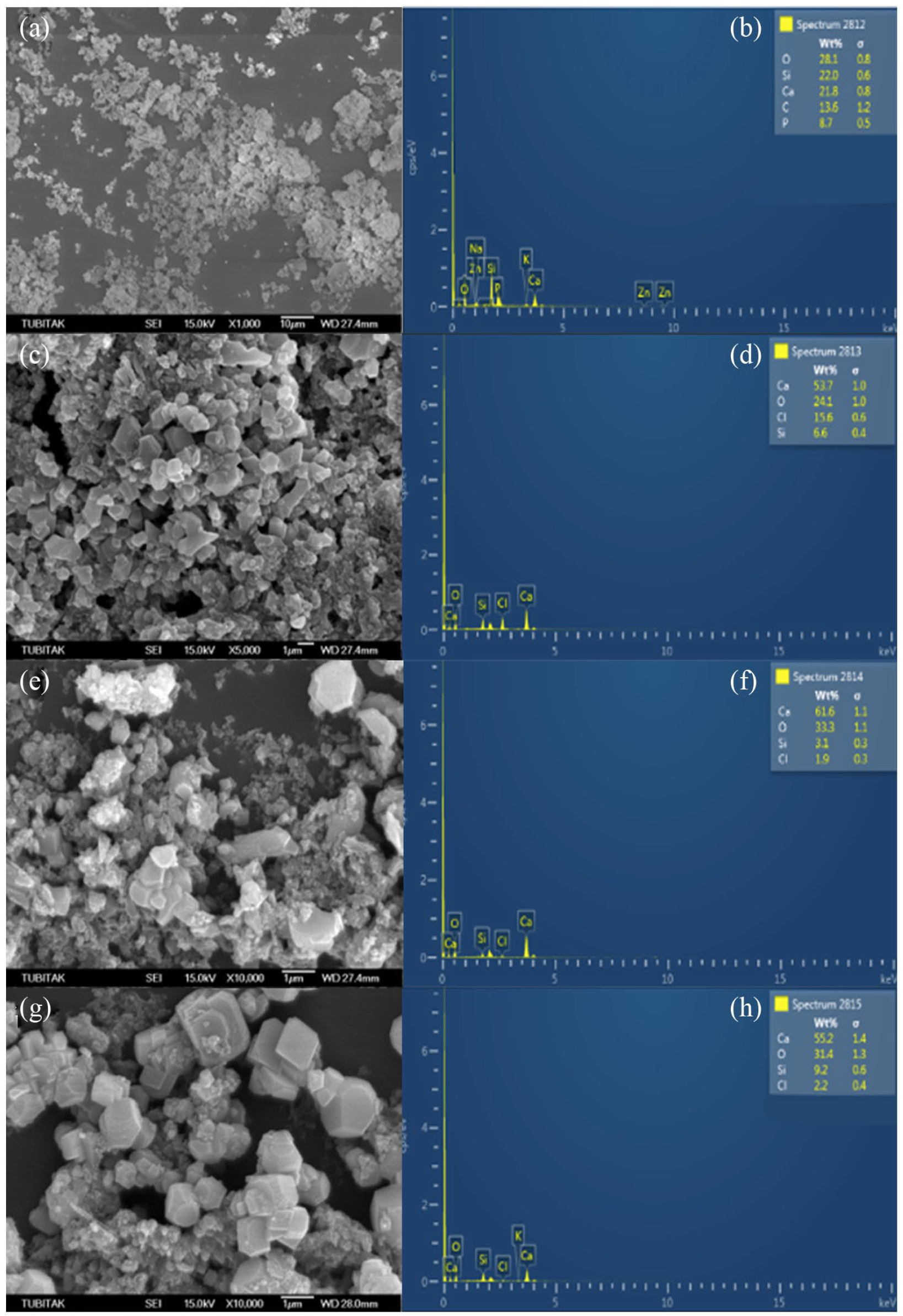
Representative SEM images of nanosized bioactive glass (a, x1000); bd (c, x5000); bd incorporating nBG (1wt%) (e, x10.000) and bd incorporating nBG (2 wt%) (g, x10.000) with EDX elemental analysis (1b, 1d, 1f and 1h).
The EDX analysis revealed that mineral deposits on nBG were mainly comprised of calcium, silicon, oxygen, chlorine, and phosphorus elements (Figure 1(b)). The elemental composition of BD was observed to be mainly calcium, silicon, oxygen, and chlorine (Figure 1(d)), with increasing calcium content on the nBG-modified cements (Figure 1(f) and (h)).
Discussion
Bioactive glasses are thought to be potential mineralizing agents in periradicular tissues due to their release of silicate, calcium, and phosphate ions into tissue fluid, which leads to apatite formation. Thus, they were proposed to be used as mineralizing agents in caries prophylaxis and as desensitizing agents by blocking open tubules in dental sensitivity therapies. 13 Similarly, the SEM–EDX results of this study showed the presence of oxygen, calcium, silica, and phosphorous ions in the nBG content.
Corral-Nunez et al. added nBG to BD and glass ionomer cement materials and observed changes in their bioactivity. 9 nBG was reported to have increased the bioactivity of BD and the formation rate of crystalline apatite on the material surface after contact with tissue fluids. In that study, the nBG (size 40–70 nm) produced using the sol–gel method was added to BD at 1%–2% by weight. Following the same synthesis protocol, nBG of about 28–55 nm diameter was produced and added to the BD material at 1%–2% by weight in this study. To the best of our knowledge, this is the first study to evaluate the physicomechanical properties of modified cement with 1%–2% by weight of nBG compared with the control group of unmodified BD. However, the variability of nBG synthesis procedures, molar composition, and the particle size range of nBG in the literature make it difficult to compare our results with previous reports that studied the incorporation of nBG into various restorative dental materials.
Odontogenic differentiation and dentin formation were thought to have been induced by nBG particles. Thus, it was suggested that bioactive glass can be used as a pulp-capping material for dentin regeneration. 14 In an in vivo study, it was reported that a patty made of a mixture of bioactive glass and water increased the integrity of dental structure due to increased mineralization and other chemical changes observed in the dental tubules. 15 Therefore, the authors of that study emphasized that bioactive glass can be a promising filling material. In another study, the same researchers showed that bioactive glass can form an apatite layer at body temperature and in an environment of artificial saliva. 16
High compressive strength cement is a sought-out property, especially for that used in vital pulpal therapy and coronal barrier procedures.17,18 The results of this study showed that the addition of nBG to BD increased the compressive strength of the modified samples. Although this increase was not statistically significant, it showed that the addition of nBG, which is known for its fragile structure, did not have a negative effect on the assessed physical properties of the modified cement. This might have been due to the increase in the powder-to-liquid ratio of the modified cement when the nBG powder was added to it, because the compressive strength was highly affected by the liquid content of the cement. It was reported that compressive strength increased in Portland cement as the powder-to-liquid ratio increased. 19 However, the long-term properties of the modified cements should be determined with further studies.
Flores-Ledesma et al. added bioactive glass to an MTA-like cement and reported decrease in the compressive strength of the latter; however, this decrease was not less than 5 MPa – the expected cement resistance threshold equal to the compressive resistance of zinc oxide eugenol type-IV cement. 20 This variation in the results might have been related to factors such as the powder-to-liquid ratio, conditions of the environment (dry or moist), and durations of incubation in these environments before testing the materials.
However, Ana et al. mixed bioactive glass with resin-modified glass ionomer cement in different ratios and found a significant increase in compressive strength, which may be related to strong covalent bonds formed between the resin matrix and glass. 21 De Caluwe et al. reported that the addition of bioactive glass to glass ionomer resin increased its bioactivity and that 10% bioactive glass content did not have an effect on compressive strength values, while higher contents caused a rapid decrease in compressive strength. 22
The compressive strength of BD was reported to have been affected by conditions such as contact with irrigation solutions or whitening agents, blood contamination, and low pH environment.18,23–25 Therefore, further studies are needed to evaluate the effects of these conditions on the compressive strength values of the modified materials used in this study.
Increased porosity in a cement structure has been reported to decrease microhardness. 26 The results of this study showed that the addition of nBG significantly increased the microhardness of BD, and the highest microhardness values were obtained in samples with 2% nBG by weight.
Saghiri et al. evaluated the effects of the addition of bismuth oxide with different particle sizes on the physical properties of white MTA. 26 The results showed that while bismuth oxide with normal particle sizes had adverse effects on compressive strength and microhardness of MTA, the addition of nanoparticle-sized bismuth oxide increased the physical properties of MTA. The authors argued that this might have been related to the closure of open pores in MTA by nanosized bismuth oxide powder, which strengthened the structure of the cement. Similarly, in this study, nBG particles might have filled the small pores between hydrated calcium silicates and decreased the porosity of the cement, which caused an increase in the microhardness values.
The setting time of the materials is an important issue in clinical applications. Cements with long setting times have higher solubility due to the “wash-out effect” while the cements are in contact with tissue fluids. In contrast, the manipulation and clinical usage become difficult in cement types with short setting times. An ideal cement should be set within a period that is appropriate for clinical application. 12
According to the results of this study, the addition of nBG to BD significantly decreased the initial setting time to 12 min in the modified BD compared to 14 min in the original material. This might be associated with the hydrophilic structure of bioactive glass. Smaller sizes of bioactive glass particles compared to calcium silicate particles and higher surface sizes occurred and might also have shortened the setting reaction. Therefore, calcium silicates might have turned into calcium silicate hydrates in a shorter time due to faster reaction.
Increased solubility of cement can decrease its sealing ability, which would increase bacterial filtration. 27 Solubility is not a desired feature of cement materials. Increased ion release rates and the consequent occurrence of mineralization are reported in materials with bioactive glass addition. 7 Ion release means dissolution of the cement material. 20 Contrary to the expectations of the researchers of this study, solubility was observed to have been significantly decreased in the groups with bioactive glass added compared to the BD group. Because the amount of BD powder was kept constant in all the groups, the powder ratio increased in the groups with bioactive glass additions. Solubility is related to the powder-to-liquid ratio of the cement. A higher powder ratio could have caused the reduced solubility in the experimental groups in which bioactive glass was added. However, the effect of the longer maturation of the modified cements in a humid environment should be further studied for solubility properties.
Flores-Ledesma et al. discovered a relationship between setting time and the solubility of different types of cement. 20 In this study, a similar relationship was found. Groups with a decreased setting time were also observed to have reduced solubility as well; however, a limitation of this study is that solubility was evaluated as changes in weight of the specimens. Because powder ratios were increased in the groups in which nBG was added, the lower solubility observed in these groups might have been due to the increased weights, as different types of cement with increased powder ratios have higher water absorption rates. 27 This might have led to a false perception that modified cements had lower solubility in distilled water. For example, Gandolfi et al. reported that the solubility of bioactive glass that contained GuttaFlow bioseal root canal filling patty was low; however, the material had a high rate of water absorption. 28 The authors argued that the material was expanded, and then ion releasing occurred; however, the solubility was masked by water absorption. Therefore, the ion release rates of the modified cement samples in this study should be determined, and possible volume expansions or contractions should be measured using micro-CT analysis in further studies.
Because there are limited data in the literature, future studies should be performed to understand the optimal particle size of nBG and mixing ratio with BD for the highest bioactivity and mechanical properties. Moreover, due to the limitations of in vitro studies, clinical studies should be performed to obtain more relevant clinical data about the incorporation of nBG into BD.
Conclusions
Within all the limitations of this in vitro study, it can be concluded that the addition of nBG did not change the compressive strength of BD. However, nBG increased the microhardness, shortened the setting time, and decreased the solubility of BD.
Footnotes
Acknowledgements
The authors would like to thank Mrs. Ebru Osmanoglu Akyol from Variance Statistical Consultancy, who performed the statistical analysis. The authors would also like to thank Dr. Belma Zengin Kurt and Dr. Aydan Dag for producing nanosized bioactive glass at Bezmiâlem Vakif University, Faculty of Pharmacy, Department of Pharmaceutical Chemistry.
Contributorship
Planning: MBG, AUE
Laboratory: MBG, TYO, ANDS, BAU
Writing of the publication: MBG, AUE
Statistical analyses, preparation of tables and figures: MBG, AUE
Declaration of conflicting interests
The author(s) declared no potential conflicts of interest with respect to the research, authorship, and/or publication of this article.
Funding
The author(s) disclosed receipt of the following financial support for the research, authorship, and/or publication of this article: This research was supported by the Scientific and Technological Research Council of Turkey (TUBİTAK) with grant number 218S868.
Ethical statement
This study had no human participants.
Guarantor
Mehmet Burak Guneser