
Abstract
The rapid and tailored biofabrication of natural materials is of high interest for the field of tissue engineering and regenerative medicine. Scaffolds require both high biocompatibility and tissue-dependent mechanical strength to function as basis for tissue-engineered implants. Thus, natural hydrogels such as fibrin are promising but their rapid biofabrication remains challenging. Printing of low viscosity and slow polymerizing solutions with good spatial resolution can be achieved by freeform reversible embedding of suspended hydrogels (FRESH) bioprinting of cell-laden natural hydrogels. In this study, fibrin and hyaluronic acid were used as single components as well as blended ink mixtures for the FRESH bioprinting. Rheometry revealed that single materials were less viscous than the blended bioink showing higher values for viscosity over a shear rate of 10–1000 s−1. While fibrin showed viscosities between 0.1624 and 0.0017 Pa·s, the blended ink containing fibrin and hyaluronic acid were found to be in a range of 0.1–1 Pa·s. In 3D vascularization assays, formation of vascular structures within the printed constructs was investigated indicating that the printing process did not harm cells and allowed formation of vasculature comparable to moulded control samples. Best values for vascularization were achieved in bioinks consisting of 1.0% fibrin-0.5% hyaluronic acid. The vascular structure area and length were three times higher compared to other tested bioinks, and structure volume as well as number of branches revealed almost four times higher values. In this study, we combined the benefits of the FRESH printing technique with in vitro vascularization, showing that it is possible to achieve a mechanically stable small-scale hydrogel construct incorporating vascular network formation.
Introduction
Biofabrication of hydrogel scaffolds is key in tissue engineering allowing the manufacturing of custom-designed implants. 1 One of the most popular fabrication techniques is 3D-bioprinting which can be used to produce constructs based on patients’ needs and requirements such as anatomical features.1,2 Patient derived computer tomography data can be used to create a tailored implant structure, which can be fabricated based on these data. 3 There are various bioprinting techniques to generate tissue and organ constructs such as pressure-driven additive manufacturing, inkjet or droplet-on-demand as well as thermal and laser-induced techniques, to name the most common methods. 4 One of the challenges in creating 3D-bioprinted cell-laden constructs is the choice of scaffold materials compromising shape fidelity as well as suitability for cell embedding, proliferation, and tissue maturation. 5 The increase of material stiffness guarantees shape retention and is known to have an impact on pore size, diffusion properties and moreover, cell viability, and spreading.5,6 These parameters influence the nutrition of cells in tissue-engineered constructs and as well the formation of a functional vascular network, which is necessary especially for fabrication of larger implants.7,8 Different strategies are described for vascularization in vitro. It can be achieved by co-culture of endothelial cells with supporting cells acting as pericytes. The cells can form a delicate capillary vessel network in suitable scaffolds based on sprouting of endothelial cells supported by alignment of supportive cells such as fibroblasts.9,10 This process is initiated by a suitable culture medium and growth factors secreted by both cell types. Vascular endothelial growth factor (VEGF) is one of the key cytokines related to vascularization. 11 Thereby, the cell survival rate and integration of the constructs after transplantation can be improved. A homogenous distribution of cells and thus, capillary structures is essential to provide a sufficient nutrient supply over the whole scaffold. In previous studies we have shown, that low viscous fibrin control groups revealed significantly better vascularization characteristics compared to printable agarose-collagen samples due to a lower cross-polymerization, increased pore size, and the presence of adhesion motifs.5,10 Fibrinogen combines these properties, making it attractive for cell migration, proliferation, and vascularization. Thus, a method for rapid fabrication of such low viscosity fibrin solutions is required to create 3D-printed tissue-engineered constructs. In a study by Hinton et al., 12 an innovative freeform reversible embedding of suspended hydrogels (FRESH) technique was described, allowing the printing of cell-laden biological hydrogels such as collagen, fibrinogen, or alginate with an elastic modulus of less than 500 kPa. This inkjet printing method utilizes a temporary, thermoreversible supportive gelatine bath, which allows the gradual build-up of the hydrogel, nozzle guidance, and guarantees post-printing stability. The supportive bath consists of small particles such that it has a slush-like consistency; in which the printer nozzle can be moved freely and the bioink can be ejected. Thereby, highly complex 3D structures can be printed with diverse low viscosity bioinks. Besides construct fidelity, this technique is known to reveal a cell viability of 99.7%. 12
In this study, we focussed on 3D biofabrication of a vascularized fibrin-hyaluronic acid (FIB-HA) construct in vitro. Vascularization was achieved by the co-cultivation of human endothelial cells derived from umbilical cord veins and human dermal fibroblasts. The alignment of fibroblasts at sprouting endothelial cells leads to mechanical support of delicate vessels and is initiated by growth factor gradients secreted by both cell types. Vascular endothelial growth factor (VEGF) is one of the key cytokines related to vascularization. 11
The novelty lies in the combination of (i) bioink formulation, (ii) FRESH printing method, and (iii) vascularization. To our knowledge, there are no studies showing successful vascularization in 3D-printed HA-fibrin formulations using the FRESH printing method. We hypothesize, that FRESH printing allows us to avoid previously named compromises regarding bioink composition that have been necessary so far and to enable both printability and vascularization.
Materials and methods
Cell isolation and culture
Cell isolation from all tissues was approved by the ethics committee of the Faculty of Medicine of RWTH Aachen University (EK 218/14).
Human umbilical venous endothelial cells (HUVECs) were isolated from human umbilical cord veins provided by the Clinic for Gynaecology and Obstetrics (RWTH Aachen University Hospital). HUVECs were isolated according to an established and described protocol. 13 In brief, umbilical cord veins were flushed with phosphate-buffered saline (PBS, Gibco) and incubated with type II collagenase (1 mg/mL in Hank’s Balanced Salt Solution, HBSS (with Mg + Ca), Gibco) for 30 min at 37°C and 5% CO2. Detached endothelial cells were centrifuged at 500g for 5 min followed by seeding cells on pre-coated cell culture flasks (with 2% gelatine, type B, Sigma-Aldrich). Experiments were conducted with cells up to passage 4.
Human dermal fibroblasts (HDFs) were isolated from juvenile foreskins provided by the Urology Clinic (RWTH Aachen University Hospital) following a protocol described before.14,15 In short, the dermal tissue was cleaned shortly in 70% ethanol (VWR) followed by three washing steps in PBS (with 1% antibiotics/antimycotics (ABM), Gibco) for 5 min each step. After removing fatty tissue and blood residues, the dermal tissue was incubated in 25 U/mL dispase (Gibco) over night at 4°C. Afterwards, the dermal tissue was incubated for another 60 min at 37°C and 5% CO2. Then, the epidermal layer and the dermis were separated and the latter was minced and digested in 100 U/mL collagenase type II (in HBSS (with Mg + Ca), Gibco) for 3 h at 37°C and 5% CO2. After filtration and a centrifugation step at 500g for 5 min, isolated cells were cultured in Dulbecco’s Modified Eagle Medium (DMEM, +10% foetal calf serum (FCS), both Gibco). Experiments were conducted with cells up to passage 6.
FRESH 3D printing process
Preparation of gelatine support bath
The gelatine support bath was prepared according to the kit’s manual (FRESH kit, Allevi) adapted from Hinton et al. 12 In short, 10 g gelatine (Type A from porcine skin, Allevi, 300 bloom) were dissolved in 250 mL distilled water at a maximum temperature of 40°C–45°C, 0.4 g CaCl2 were added to hold final concentrations of 4.5% (gelatine) and 11 mM (CaCl2). CaCl2 is needed to promote the polymerization of fibrinogen during printing process. The gelatine solution was cooled at 4°C for 24 h in a mason jar (Ball Inc.) to solidify it. The jar was then filled to the top with cooled CaCl2 solution (4°C), the solution was stored for 1 h at −20°C followed by blending thoroughly at highest speed (Osterizer MFG, speed III) for 1 min. After blending, the slurry was transferred to 50-mL centrifugation tubes, mixed with cold CaCl2 in a ratio 4:1 and centrifuged five times at 3800 g and 4°C for 4 min each. After each centrifugation step, the supernatant (white foam on top of supernatant) was removed from the gelatine slurry fraction and replaced by new CaCl2 solution. After five centrifugation cycles, no foam or bubble occurred anymore. This slurry was stored at 4°C until needed. Directly before use, the slurry was finally centrifuged at 230 g for 5 min, the supernatant was discarded and the slurry poured into 24 well plates (500 µL/well, VWR). For printing fibrinogen-containing inks, the gelatine matrix was enriched with 375 µL thrombin for a final thrombin concentration of 5 U/mL.
Preparation of bioinks and 3D printing system
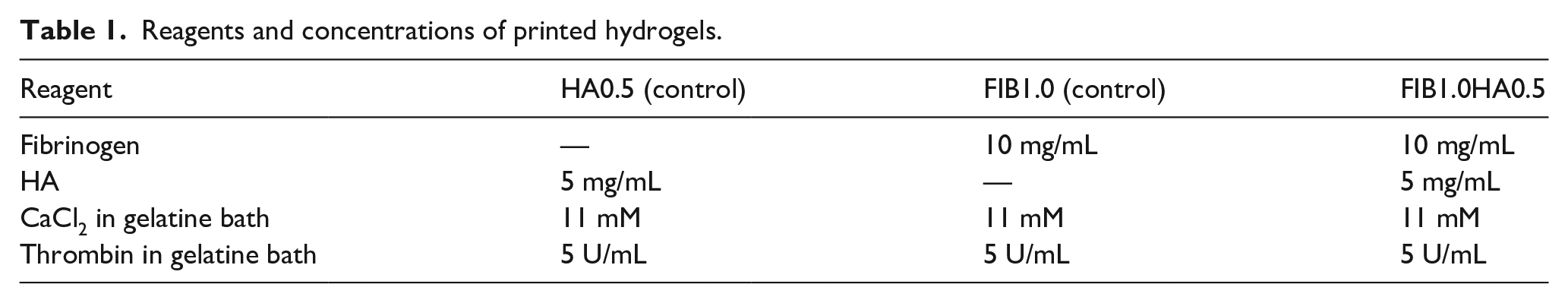
The preparation of fibrinogen-hyaluronic acid ink (FIB1.0HA0.5) was adapted from Hinton et al. 12 In short, the ink was mixed fibrinogen (from human plasma, Merck Millipore) and hyaluronic acid (high molecular weight, ~1.5–1.8 × 10⁶ Da, from Streptococcus equi, Sigma). For this, the fibrinogen stock solution (for preparation see Tschoeke et al. 16 was diluted with TRIS-buffered saline (TBS) to the desired concentration (5 or 10 mg/mL for FIB0.5 or FIB1.0, respectively)) and the HA powder (2.5 or 5 mg/mL for HA0.25 or HA0.5, respectively) was added directly to the fibrinogen solution. To fully dissolve the HA, the fibrinogen-HA solution was incubated for 48 h at 37°C prior use. For better visualization of non-cell-laden constructs, the inks were dyed with 1% black china ink. Further, a fibrinogen-only ink was prepared using 10 mg/mL FIB1.0 as control sample. The HA was dissolved in TBS buffer to reach a final concentration of 5 mg/mL in control samples (see Table 1).
Reagents and concentrations of printed hydrogels.
Preparation of cell-laden inks
Inks were prepared as described above (FIB1.0HA0.5). Cells were detached from cell culture flasks and suspended at a concentration of 3 × 10⁶ cells/mL ink for each cell type. Inks and cells were thoroughly mixed by pipetting and transferred to 10 mL Luer-lock syringes (BD). Cell-laden inks contained both cell types with an equal number of cells each type.
3D bioprinting system
Printed constructs were fabricated with the Biobots 2 3D printer (Allevi) (see Figure 1(a)). The used printer is a pressure driven extrusion bioprinter, equipped with a pneumatic air compressor (California Air Tools), which allows pressure ranges between 0 and 68.9 bar. Further, the printer contains two heated extruder heads providing temperatures between room temperature and 160°C. The Computer-Aided Design (CAD) models of printed constructs were designed with SolidWorks 2016 (Dassault Systèmes). The CAD models were retrieved from GrabCAD. Further processing was performed with Repetier-Host software (Hot-World GmbH & Co).

Preparation steps of the gelatin bath (a), Allevi 3D-bioprinter setup (b, c), CAD model (d), stereolithography file (e), and g-code file (f) for printing.
Here, the digital 3D models were converted to G-code files (see Figure 1(c)–(e)). The Repetier software allowed adjustment of following printing parameters: nozzle size, filament diameter, velocity of the printer, infill density, layer height, and fill pattern. Experiments were conducted with nozzles with 12.7 mm length, 30-gauge, and 150 µm inner diameter (EFD Nordson). The syringe loaded with the ink was placed in the printer’s syringe holder and the nozzle height was adjusted to be immersed in the gelatine slurry. Fibrinogen-HA constructs were printed in a cylindrical shape with a height of 2 mm and a diameter of 8 mm with a printer velocity of 8 mm/s, a fill density of 1.5 mL, a layer height of 0.2 mm, and a grid fill pattern. All printed constructs were allowed to polymerize for 20 min after printing at room temperature.
Optimal printing parameters were chosen based on preliminary trials for best shape fidelity (data not shown).
To remove the surrounding gelatine slurry from the printed constructs, gels were incubated at 37°C for 20 min to melt the gelatine (fibrinogen inks). Finally, constructs were rinsed with PBS and cultured in EGM-2 medium with 0.16% tranexamic acid (Carinopharm) at 37°C and 5% CO2.
Rheological validation
Viscosity of inks was evaluated using a Kinexus Ultra + rheometer (Malvern) fitted with a cone and plate geometry. The following inks were pipetted on the rheometer plate and measured: distilled water (control), FIB1.0, HA0.5 (5 mg/mL), and FIB1.0HA0.5. For each combination and measurement three samples were measured (n = 3). The FIB1.0HA0.5 ink was tested without cells as well as cell-laden, testing three biological replicates. For cell-laden samples, HUVECS and HDFs were embedded in the bioinks with a concentration of 3 × 10⁶ cells/mL for each cell type. The shear rate was increased from 10 to 1000 s−1over a ramp time of 20 min. Measurements were performed at 25°C and inks were allowed to stabilize for 5 min before rheological characterization.
Cell experiments
Moulded hydrogels for 3D vascularization assay
Comparing the effect of 3D-printing on vascularization, control fibrin gels (FIB0.5) were moulded following a protocol described previously.5,10 In brief, cells were trypsinized (Trypsin EDTA, Pan Biotech) and embedded into hydrogels with a concentration of 3 × 10⁶ cells/mL hydrogel for each cell type. Therefore, cell pellets were suspended in warm medium, counted, diluted adequately with TBS solution to reach the aimed concentration, and pipetted into the wellplate moulds. Then, the single hydrogel reagents were added separately. Fibrinogen is the last component which was added, to avoid early polymerization. Hydrogels were moulded with following reagents and concentrations (see Table 2).
Reagents and concentrations of moulded hydrogels.

Hydrogels were polymerized at room temperature for 20 min, followed by incubation at 37°C for 20 min. Gels were cultured in EGM-2 medium supplemented with 0.16% tranexamic acid at 37°C and 5% CO2.
FRESH printed hydrogels for 3D vascularization assays
Cell-laden hydrogels were prepared as described above (see Table 3) and HUVECs and HDFs were added with following concentrations: 3 × 10⁶, 4 × 10⁶, or 5 × 10⁶ cells/mL each cell type. Cylinders with a height of 2 mm and a diameter of 8 mm were printed with a nozzle length of 12.7 µm at a pressure of 0.38–0.41 bar and a printing velocity of 8 mm/s in a gelatine slurry. Printing parameters were determined based on preliminary printing parameters optimizations (data shown in Supplemental Tables 1 and 2). After removing the gelatine support material, constructs were cultured for 14 days at 37°C and 5% CO2 in EGM-2 medium.
Composition of printed hydrogel ink FIB1.0HA0.5 and tested cell concentrations.

Evaluation of vascularization assay
Immunohistochemistry
Hydrogels were fixed in ice-cold methanol for 30 min at room temperature. After removing the methanol, gels were washed in PBS and stained afterwards with mouse anti-human CD31 primary antibody (1:100, Sigma) for 48 h at 37°C and 5% CO2. Then, the antibody solution was removed and gels were washed several times with PBS, followed by secondary antibody application (goat anti-mouse Alexa Fluor 594, 1:400, life technologies) for 48 h at 37°C and 5% CO2. Nuclei were stained with 4′,6-diamidin-2-phenylindol (DAPI, 1.5 µg/mL, Roth) followed by three washing steps with PBS.
Vascularization was visualized by two-photon laser scanning microscopy (TPLSM; Olympus Fluoview 1000 MPE) equipped with a 25x water immersion lens (NA 1.05, Olympus Optical) and a MaiTai Deep-See Titan-Saphir laser (Spectra Physics). Images were taken at random spots with a stack depth of 140 µm using the software Fluoview FV 10 4.2. Image processing and determination of vascularization parameters vessel volume, length, surface area, and branches were performed using Imaris software (Bitplane Inc.). The vessel network was therefore covered with a virtual isosurface excluding cell debris and calculating automatically all parameters. 10
Statistical analysis
Statistical analysis was performed using GraphPad Prism 7 (GraphPad Software), determining p-values by one-way ANOVA tests followed by Dunnett’s post-hoc test. A value of p < 0.05 was considered statistically significant.
Results
Rheological evaluation
Bioinks were rheologically characterized by measuring viscosities with shear rate ramps between 10 and 1000 s−1 (see Figure 2). Distilled water was chosen as control, since shear rates do not affect the viscosity of water (Newtonian fluid). The mean value for water obtained in the linear section was found to be 0.0009 Pa·s. Measured inks showed a shear-thinning behavior, especially for FIB1.0 samples. FIB1.0 showed viscosities varying between 0.1624 Pa·s at low and 0.0017 Pa·s at high shear rates. Interestingly, viscosity of FIB1.0HA0.5 ink was considerably higher than for the single components FIB1.0 and HA0.5. Compared to cell-free ink, cell-laden FIB1.0HA0.5 showed differences in the viscoelastic properties. With cells, a lower viscosity was observed and the shear-thinning effect was decreased.

Dynamic viscosity of the FIB-HA bioinks and the single materials. Dynamic viscosity of distilled water was used as control sample. The diagram shows the mean value ± standard error of mean for each measured shear rate point (n = 3).
Cell experiments
Vascularization assay
Moulded hydrogels
The effect of material properties on vascularization was first evaluated in moulded inks, omitting the influence of the printing process. All inks were compared to each other and the control (FIB0.5, see Figure 3). In TPLSM images, the control samples showed formation of vascular structures throughout the hydrogel. Only a few cell clusters or rounded cells were observed. The FIB1.0HA0.25 ink showed the least complex vascularization, however, with increasing HA content, the FIB1.0HA0.5 samples revealed a more complex vasculature, which was even more developed compared to the FIB0.5 control samples. The observations from TPLSM were confirmed by quantitative evaluation of parameters such as capillary structure volume, surface area, length, and branches. For all parameters except the capillary structure length, FIB1.0HA0.5 ink showed significantly higher values than other tested inks or controls. In FIB1.0HA0.5, capillaries revealed a 28.6105 ± 8.0768 µm2 structure area while control samples showed a mean value of 10.000 ± 2.6766 µm2. The mean volume of structures revealed the same tendency: capillary structures in the FIB1.0HA0.5 ink had a mean volume of 78.4656 ± 14.6654 µm3 compared to the FIB0.5 control with 26.5656 ± 9.4988 µm3 structure volume. A four times higher value of structure length was detected for FIB1.0HA0.5 inks compared to the FIB0.5 control, though not statistically significant. Furthermore, a significantly higher branching number was seen for FIB1.0HA0.5 samples compared to the FIB0.5 samples.

Vascularization in moulded FIB0.5 (a), FIB1.0 (b), FIB1.0HA0.25 (c), and FIB1.0HA0.5 (d) hydrogels. TPLSM images and cross-sectioned were obtained and analyzed with Imaris visualizing formation of vascular structures and lumen (yellow arrows). Vascularization was evaluated by parameters surface area, capillary volume, capillary structure length, and branches of vascular structures. A value of p < 0.05 was considered to be significant. For the three tested parameters area, volume, and number of branches, the FIB1.0HA0.5 bioink showed significantly higher values compared to other tested hydrogels (FIB0.5, FIB1.0, and FIB1.0HA0.25). Scale bars: 100 µm.
FRESH printed inks
Cell experiments with 3D-printed samples were performed based on preliminary studies focussing on the influence of fabrication and culture conditions as well as cell embedding on hydrogel behavior. Neither 3D printing nor cell embedding had a significantly harmful effect on hydrogel stability 14 days post-printing. Cell viability (⩾87.3%) was not harmed by the 3D printing process either. For cell proliferation, significant differences between moulded control samples and 3D-printed bioinks were seen by days 4 and 7 post-printing. Moulded samples showed a twice as high proliferation compared to printed samples. By day 14 post-printing, cell proliferation was similar for both fabrication methods and revealed no significant difference (cell viability and proliferation results are shown in the Supplemental Data).
Further experiments were conducted on 3D-printed FIB1.0HA0.5 inks with three different cell concentrations (3 × 10⁶, 4 × 10⁶, and 5 × 10⁶ cells/mL of each cell type, see Figure 4). Images from TPLSM revealed that mainly samples with 4 or 5 × 10⁶ cells/mL had a different structure formation compared to the lower concentration: Instead of single structures or delicate vessel networks, a monolayer-like network of HUVECs, mainly located on the gel surface, could be observed. Furthermore, cells seemed to be less homogenously distributed in bioprinted blends compared to cells in moulded hydrogels. From the qualitative evaluation, a lower cell concentration of 3 × 10⁶ cells/mL revealed best formation of a capillary network. No monolayers were formed. No images showing a cell monolayer were used for evaluation. Only capillary-like structure formation was taken into account to achieve a comparable quantification of the formed structures. Values for structure surface area, volume, length, and branching points revealed no significant difference comparing the three tested cell concentrations. The vasculature surface area showed comparable values for all three tested cell concentrations. With regard to structure volume, the highest values were noted for the highest cell concentration of 5 × 10⁶ cells/mL per cell type. However, the highest mean structure length was seen for samples seeded with 4 × 10⁶ cells/mL per cell type, though not significant. The total number of branches was comparable in all three tested conditions. Cross-sections of capillary-like structures revealed a high number of hollow capillaries with a luminal diameter of 4–21 µm (see Figure 4).

Vascularization in printed FIB1.0HA0.5 bioinks with varying cell concentrations of 3 × 10⁶ (a), 4 × 10⁶ (b), and 5 × 10⁶ cells/mL (c). TPLSM images and cross-sections were obtained and analyzed with Imaris visualizing formation of vascular structures and lumen. Vascularization was evaluated by parameters area, volume, length, and branches of vascular structures. A value of p < 0.05 was considered to be significant. For all tested vascularization parameters, no significant differences were observed for the varying cell concentrations. Cross-sections of capillary-like structures revealed a luminal diameter of 4–21 µm. Scale bars: 100 µm.
The comparison between moulded and FRESH-printed FIB1.0HA0.5 samples seeded with HUVECs and HDFs in a concentration of 3 × 10⁶ cells/mL of each cell type revealed no significant difference for either vascularization parameters. In TPLSM images, the vascular network in moulded samples occurred to be more branched and differentiated than in printed hydrogels. The evaluation confirmed this observation with a higher value for the total number of branches in moulded hydrogels (19.3 ± 5.2; not significant). Other parameters, such as vascular structure area, volume, and length were comparable for both fabrication techniques (see Figure 5).

In preliminary experiments, the bioink FIB1.0HA0.5 was printed in circular and y-shaped (see Figure 6(a) and (b)) using the FRESH printing technique. For cell-laden constructs, both cell types, HUVECs and HDFs were embedded with a concentration of 3 × 10⁶ cells/mL of each cell type (see Figure 6(c)). The average inner diameter of circular constructs was 1.15cm. The average height of the y-shaped constructs was 1.2 cm.

Cell-laden, FRESH bioprinted FIB1.0HA0.5 inks after melting of the gelatine support bath. Circular (a) and y-shaped (b) hydrogel inks with a cell concentration of 3 × 10⁶ cells/mL for HUVECs and HDFs each (c). (a, b) G-code models used for printing. Scale bar: 1 cm.
Discussion
To date, the development of bioinks suitable in terms of printability, resolution, and stability, which are able to promote cell migration, proliferation, and differentiation is still a major challenge.5,17 The use of natural polymer-based printing materials is challenging with regards to processing and fabrication via the mentioned printing technologies. One of the main challenges is the viscosity of materials, which makes clean processing and true-to-shape printing more difficult. Here, a synthetic material could offer more handling possibilities. However, the main difference is that natural polymers provide an environment with a higher cell-compatibility. Furthermore, the potential of vascular network formation is of highest interest, to ensure nutrition of tissue-engineered constructs and in vivo integration. Thus, the use of a natural polymer seems to be a prerequisite for promising cell compatibility as shown by studies of Lee et al. 18 as well as Contessi Negrini et al. 19 Both studies used biological materials for their vascularization approach. The authors described, that sacrificial materials were used to create hollow channels in a polymer construct, which were removed and finally seeded with endothelial cells to create a vascular network. The size, structure, and composition of these printed vascular structures is not comparable with vascular structure formation initiated by endothelial cells and recruitment of supportive cell types such as fibroblasts; with the later forming capillaries with physiological diameters. Currently, no available bioprinting method can achieve resolutions of 5–10 µm for printing sacrificial materials acting as channels for vascular structures. Furthermore, the surrounding of the vasculature is mainly created and designed by the supportive cell types strengthening the extracellular matrix and thus, vasculature, which is also missing in approaches with printed channels. In this study, we aimed to combine printability and in vitro vascularization by using the FRESH printing technique. In general, the printability is mainly influenced by rheological properties of the bioinks. It comprises two key facts, the intrinsic dispensability of the ink regulated by the viscosity as well as the print fidelity which is more associated to the mechanical strength and the capacity of the printed construct to sustain its 3D shape. For printing techniques such as inkjet, laser-assisted, or extrusion-based bioprinting, only high viscous inks can be fabricated rapidly and with high shape fidelity. Low viscous inks do not have an adequate mechanical strength to be fabricated properly by one of these printing techniques. For FRESH printing, the use of a supportive gelatin bath enhances the construct stability as well as shape fidelity. Furthermore, the support bath enables printing of low viscous materials such as fibrinogen, which is used in this study, as it shows high cell compatibility and supports vascularization.
Mechanical properties of bioinks consisting of fibrin and hyaluronic acid were rheologically tested, revealing that the blended ink has a higher viscosity compared to its single components. External salts seem to have a decisive effect on the interaction of fibrinogen and hyaluronic acid promoting a viscosity increase. 20 Furthermore, the total polymer concentration in the hybrid structures is increased and therefore achieved higher viscosity levels. 21 In contrast, the incorporation of cells leads to a noticeable decrease of viscosity. This is in accordance to Billiet et al. 22 showing that cell densities up to 1.5 × 10⁶ cells/mL reduce the viscosity of about 50% in gelatine methacrylamide bioinks. In this study, the dilution with cell suspensions might have an impact on bioink viscosity and thus, printing resolution and dispensability. However, the viscosity of biological hydrogels only plays a minor role in the use of FRESH printing technology. Due to the supporting gelatine bath, it is also possible to 3D print low-viscosity materials with a high degree of dimensional stability.
In terms of cell viability, parameters such as printing pressure and nozzle type can have a noticeable influence.23,24 In this study, the obtained cell viability of at least 87% is lower compared to the study presented by Hinton et al. 12 but is still within expectations shown in literature for extrusion bioprinting and above average considering the inner nozzle diameter to be 150 µm.23,25 While Hinton et al. investigated cell viability within flat sheets, this study focussed on the fabrication of a thicker, more complex construct structure. In general, neither the gelatine support bath nor the FRESH printing process had a significant impact on cell viability making the printing technique suitable for further cell-laden approaches.
A further limiting factor for the printing process might be the layered addition and alignment of cells, allowing a strong growth of cell structures in the x-y-orientation. In the moulding process, cell distribution was shown to be more random and thereby equally. The formation of a vascular network was proven both in moulded hydrogels and printed bioinks. For moulded hydrogels, a high donor dependency was seen with regards to standard deviation of the parameters structure volume, length, area, and number of branches. This donor variability is an intrinsic issue that always needs to be taken into consideration in studies using primary cells. 26 In moulded hydrogels, we observed that the addition of hyaluronic acid to fibrin gel bioinks has an angio-inductive effect on capillary-like network formation which was previously described to occur due to an increased matrix stiffness. 27 The inclusion of growth factors is ensured by the use of growth factor containing medium (EGM-2, Lonza supplemented with FBS, VEGF, hydrocortison, heparin, rhFGF, R3-IGF-1, ascorbic acid, hEGF, and GA-1000). These cytokines and growth factors support endothelial cell growth as well as the formation of vascular structures. Throughout all vascularization studies in our group, this additional treatment with growth factor enriched medium was performed and a frequent medium exchange guaranteed a constant level of all growth factors such as vascular endothelial growth factor, which was added by Zhang et al. in the form of microspheres releasing VEGF constantly for their in vivo experiments. Therefore, the main principle of growth factor inclusion is comparable with the approach proposed by Zhang et al. 28
In a previous study it was shown that a higher concentration of the blended hydrogel polymers increased the density of the network and allowed vascularization.5,10 Even though high molecular weight hyaluronic acid is known to have rather anti-angiogenic potential,29,30 a recent study stated that this mechanism is highly depending on the experimental conditions and HA molecular size. 31 It has been shown that high molecular weight HA seems to have anti-inflammatory and immunosuppressive properties. However, the low molecular weight HA is displayed as proinflammatory. Cross sections of capillary-like structures revealed a high number of hollow capillaries with a luminal diameter of 4–21 µm which is within the values for capillaries reported in literature.5,10,32 For FRESH printed bioinks, we observed that the lowest cell concentration obtained the best results for vascularization. Further, printed constructs revealed lower values compared to the moulded equivalents and some samples showed monolayer-like structures of HUVECs. These monolayers might have occurred due to the layer-by-layer printing process or by the higher cell concentrations (4 and 5 × 10⁶ cells/mL) used for the vascularization assay.
In general, biofabrication of vascularization-supportive hydrogel scaffolds was proven in this study but limitations still need to be addressed in future applications. The extrusion process of cells needs to be optimized with regard to cell distribution to avoid formation of cell clusters or cell “carpets” by adapting the layer-by-layer extrusion pattern. The overall development of bioinks for 3D printing in terms of printability, resolution and stability, promoting cell adhesion, migration, and differentiation remains challenging.
In this study, we showed that a combination of 3D-printability of fibrin gel matrices and vascularization are not mutually exclusive. Fibrin-HA bioink is a suitable support material for the purpose of 3D creation of tissue-engineered constructs to allow vascularization of the material while providing suitable mechanical stability on long-term. Moreover, this method can be used for soft tissues with a complex/tubular geometry, for example tissues of the pulmonary or gastrointestinal tract. A key benefit of this FRESH printing application is the inclusion of the patient’s own cells and the use of patient-derived CT data. This allows personalized treatment and reduces the risk of implant rejection and implant tissue death. In preliminary experiments, cell-laden FIB1.0HA0.5 inks were used to create y-shaped tubular structures as well as circular shaped constructs. They maintained high shape fidelity compared to the respective CAD designs and can be used as foundation for further studies.
With the FRESH printing technique, it is possible to prevascularize 3D-printed constructs which is a promising outlook toward the development of complex tissue engineered implants.
Supplemental Material
sj-pdf-1-jbf-10.1177_22808000211028808 – Supplemental material for FRESH bioprinting technology for tissue engineering – the influence of printing process and bioink composition on cell behavior and vascularization
Supplemental material, sj-pdf-1-jbf-10.1177_22808000211028808 for FRESH bioprinting technology for tissue engineering – the influence of printing process and bioink composition on cell behavior and vascularization by Franziska Kreimendahl, Caroline Kniebs, Ana Margarida Tavares Sobreiro, Thomas Schmitz-Rode, Stefan Jockenhoevel and Anja Lena Thiebes in Journal of Applied Biomaterials & Functional Materials
Supplemental Material
sj-tif-2-jbf-10.1177_22808000211028808 – Supplemental material for FRESH bioprinting technology for tissue engineering – the influence of printing process and bioink composition on cell behavior and vascularization
Supplemental material, sj-tif-2-jbf-10.1177_22808000211028808 for FRESH bioprinting technology for tissue engineering – the influence of printing process and bioink composition on cell behavior and vascularization by Franziska Kreimendahl, Caroline Kniebs, Ana Margarida Tavares Sobreiro, Thomas Schmitz-Rode, Stefan Jockenhoevel and Anja Lena Thiebes in Journal of Applied Biomaterials & Functional Materials
Supplemental Material
sj-tif-3-jbf-10.1177_22808000211028808 – Supplemental material for FRESH bioprinting technology for tissue engineering – the influence of printing process and bioink composition on cell behavior and vascularization
Supplemental material, sj-tif-3-jbf-10.1177_22808000211028808 for FRESH bioprinting technology for tissue engineering – the influence of printing process and bioink composition on cell behavior and vascularization by Franziska Kreimendahl, Caroline Kniebs, Ana Margarida Tavares Sobreiro, Thomas Schmitz-Rode, Stefan Jockenhoevel and Anja Lena Thiebes in Journal of Applied Biomaterials & Functional Materials
Footnotes
Acknowledgements
We thank the Clinic for Gynaecology and Obstetrics and the Clinic for Dermatology and Allergology, RWTH University Hospital, for providing the umbilical cords and the dermal tissue. This work was supported by the Core Facility “Two-Photon Imaging,” Interdisciplinary Centre for Clinical Research (IZKF Aachen) within the Faculty of Medicine at RWTH Aachen University. We thank Dr. Michael Vogt, RWTH Aachen University Hospital for his help with the two-photon microscopy analysis. Part of this study was previously published as Master’s thesis by the University of Lisboa.
Declaration of conflicting interests
The author(s) declared no potential conflicts of interest with respect to the research, authorship, and/or publication of this article.
Funding
The author(s) disclosed receipt of the following financial support for the research, authorship, and/or publication of this article: This work was supported by the German Research Foundation (Deutsche Forschungsgemeinschaft, DFG, Bonn, Germany; grant number JO 764/4-1+2).
Supplemental material
Supplemental material for this article is available online.
References
Supplementary Material
Please find the following supplemental material available below.
For Open Access articles published under a Creative Commons License, all supplemental material carries the same license as the article it is associated with.
For non-Open Access articles published, all supplemental material carries a non-exclusive license, and permission requests for re-use of supplemental material or any part of supplemental material shall be sent directly to the copyright owner as specified in the copyright notice associated with the article.