
Abstract
In this study, ordered and uniform TiO2 nanotubular structures were obtained on the surface of the Ti15Mo alloy by anodic oxidation. The amorphous state of TiO2 nanotubes formed under different anodization conditions was investigated. Crystallization of TiO2 into anatase phase occurs during annealing at temperatures of around 400°C, whereas anatase to rutile transformation starts around 500°C and is completed at 800°C. Phase transformations in annealed samples led to morphological changes of tubular nanostructures, suggesting that the oxide layer formed at the nanotube/substrate interface serves as nucleation sites for more stable phases of TiO2. The proliferation of fibroblasts cells under annealing conditions of 450°C, and of untreated samples (control group), was evaluated after 1, 4, and 7 days in cell culture using fluorescence microscopy images. A gradual increase in the number and size of cells was observed, indicating a non-toxic alloy. There was also better surface coverage on anodized samples compared with the untreated group; as well as increased development of the cytoskeleton in samples after anodization. The results of this study showed that the growth of TiO2 nanotubular structures associated with annealing allow better cell adhesion on the Ti15Mo alloy surface.
Keywords
Introduction
Among metallic materials, commercially pure titanium and titanium alloys have attracted great attention from the scientific community working in biomaterials, as these alloys exhibit high corrosion resistance, biocompatibility, and suitable mechanical properties, including low elastic modulus. However, for long-term usage, their bioactivity must be improved. In this regard, to enhance biological actions such as cell response or osseointegration in Ti-based biomaterials, surface modification through physical or chemical treatments may lead to rougher bioactive surfaces, which typically improves these aspects. 1 Similarly, the development of nanostructured materials has revealed the potential to develop new kinds of materials with different properties as compared with bulk materials. Thus, Ti-based nanostructures such as TiO2 nanotube (TNT) arrays are receiving great attention in different areas of materials science, and have versatile properties that lead to numerous applications. 2
Highlighted functional applications of nanotube layers include photocatalysis; 3 water splitting for the generation of hydrogen, pollution degradation or the reduction of CO2; coating of biomedical implants in the field of biomedicine,4,5 and drug delivery systems; besides applications as dye-sensitized solar cells, 6 ion-insertion batteries, 7 and electrochromic materials. 8
In particular, in the biomedical area, a nanotube layer can make a surface bioactive, and, as reported in the literature, may stimulate cell growth, thus promoting tissue regeneration as well as promoting differentiation of osteoblasts and bone matrix production.9–11 Nevertheless, depending on the synthesis method, which can include electrochemical anodization, hydrothermal processes, and sol-gel among others, unique features of TNTs may be modified. 12 Anodization, which is one of the most widely used processes, can yield anything from highly porous nanotubular structures to highly ordered layers. Moreover, adequate control of parameters such as applied potential, anodization time, electrolyte type, and annealing temperature, can affect the geometry, crystallography, and topography features of the resulting nanotubes.12–16
In this context, the potential applied influences both nanotube diameter and thickness. At low applied voltages, nanotube length is a few hundred nanometers, with diameter reaching some tens of nanometers. Anodization time can also influence formation mechanisms: with short times sometimes no nanotubes form, but with increasing time highly ordered nanotubes can be formed. Since different electrolytes may produce electric fields of different intensities, as well as their being differences in chemical dissolution rate of the oxide layer, different titanium oxide morphologies may be obtained, such as compact oxide, disordered porous, highly ordered porous, and highly self-organized nanotubular layers. 14
On the other hand, some authors have shown that the tubular nanostructures formed show some dependence on features of the substrate,17–19 and, since TiO2 can exist in three different allotropic forms—anatase, brukite, and rutile—phase transformation between these forms can also affect nanotube formation.20–22 Thus, since nanotubes, as currently prepared, present an amorphous structure, appropriate annealing allows structures made up of defined proportions of anatase and rutile to be prepared. 16
Therefore, in this study, we studied the effect of annealing on morphology and structural phase transformation of TNTs grown on Ti15Mo biomedical alloy. We also evaluated cell responses to the better morphologies obtained.
Materials and methods
Processing of the alloy
Ingots of Ti15Mo alloy (wt.%) were produced in an arc furnace under an argon atmosphere. They were submitted to heat treatment at 1000°C for 24 h under vacuum and cold-worked by rotary swaging at room temperature producing rods with a 10 mm diameter. Bars were treated at 950°C for 2 h and quenched in water. From the bars, 4-mm-thick discs were obtained, and, after polishing and ultrasonic cleaning, these discs were used as substrates for the growth of TNTs.
Production and characterization of TNTs
TNTs were produced by the anodization process, at room temperature, with a constant potential of 20 V for 24 h in an electrolyte containing glycerol in combination with ammonium fluoride (NH4F). The samples were annealed in air at different temperatures 200°C, 400°C, 450°C, 500°C, 600°C, and 800°C for 1 h to promote the transition amorphous-anatase- rutile in TNTs. The samples prepared at different temperatures were referred to as TNT200, TNT400, TNT450, TNT500, TNT600, and TNT800, respectively.
The surface morphologies of the samples were characterized by means of scanning electronic microscopy (SEM) (FEI Magellan 400 L). The crystalline structure of TNTs was analyzed through X-ray diffraction (XRD), in a Rotaflex RU200B diffractometer, using Cu-Kα radiation (λ = 1.54056 Å) with an incident beam angle of 2°. Vibrational characteristics were investigated by Raman scattering; the spectra were collected at room temperature under ambient conditions using Jobin-Yvon T64000 spectrometer, using the line 514.5 nm of an Ar+ laser as excitation source. The scattered light was collected in a backscattering configuration, and spectra were recorded at 2.0 cm−1 resolution.
In vitro study—cell culture
Cell cultures were evaluated for better condition after annealing. For this study, human fibroblast cells at fourth passage were used. The cells were cultured in 13 ml of culture medium (Medium 106, Invitrogen) with low serum growth supplement (LSGS), and incubated in a culture flask of 75 cm2 at 37°C under 5% CO2 atmosphere. Human dental fibroblasts were seeded over the surfaces and samples were incubated at 37°C under 5% CO2 atmosphere for cell growth.
Cell adhesion and proliferation were investigated using fluorescence microscopy. The cells were stained using calcein-AM (Invitrogen) and 4′6-diamidino-2-phenylindole dihydrochloride (DAPI, Invitrogen). The samples were placed on a well-plate and incubated with 3 µL Calcein-AM solution in PBS at room temperature. After 20 min, the samples were washed in PBS and transferred to a new plate filled with DAPI stain (1:50 dilution) with bovine serum albumin (2% in PBS). The samples were kept in the solution for 5 min.
Cell morphology was investigated using SEM. The cells were fixed and the surfaces of the samples dehydrated. The samples were incubated in a secondary fixative for 10 min. Subsequently, they were dehydrated in successive solutions of increasing concentrations of ethanol for 10 mins each, and finally dehydrated for 10 mins. Prior to analysis, a gold layer was deposited on the sample surface.
Results and discussion
TNTs characterization
Figure 1(a) shows the surface morphology of the Ti15Mo alloy, which consists of a single β-phase formed by equiaxed grains. Figure 1(b) shows the same surface after the anodization process. An uniform and ordered tubular nanostructure is observed in which most of the pores are open at the top of the layer in the as-anodized sample, similar to that observed in our previous studies for Ti7.5Mo alloy. 13 The average pore diameter and wall thickness, estimated from the micrographs, were ~65 nm and ~20 nm, respectively.
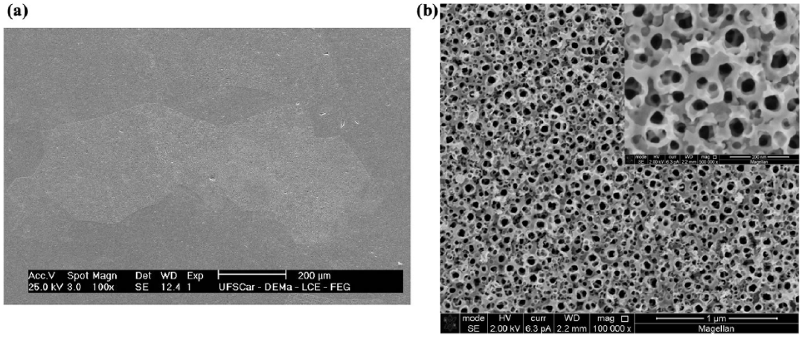
SEM micrographs showing the surface morphology of Ti15Mo alloy (a) before and (b) after the anodization process. SEM: Scanning electron microscopy.
Figure 2 shows the evolution of the surface morphology of samples anodized and annealed at different temperatures as observed by SEM. For the sample annealed at 200°C, it is possible to observe changes in the pores on the tubular nanostructure (Figure 2(a)). As the annealing temperature was raised from 400°C to 450°C, there was an increase in wall thickness that led to the closure of some pores, as observed in Figure 2(b) and (c). The effect of annealing temperature was more evident above 500°C since an initial state of coalescence is observed. The tubular nanostructure tends to vanish and some pores are retained (Figure 2(d)). Coalescence increased at 600°C, causing a full collapse of the nanotube arrays with subsequent growth of TiO2 particles at 800°C (Figure 2(e) and (f)).
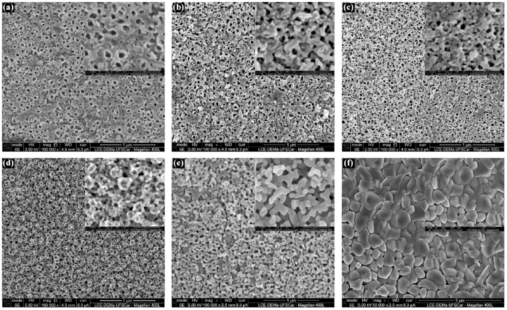
SEM micrographs showing evolution of the surface morphology of samples anodized and annealed at (a) 200°C (b) 400°C (c) 450°C (d) 500°C (e) 600°C, and (f) 800°C.
Structural and vibrational characteristics were obtained using XRD and scattering Raman and the results are present in Figure 3. XRD patterns for TNT-as anodized and TNT-200 samples were found to be amorphous as only diffraction peaks related to substrate Ti15Mo were observed.
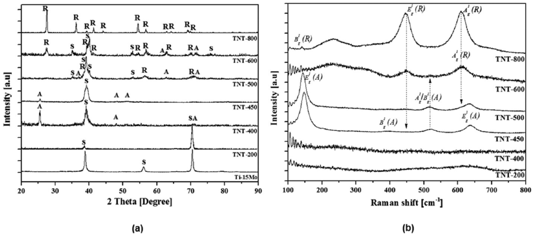
(a) XRD pattern of TNT anodized and annealing at temperatures between 200°C and 800°C, showing the evolution phases, anatase (A) and rutile (R), besides peaks related to the substrate (S). It is worth noting that, for temperatures above 450°C, substrate changes and peaks related to the hexagonal phase appear. (b) Raman spectra of TNT anodized and annealing at temperatures between 200°C and 800°C, showing the evolution phases, (A) anatase and (R) rutile. XRD: X-ray diffraction; TNT: TiO2 nanotube.
Such a statement is consistent with the Raman spectrum since no peak was observed, and only weak and broad peaks were present, which may suggest a lack of crystallinity and/or local lattice imperfections. 23 After annealing at 400°C, diffraction peaks associated with a single anatase phase appeared, although they were not seen in the Raman spectrum. This behavior may be related to the average structural information given by the long-range order of the XRD pattern, which differs from local analysis obtained by Raman spectroscopy. However, the Raman spectrum showed apparent broad and overlapping peaks around 620 cm1 and below 350 cm–1. Literature reports state that, in Ti foils, anodized bands near 680 cm–1 may be assigned to Ti–O bonds in the TiO68– octahedron, and bands around 350 cm–1 and 250 cm–1 may be related to O–O interactions. 24 As the annealing temperature was raised to 450°C, the diffraction peak of anatase phase around of 25.5o corresponding to the (101) plane became more intense, indicating an increase in crystallinity.
Regarding vibrational properties, from the space group
Thus, the Raman spectrum for TNT-400 exhibited a sharp peak at 147 cm–1, as well as peaks at 516 cm–1 and 634 cm–1 associated with
For sample TNT-500, the XRD pattern showed diffraction peaks of the anatase phase along with low intensity peaks due to reflections of the rutile phase. In the Raman spectrum for this sample, vibration modes of the anatase phase were observed; however, the rutile phase may be present, since a shoulder at 615 cm−1 and a weak peak at 443 cm–1 were detected, corresponding to the
At the annealing temperature of 600°C, the anatase–rutile transformation is more evident as diffraction peaks associated with the rutile phase appear, whereas more intense anatase diffraction peak (θ=25.5o corresponding to the (101) plane), verified at 400°C and 450°C disappears.
From the Raman spectrum, the
A complete transformation to rutile phase was observed at 800°C since the XRD pattern and Raman spectrum showed diffraction planes and peaks associated with the vibrational modes characteristic of the rutile phase, respectively.
In this sense, the phase transformation of TiO2 nanotubes from amorphous state to anatase phase occurred around 400°C, and from anatase to rutile occurred at 500°C. Consequently, atomic rearrangements due to phase transformation led to changes in tubular nanostructure characterized by an increase in wall thickness, closure of pores, and subsequent collapse.
Impurities in interstitial and oxygen vacancies in the nanotubes/substrate interface are reported to play an important role during the crystallization and phase transformations of TNTs.20, 24 In this sense, impurities coming from the reduced states of titanium increase the number of oxygen vacancies in the nanotube/substrate interface, so, during annealing, an oxide layer of rutile phase probably forms on the interface due to due to thermal oxidation of the substrate—a fact that contributes to nanostructure collapse.
It has been suggested that, among factors that may further the bioactivity and bone growth in materials for biomedical applications, roughness and the crystalline phase play important roles.10, 27 Thus, in nanotubular structures, cells may grow into the nanotube pores, producing an interlocked cell structure.
On the other hand, reports also indicate that the anatase phase is much more beneficial for bone growth than the amorphous and rutile phases, presumably because of the better lattice match with hydroxyapatite. 11
In this sense, since annealing at 450°C led to crystallization of the nanotubes into anatase phase, thus retaining the tubular nanostructure, the cell culture experiments were performed under this sample condition.
In vitro study—cell culture
The proliferation of fibroblast cells in anodized samples was evaluated at room temperature, with a constant potential of 20 V for 24 h in an electrolyte containing glycerol in combination with ammonium fluoride (NH4F) and annealing at 450°C after 1, 4 and 7 days in cell culture using fluorescence microscopy images. Samples no treated were used as control group.
Analyzing Figure 4, we can notice the gradual increase in the number and size of cells, indicating no toxic alloy. Better spreading on anodized samples compared with the control group was verified, as well as higher development of the cytoskeleton in the anodized samples.
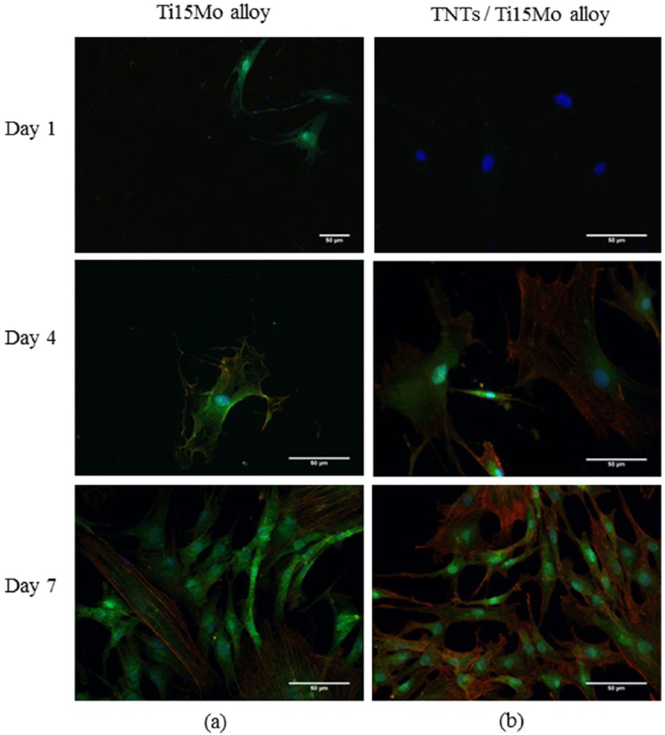
Morphology of human fibroblast cells over samples of Ti15Mo (a) not treated and (b) after anodization at room temperature, with a constant potential of 20 V for 24 h in an electrolyte containing glycerol in combination with ammonium fluoride (NH4F) and annealing at 450°C.
SEM images were taken for the same groups and periods of cellular growth. The images confirmed the results obtained by fluorescence images, showing higher proliferation over surfaces of anodized samples.
In Figure 5, we can see that cells grown on anodized samples have better confluence over the surface, resulting in a greater cell volume after the 7th day of culture. These results agree with studies reported in the literature for surfaces containing TNTs.4,9,28 This behavior can be attributed to the hydrophilic character of the anodized samples and cell expansion inside the nanotube.
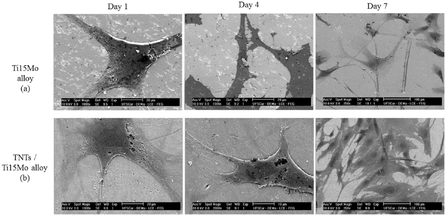
Scanning electron microscope images of human fibroblast cells samples on Ti15Mo alloy surface after day 1, day 4, and day 7 (a) not treated and (b) after anodization at room temperature, with a constant potential of 20 V for 24 h in an electrolyte containing glycerol in combination with ammonium fluoride (NH4F).
These results show the most favorable cell behavior and the highest activity after the 7th day of cells growth. This statement is based on the greater amount of protein found in the cells of anodized surfaces, as seen in the immunofluorescence images, represented in Figure 6.
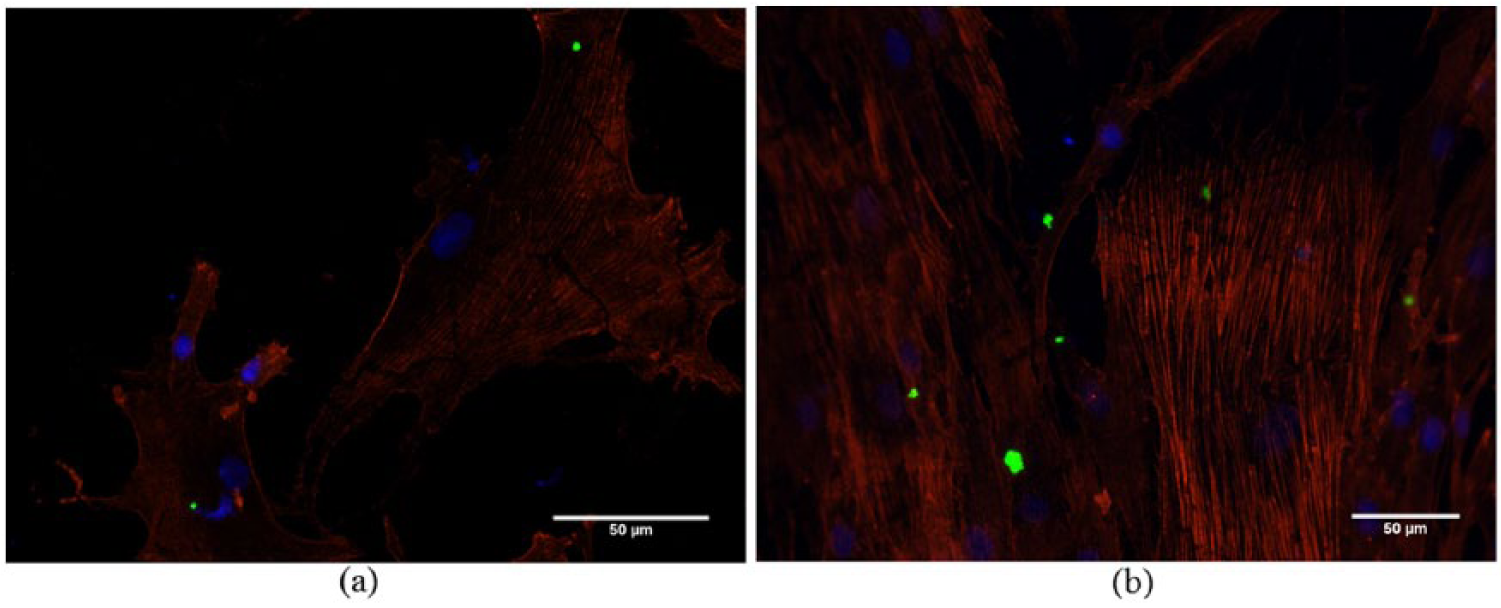
Immunofluorescence images of control (a) and anodized groups (b) showing (in green) proteins synthetized in the cells after the 7th day of cell growth.
Conclusions
This work showed that it is possible to grow an ordered and uniform TiO2-based tubular nanostructure on the surface of Ti15Mo alloy. In the as-anodized condition, the nanostructure presented an amorphous state, as observed from Raman spectra and XRD pattern analysis, with anatase phase formation after annealing at 450°C. At higher temperatures, the transformation to rutile phase is induced, which is completed around 800°C. SEM micrographs showed that the phase transformations during annealing affect the nanostructure, since the oxide layer and subsequent oxygen diffusion, which frequently occur in the interface nanotube/substrate, serve as nucleation sites of more stable phases of TiO2 (anatase and rutile). Therefore, with particle growth, the tubular nanostructure tends to disappear and finally collapse. The cell growth was evaluated for the best condition of annealing (450°C) after anodization. After the 7th day of culture, the highest spreading and best interaction between the cells with the surfaces were verified for this condition when compared with a control group. These results suggest that this surface treatment could improve the performance of this alloy in biomedical applications.
Footnotes
Declaration of Conflicting Interests
The authors declare no potential conflicts of interest with respect to the research, authorship, and/or publication of this article.
Funding
The authors disclose receipt of the following financial support for the research, authorship, and/or publication of this article: The authors gratefully acknowledge the Brazilian research funding agencies FAPESP (grant 2013/00317-4) and CNPq (grant486352/2013-7) for financial support.