
Abstract
Study design
Experimental spinal cord lesion study.
Objectives
To evaluate the effects of erythropoietin at different doses on neural regeneration in rats undergoing spinal cord injury.
Methods
Anesthetized Wistar rats were submitted to standardized spinal cord injury and randomized into eight groups, receiving different magnitudes of trauma and single or repeated doses of intraperitoneal erythropoietin (500 or 5000 IU/kg of body weight). We evaluated motor function using BBB scores and sensorimotor behavior by observing the rats walking on a horizontal ladder (at 2, 4, and 6 weeks) and performed histological analysis of the spinal cord after euthanasia. We compared the scores between groups using analysis of variance (ANOVA) and Bonferroni multiple comparisons.
Results
The experiments were conducted with 10 animals per group (n = 80), none of which died or were excluded. BBB scores increased over time (meaning recovery) in all groups (P < 0.001 for all). From the fourth week, animals receiving lower trauma and higher erythropoietin doses had higher BBB scores than those receiving lower doses. The total number of steps and correct steps taken on the horizontal ladder increased, and slips decreased over time with treatment in all groups. Although the number of errors was different between moments (P < 0.001), it was not different between groups (P = 0.707). Rats receiving higher impact lesions had more spinal cord necrosis and worse recovery of neuronal fibers than the rest.
Conclusions
Animals receiving a higher dose of erythropoietin and suffering minor trauma showed better and faster neurological recovery. Repeating erythropoietin after a week showed no benefit.
Introduction
The role of erythropoietin as a protective agent in nervous system injuries was discovered in the early 1990s when erythropoietin mRNA was found in brain cells. Studies showed it increased in hypoxia-related situations. 1 Later, it was shown that astrocytes and neurons could express erythropoietin in their receptors 2 and that this substance has a trophic effect on cholinergic neurons of the central nervous system. 3 Classically, the blood-brain barrier is impermeable to the passage of larger molecules. However, certain larger molecules can be transported through the capillary endothelium,4-6 which makes their administration via the peripheral route possible. This discovery prompted researchers to investigate the therapeutic use of erythropoietin in nervous tissue injuries. Studies have found that the death of glial cells and neurons in the spinal cord after traumatic injuries occurs through a secondary apoptotic mechanism. Therefore, the inhibition of this process would provide better neurological recovery.7-13
Recombinant human erythropoietin is a glycoprotein produced by recombinant DNA technology with the same amino acid sequence and biological function. It is approximately 80% homologous to rodent erythropoietin and has been shown to be biologically active for erythroid and neurotrophic functions.14,15 This suggested experimental trials using erythropoietin in ischemic and traumatic spinal cord injuries. In the vast majority of them, its neuroprotective effect was confirmed, with better functional and histological results being observed in animals subjected to treatment with erythropoietin when compared to control groups with other substances and placebo solution.16-19
What we know about the cytoprotection dose-response curve of erythropoietin is that only providing a higher dose will not directly result in a superior effect.11,20,21 Many other variables still need to be better evaluated and questions to be answered about the effects of erythropoietin and spinal cord injuries, such as the energy of the trauma suffered or intervals between doses, which can affect the results at the same time. The present study aims to research the neurological and histological effects of erythropoietin in different doses and magnitudes of experimental spinal cord trauma suffered in rats, contributing to a better understanding and possible use of this substance in this injury.
Objective
To evaluate the effects of erythropoietin at different doses and in different magnitudes of trauma on the neural regeneration in rats submitted to a standardized and reproducible model of blunt spinal cord injury. We hypothesized that the drug effects might be mediated by the level of trauma, dosage and posology.
Methods
Ethics
In this study, all institutional regulations, and all local governmental and international guidelines for the care of experimental animals and pain control were followed. The protocol of this experimental study was approved by the university ethical board on February 5th, 2020, which approved the use of animals in this study. This study is reported according to the ARRIVE guideline. 22
Study Design and Sample Size
This is an experimental spinal cord lesion study with eight groups of healthy Wistar rats, all receiving different dosages of erythropoietin after different levels of spinal contusion. Neurological recovery was evaluated individually by a standardized motor neural function system as recommended by the MASCIS (Multicenter Animal Spinal Cord Injury Study) protocol.23,24 After euthanasia, quantitative and qualitative histological analyses of spinal cord cross-sectional cuts was conducted. This study had no control group, as the viability of mechanical contusion 25 and the effects of erythropoietin had already been widely proved in prior research by other groups and ours,17,19,26-30 always showing superior results compared with sham, saline solution, or both.28,30-36 Spinal injuries, pharmacological treatment and functional and histological analyses were all conducted in the same university laboratory and using the same equipment and materials.
Level of Experimental Spinal Cord Injury Produced by the Fall of a 10-g Weight and Dosage of Intraperitoneal Erythropoietin (Recombinant Form) Administered to Each Animal in the Study Groups.

aIn two-doses regimens, there was one-week interval between them.
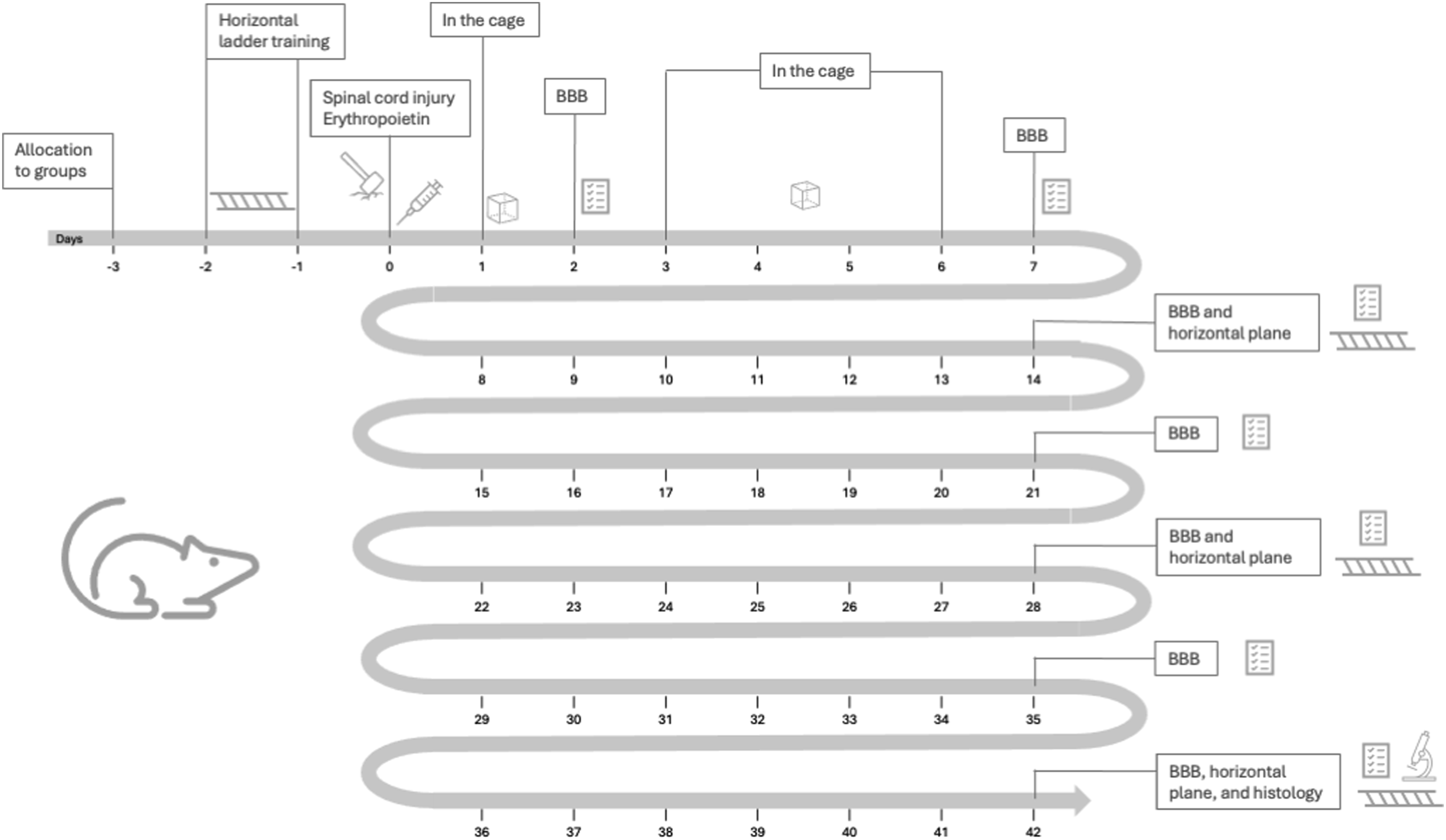
Timeline of study procedures.
Inclusion and Exclusion Criteria
We used healthy Wistar rats, 20–21 weeks old, weighing 340–450 g coming from the same university vivarium. Male rats were used as they have usually general calmer and less aggressive behavior and would not be susceptible to hormonal fluctuations. To be included in the study, the animals should present normal coat under visual inspection, good general health condition, and no signs of autophagy or mutilation behavior. They would not be included if they had anomalies observed macroscopically in the medullary region. Rats would be excluded if they died after the experimental injury; if they had persistent infection after 10 days of antibiotic treatment; or if they lost greater than 10% of body weight after injury.
Rats would not be included in this study if they did not have normal initial motor skills as assessed by the BBB scale (details on this assessment below) initially. On the other hand, they would be excluded from the study if they presented normal movement (21 points on the BBB scale) even after the experimental injury.
Spinal Cord Lesion
The NYU Weight-Drop Impactor (John A Gruner, Department of Neurosurgery, New York University Medical Center, New York, NY, USA) was used to create medullar lesion in all rats, according to methodology used in our lab.25,39-42 All rats were sedated and under anesthesia before the procedure.
Inside an acrylic box, animals were first slightly sedated using isoflurane (1.5-2.0 vol%) in 100% oxygen for around 2 minutes. A mask was then placed to cover the animal’s entire snout, taking it to a deeper anesthetic plane. The absence of corneal reflexes and of reaction to compression of the paws and tail confirmed the establishment of a deep anesthetic plane. The coat was then shaved from the rat’s back and the dorsal region was cleaned. Supplementary Figure 1 shows these steps.
The rats were then submitted to anesthesia by subcutaneous injection of tramadol hydrochloride (3 mg/kg) and pentabiotic (24,000 IU/kg). A 2-3-cm long incision was made in the dorsal midline to expose the posterior arches of the spine, with subperiosteal dissection of the spinous processes and laminae from T9 to T11 (as shown in Supplementary Figure 2). The spinous process and lamina of the T10 vertebra and the distal half of the T9 spinous process were removed until the dural sac was completely exposed to receive the impact by the NYU equipment.
The rat was then placed in the NYU Weight-Drop Impactor to receive the spinal cord blunt trauma. The tip of the computerized weight drop impact equipment’ nail was positioned (Figure 2). The 10-g rod was graduated to drop from a 12.5-mm or 25-mm height depending on which group the animal belonged to. Procedures for spinal cord blunt trauma using the NYU weight-drop impactor. (A) The Wistar rat is placed on the equipment with the spinal cord exposed. (B) The equipment’s nail is positioned over the spine for controlled weight drop on it. Two adjustable claws help fix the spine to the equipment, reducing deformations of the animal’s body and movement of the spine when the weight falls, reducing errors. (C and D) After the trauma, sutures of muscular, facial and skin planes with single stitch suture with 2.0 monofilament nylon thread closed the injury.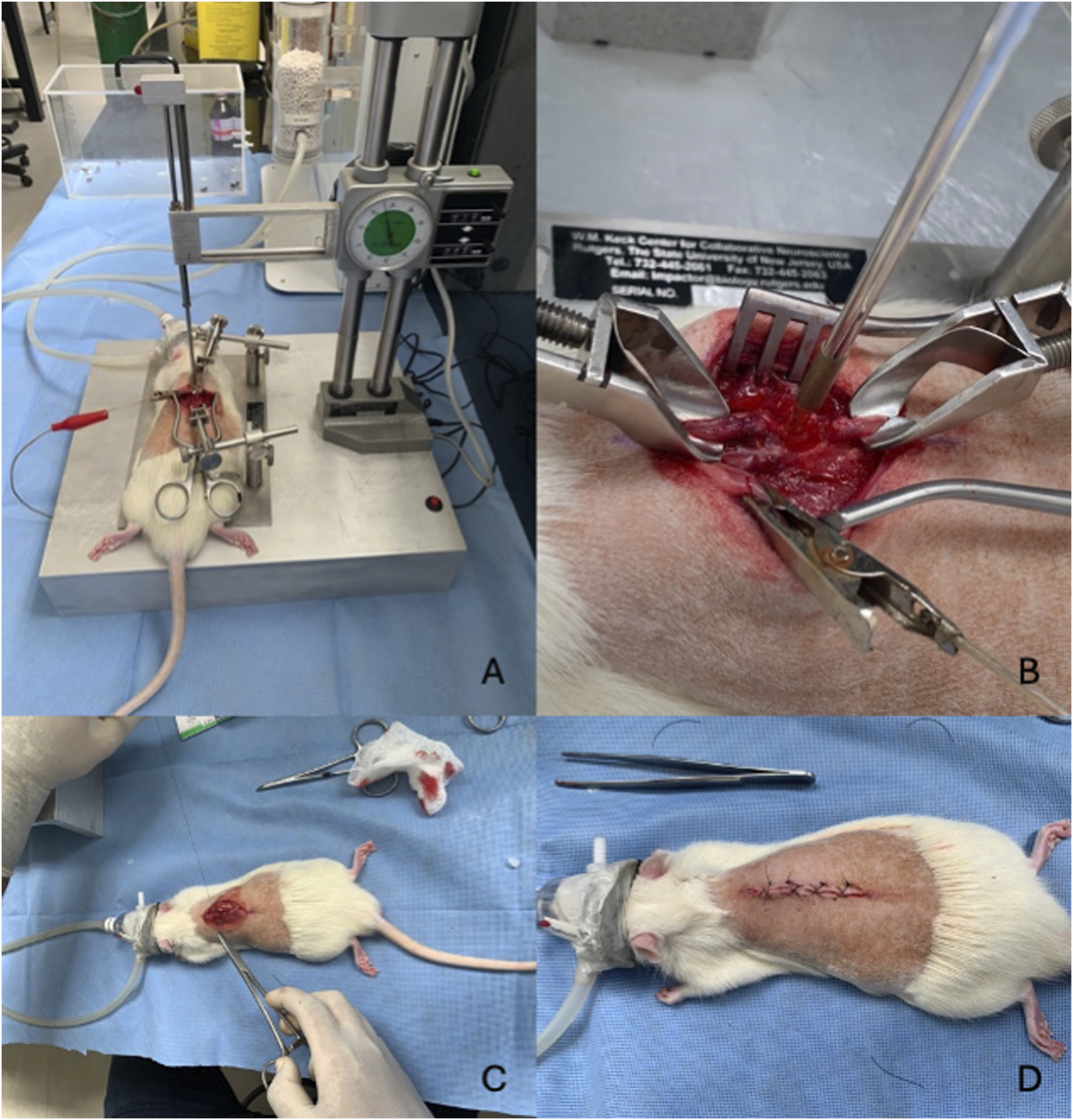
Care after Surgery
After suturing the skin, the rat was placed in an environment with temperature control for thermal comfort. Tramadol hydrochloride was administered (3 mg/kg every 8 hours) for 5 days, pentabiotic (24,000 IU/kg every 12 hours) for 7 days, meloxicam (5 mg/kg once a day) for 3 days. All animals received prophylactic antibiotic therapy in a single dose (cefazolin 2 mg/100 g, intraperitoneally).
As the animals lost their urination reflexes after the spinal cord injury, maneuvers were performed daily to extract urine from the bladder by manual pressure. The degree of dehydration was assessed daily by looking at skin turgor. The presence of blood in the urine was also checked daily as a marker of infection, as the protocol provided for antibiotic therapy (levofloxacin, 2.5 mg/kg, for 10 days) if present. If the animal continued to present blood in the urine after therapy, the infection would be considered intractable, and the animal would have painless death induced.
The animals were returned to their cages and the same pre-operative environment was preserved: ad libitum feeding and daily checking of hygiene conditions. During the 42 days of the study, the presence of exclusion criteria, infection and mutilation, was investigated daily among the rats.
Erythropoietin Administration
All animals in this study received erythropoietin manufactured by Janssen-Cilag (Eprex-alfaepoetina, Schaffhausen, Switzerland) from ampoules with doses of 10,000 IU and a volume of 1 mL. Doses per rat (Table 1) could be 500 IU/kg or 5000 IU/kg depending on the group allocation, and a single dose or two doses, one-week apart, were planned. This allocation system allowed the 80 rats to receive all possible combinations of erythropoietin doses, regimens and different standardized spinal cord injury grades, with the impact from a weight falling from 12.5 mm or 25 mm.
Functional Analysis: BBB Scale
The first functional evaluation using the Basso, Beattie and Bresnahan (BBB) scale took place 48 hours (Day 2) after the spinal cord injury to check the effectiveness of the procedure. The next evaluations happened at Days 7, 14, 21, 28, 35 and 42 after surgery.
Each rat was placed inside an 80 cm × 80 cm box with the border measuring 17 cm in height for the BBB scoring. Position and movement of the rat’s hips, knees, ankles, as well as trunk, abdomen, tail and hind legs. The rats’ capacity of cleaning the paws, predominant positioning of hind legs, trunk stability and general coordination are scored too. The scores vary from zero (no movements) to 21 (normal movements).
Two evaluators (GBS and another assessor, not an author), trained in the application of the scale, conducted the BBB functional analysis. Working simultaneously, they checked how the animals moved inside the box for at least four minutes, registering the BBB scores. In case of disagreement unsolvable by discussion, the lowest BBB score was recorded for this study. Evaluators did not know the rat allocation to a group.
Sensorimotor Behavioral Assessment: Horizontal Plane
To analyze the animals’ proprioceptive function, we observed how rats crossed a horizontal ladder43,44 100-cm long and 35-cm wide, suspended at a height of 46 cm from the floor and with 1.5 cm between each metal bar (the step). For two days before the surgical procedure, the animals were trained to move on this ladder 45 at least five times. Water and sugar were placed at the end of the route to stimulate voluntary movement.
During the evaluations for this study, while the rat walked down the stairs, the following data were recorded: the number of total steps, successful steps (the correct positioning of the paws on the metal steps), slips (when the paw fell between the steps), and errors. Two types of errors were distinguished: the dragging of the hind paws along the horizontal ladder and the poor positioning of the paws between the metal steps. The values from the three passages through the horizontal ladder were registered, and the average was calculated.
Locomotor function using the horizontal ladder was assessed at four moments: preoperatively, two, four and six weeks after spinal cord injury. The evaluations were made by the same evaluators as the BBB scale.
Euthanasia and Histological Evaluation
Euthanasia was performed on the 42nd day with pentobarbital (140 mg/kg) intraperitoneally to induce an anesthetic plane which, once confirmed, was followed by the induction of potassium chloride intravenously.
The medullary segment from the C3 level to the T10 level was surgically removed for histological analysis. The segment was adhered linearly on cardboard with the respective topographic identifications, and recording where macroscopic findings of spinal cord contusion were observed, identified as area “B”. The cephalic area was marked as “A” and the area caudal to the lesion as “C”. After identified and packaged, the marrows were fixed in 10% formaldehyde. Each area identified as injured was sectioned in the axial plane macroscopically at intervals of 2 mm, starting from the central area of the lesion, over an extension of 1 cm.
The slides containing transverse sections of the spinal cord at the perilesional level and nearby regions (medullary region B) were stained with hematoxylin-eosin. Histopathological changes were recorded as present (P) and absent (A). The presence of necrosis, hemorrhage, hyperemia, degeneration of the nervous substance and cellular infiltrate was analyzed. A score from 0 to 3 (absent, mild, moderate and intense) was assigned for each of the changes in each spinal section.
Quantitative histological analysis was performed using digital images at high magnification (100x) of the cranial and caudal medullary portions (A and C), after fixation in 2% osmium tetroxide solution and staining with 1% toluidine blue, obtaining adequate representation of neuronal cells. The pathologist (not the author) selected the two fields with the best representation of each segment. The images were evaluated using the Sigma Scan Pro5.0 software to count the fibers of the regenerated axons. Only neurons with a diameter equal to or greater than 15 μm were considered for counting. The pathologist was blind to animal allocation.
A regeneration index (IR) was calculated using the number of regenerated axons in the distal segment and the number of regenerated axons in the proximal segment: IR = (no axons in the distal segment/no axons in the proximal segment) × 100.
Statistical Analyzes
The evaluated parameters were described according to groups and evaluation moments using summary measurements (mean, standard deviation, median, minimum and maximum). Variables were compared using generalized estimating equations (EEG) with normal distribution and identity link function. We assumed an AR (1) correlation matrix between the evaluation moments for the BBB; and, for the horizontal plane test, between moments, the weight of the animals, successes, slips and errors. The number of steps was used as a data limit (offset). All analyzes were followed by Bonferroni multiple comparisons. Analysis of variance (ANOVA), followed by Bonferroni multiple comparisons was used to compare quantitative histological data between groups. Kruskal-Wallis tests followed by Dunn’s multiple comparisons were used to compare ordinal histological parameters.
Results
BBB Scores Distribution According to Groups and Moments of Evaluation (P-Group, P-moment and P-interaction <0.01*).
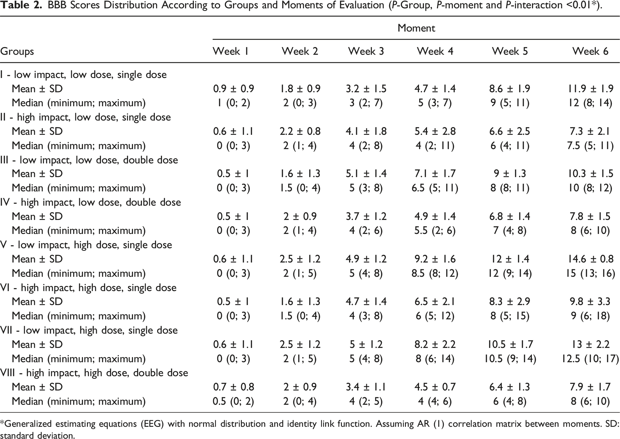
*Generalized estimating equations (EEG) with normal distribution and identity link function. Assuming AR (1) correlation matrix between moments. SD: standard deviation.
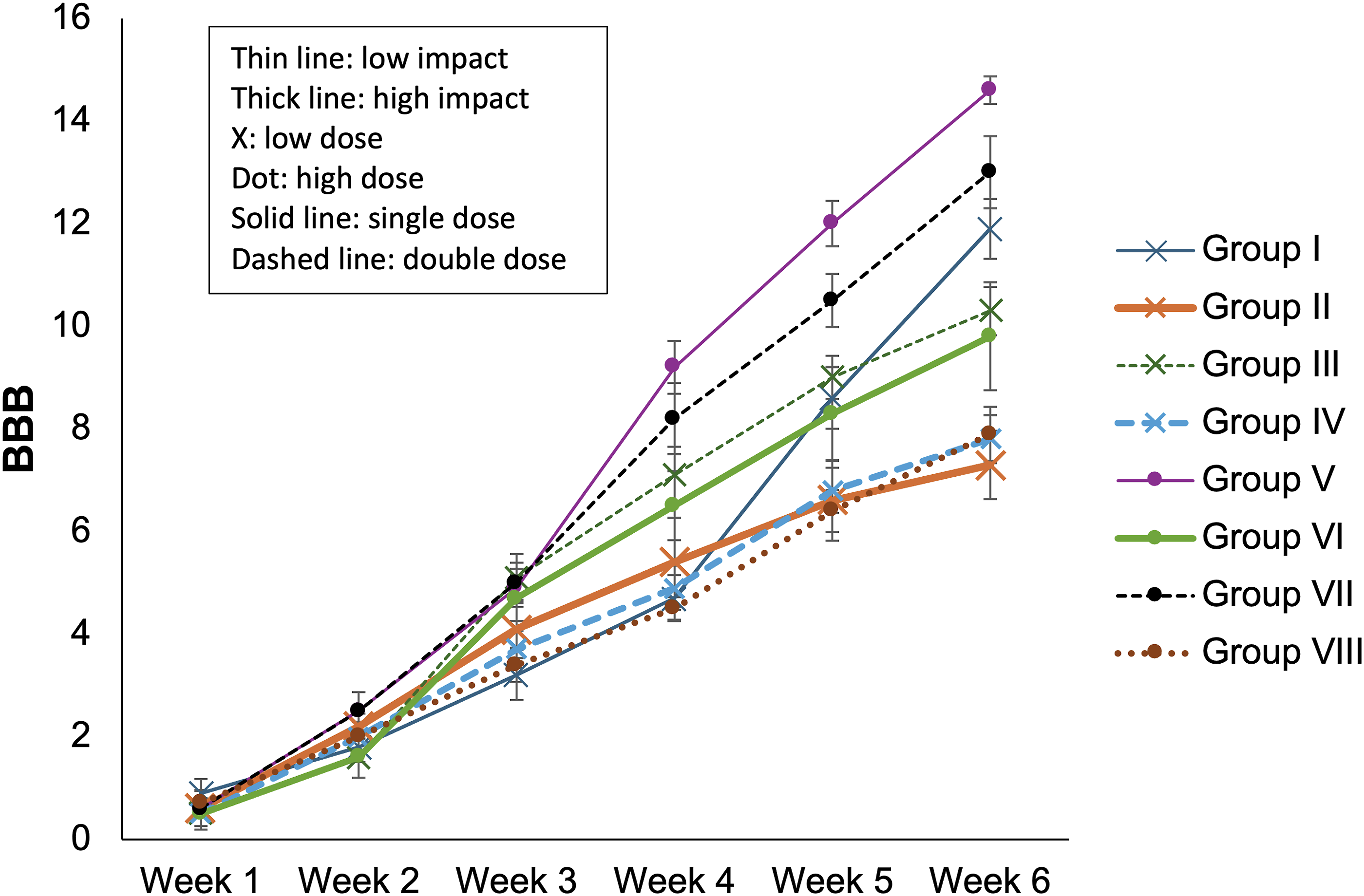
BBB scores across the moments of evaluation (higher score meaning better function).
Number of Steps and Correct Steps According to Groups and Moments of Evaluation (P-Group, P-moment and P-Interaction <0.01 in all Cases a ).
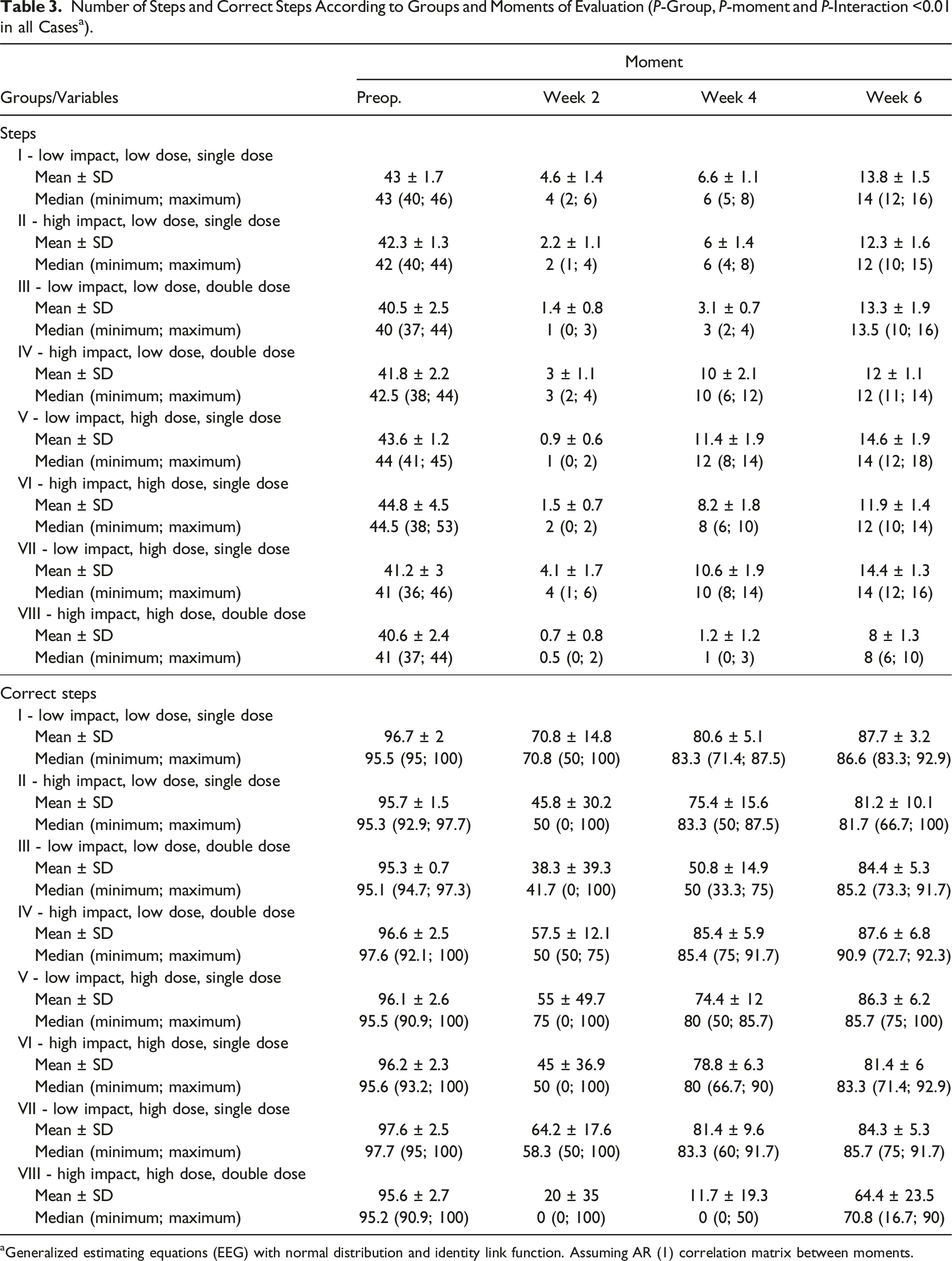
aGeneralized estimating equations (EEG) with normal distribution and identity link function. Assuming AR (1) correlation matrix between moments.
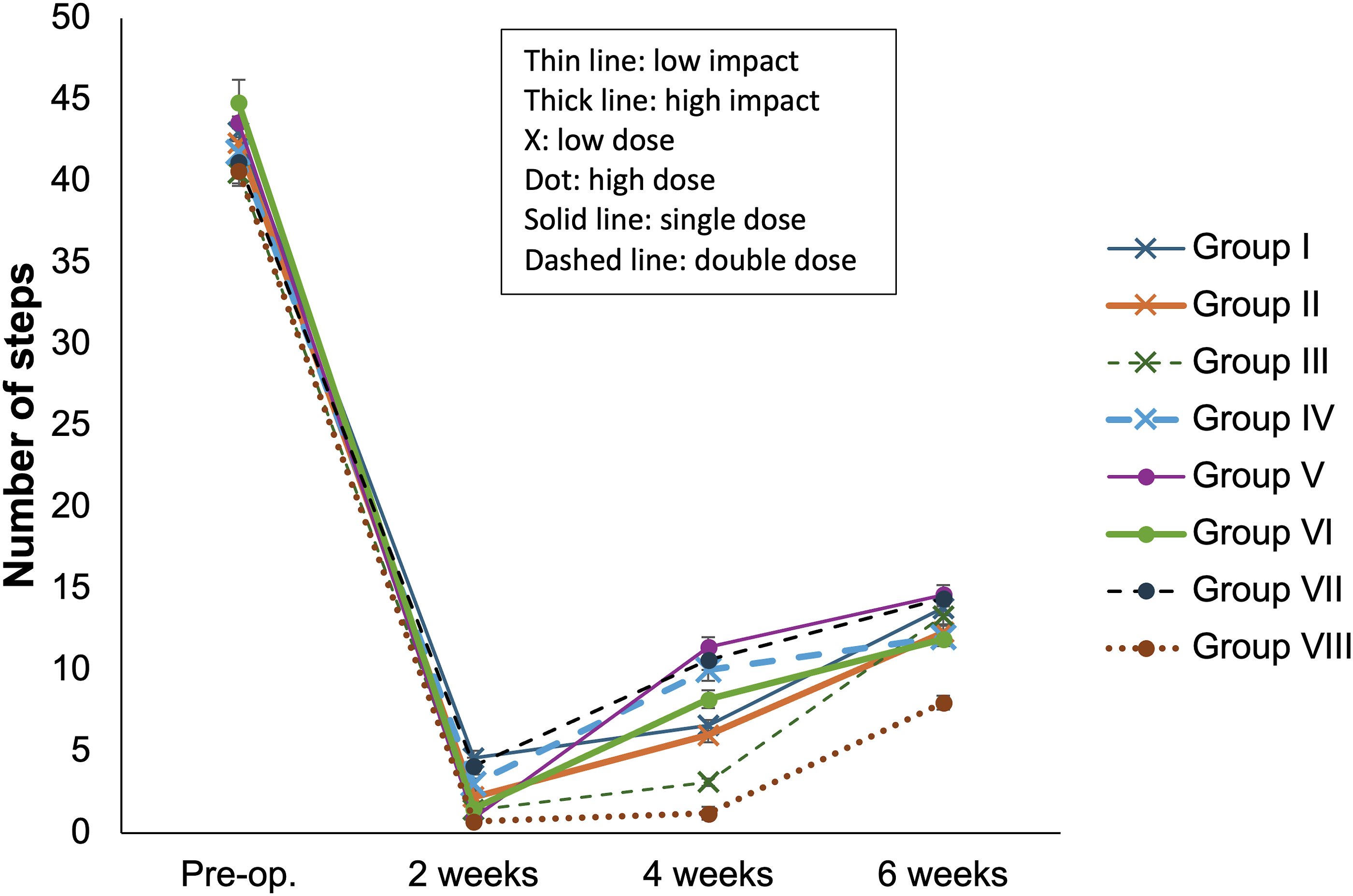
Number of steps on the horizontal ladder.
Number of Slips and Errors on the Horizontal Ladder According to Groups and Moments of Evaluation.
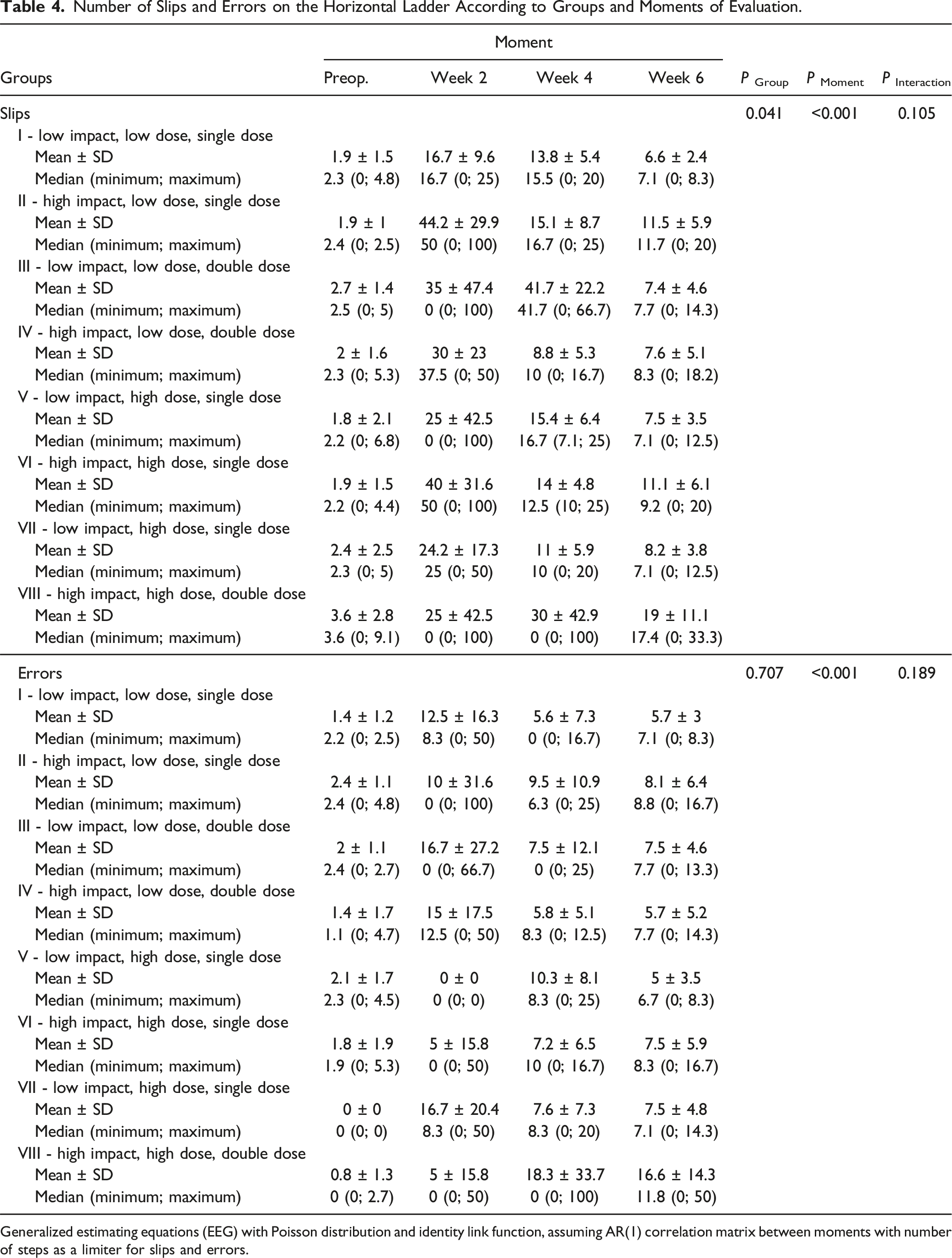
Generalized estimating equations (EEG) with Poisson distribution and identity link function, assuming AR(1) correlation matrix between moments with number of steps as a limiter for slips and errors.
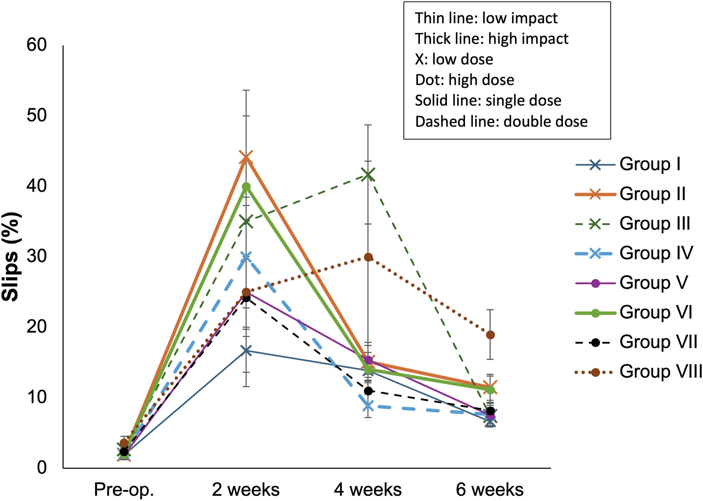
Number of slips on the horizontal ladder.
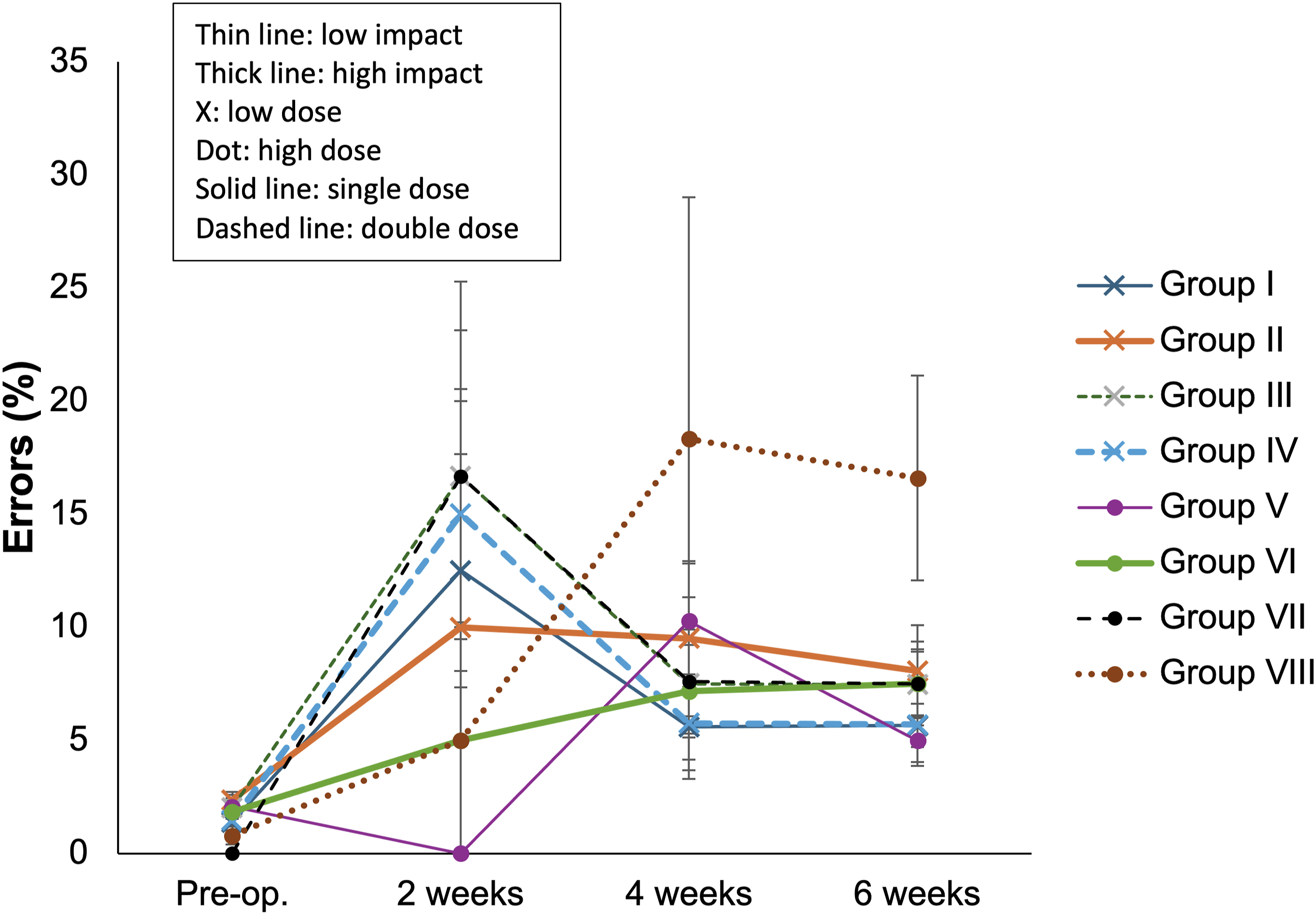
Number of errors on the horizontal ladder.
Histological Analyses Results According to Groups.
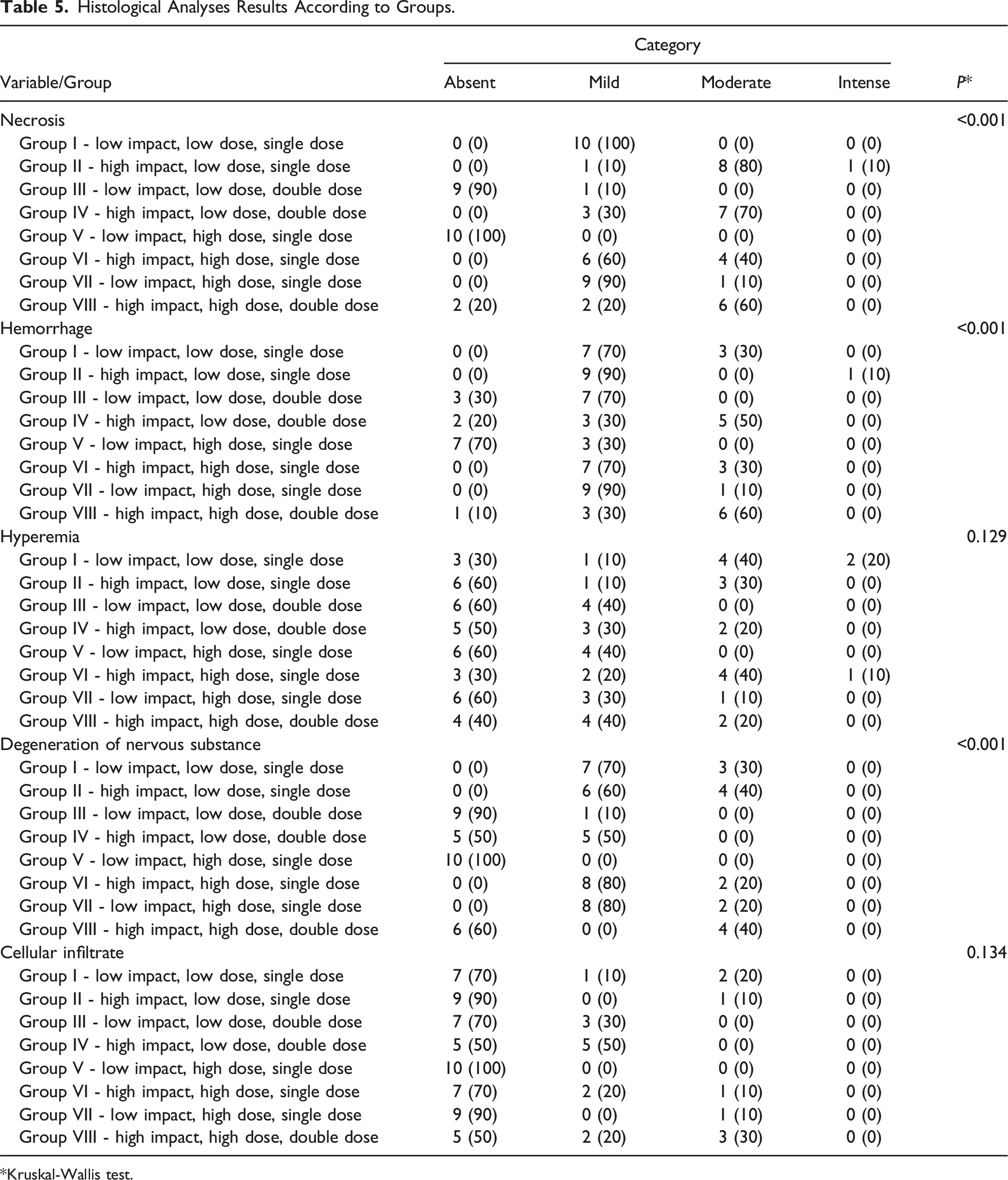
*Kruskal-Wallis test.
Comparison of the Histological Results of Regenerated Neuronal Fibers of the Cranial Medullary Portion Between Groups.
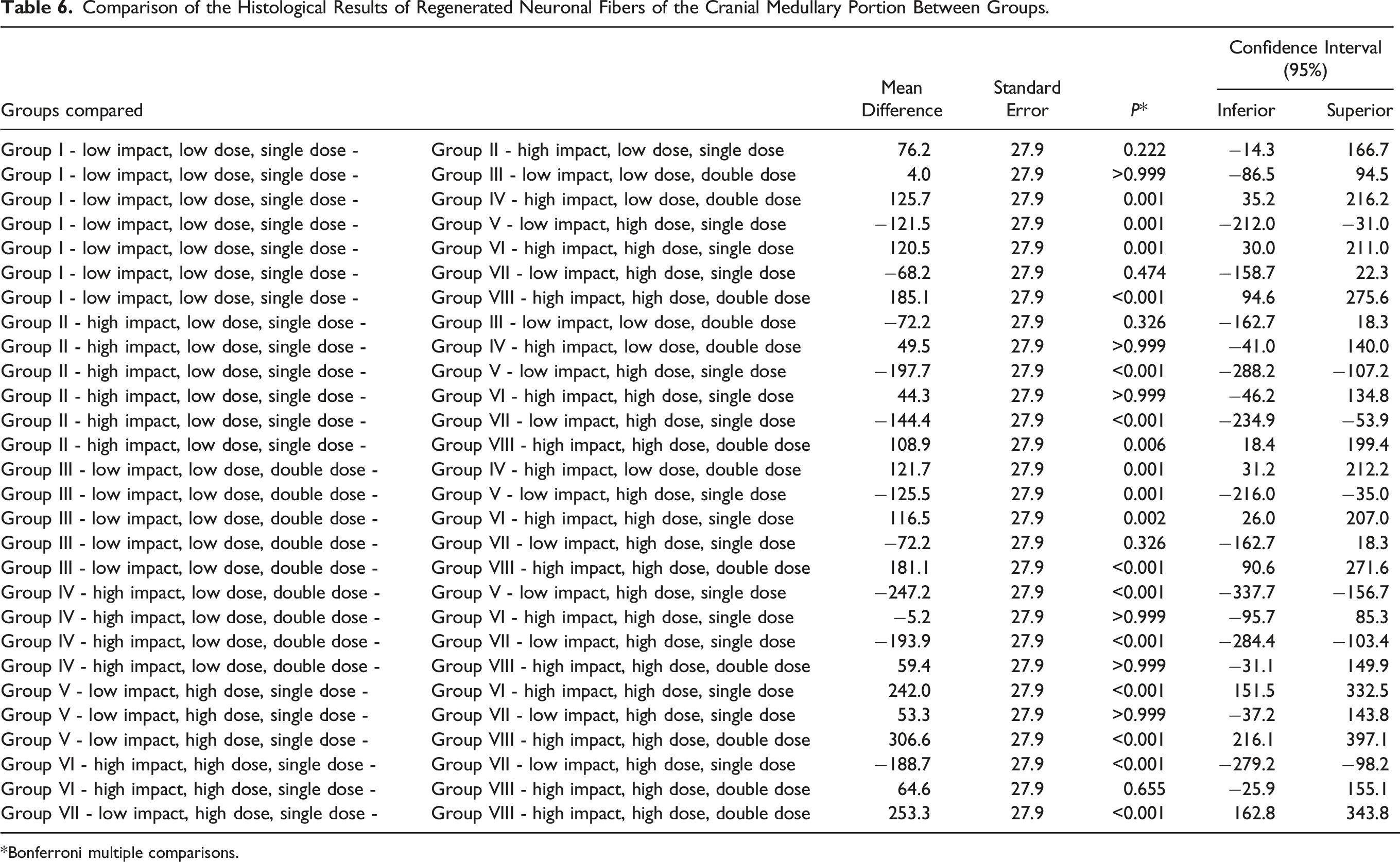
*Bonferroni multiple comparisons.
Comparison of the Histological Results of Regenerated Neuronal Fibers of the Caudal Medullary Portion Between Groups.
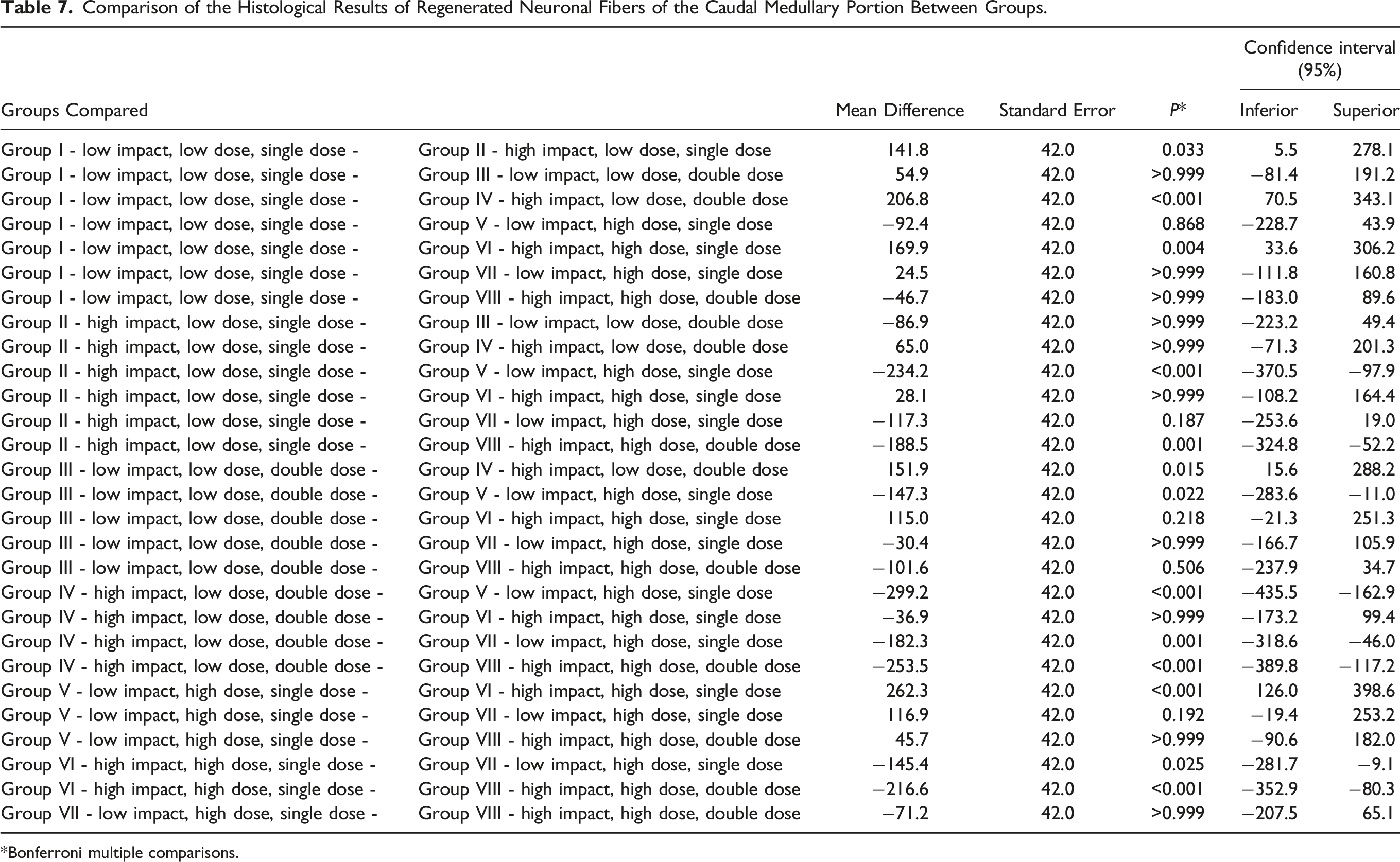
*Bonferroni multiple comparisons.
Discussion
In this study, we observed that the four groups that had less energy discharged into the spinal cord during experimental spinal cord injuries evolved with a better neurological outcome when evaluated using the BBB scale and in the horizontal plane, compared to the four groups that suffered the highest impact. This result is in accordance with logical thinking and previous literature. 46 In the BBB scale evaluation, from the fourth week onwards, among the groups that suffered low-impact injuries, the animals that received the highest dose of erythropoietin (5000 IU/ Kg) showed higher scores when compared to groups that received a dose 10 times lower. A similar pattern was also observed when evaluating the rats walking on the horizontal ladder. In the sixth week, the groups that suffered a low-impact injury and a higher dose of recombinant erythropoietin were the ones that were able to perform more steps — but the difference was not statistically significant. No differences were observed between the groups that received the same dose once or when the administration of recombinant erythropoietin was repeated after one week.
The results of the histological evaluation reflected the neurological outcome from the functional tests. In general, in the groups that suffered the greater magnitude trauma, pathological tissue changes (degree of necrosis, hemorrhage and degeneration) were statistically greater than in the groups that suffered lesser magnitude trauma. We also observed that in groups that suffered trauma of the same magnitude, animals receiving a single dose of erythropoietin presented a lower degree of necrosis than those receiving a repeated dose. The degree of hemorrhage in the group that received a lower-impact injury, and a single, high dose of erythropoietin was statistically lower than the other groups. In general, lower doses showed lower degrees of nerve substance degeneration than higher doses. The groups that suffered trauma of greater magnitude had, on average, statistically lower neuron counts in the caudal specimen than groups that suffered trauma of lesser magnitude, as expected.
These outcomes suggest some inferences. In agreement with the results found in other experimental studies,19,36 the use of erythropoietin in traumatic spinal cord injuries benefits neurological recovery, with faster and better improvement when a higher dose of the drug is used. However, repeat administration one week after injury did not show additional benefit. We understand this finding as compatible with the pathophysiology of traumatic spinal cord injury, as it is known that the cascade of biochemical reactions that occur during secondary spinal cord injury begins immediately after the trauma, and the therapeutic window of pharmacological action is short. Our results corroborate the importance of early treatment to obtain faster and better neurological recovery and avoid the emergence of sequelae, which are often catastrophic in the clinical scenario. The role that the magnitude of the trauma can influence the final result was also notable. Injuries with a high level of energy tend to have a lower response to drug treatment, making pharmacological action limited or even indifferent.
The discovery of the ability of peripherally administered erythropoietin to cross the blood-brain barrier 12 and its neuroprotective effects led to the emergence of studies with this substance for the treatment of conditions involving the central nervous system.7,11,16 Currently, several experimental trials have been published17-19,33-35,47 testing erythropoietin for treating acute injuries to the spinal cord, administered alone or in combination with other drugs. There is already evidence of its role in several situations: for example, in immune regulation, in the protection against cell apoptosis occurring during secondary spinal cord injury and in the promotion of tissue repair after ischemia. It is also known that erythropoietin can protect neurons and free radicals released in the inflammatory cascade initiated after spinal cord trauma.26,27 Additionally, there is considerable evidence that animals treated with erythropoietin after suffering spinal cord injury have better neurological outcomes and biochemical and histological changes when evaluated by the standardized motor scale.27,29,32,35,47,48
Nevertheless, despite all the advances in experimental science, many variables still need to be better clarified. More research is needed to standardize doses, routes of administration, and treatment time, among others.11,20,21,36 This work was conducted with the aim of deepening the understanding of the role of erythropoietin as a neuroprotective agent in experimental blunt spinal cord injury in rats at different doses and magnitudes of trauma. The experimental model of spinal cord injury due to weight drop, using the NYU Impactor computerized equipment, allows the standardization of procedures and comparison of results with other institutions that are part of the Multicenter Animal Spinal Cord Injury Study (MASCIS), as well as the use of BBB scale, valid, reproducible and allows interinstitutional comparisons. The NYU Impactor system has three rod height graduations (12.5 mm, 25 mm and 50 mm). In our study, we chose the heights of 12.5 mm or 25 mm only, as the 50-mm can result in complete spinal cord injury and high mortality.46,49
There are limitations to translating the results of an experimental study into clinical practice. Due to the high standardization of the mechanism of spinal cord injury and the neurological assessment scales used, there is a considerable distance between an experimental model and the patient with spinal cord injury that we encounter in our clinical practice, which presents more varied circumstances, trauma mechanisms and general health conditions, which cannot be standardized. Many factors can influence the neurological outcome of administering any drug to a human being. It is possible to argue that the effects of erythropoietin in the lab may not mirror its action in humans. However, despite considerable evidence of the benefit and safety of erythropoietin in spinal cord injury, both experimentally in rats and humans, there are still important gaps in knowledge, which motivated this study. We found that repeating the erythropoietin dosage one week after the first administration did not cause significant functional or histological effects. And despite one of the dosages tested being ten times higher than the alternative, we were unable to demonstrate significant differences between them in this study. These negative findings may inspire other researchers to investigate questions that are still pending regarding the ideal dose, treatment time, moment and route of administration.
A limitation of experimental spinal cord injury studies in general is that there is some subjectivity involved in the functional and sensitive evaluations. Although the BBB scale is international validated, the horizontal ladder for sensorimotor assessment can be observer dependent. We tried to solve this by using two evaluators and consensual rating. A limitation specific for this study is that we did not have resources for immunohistochemistry evaluation of the spinal specimens, which could have brought some light to details on the effects of the second dose of erythropoietin that the basic pathological evaluation could not.
Conclusion
In this study on the effect of intravenous erythropoietin administration after experimental blunt spinal cord injury in rats, animals that received a higher dose of the drug and suffered minor trauma showed better and faster neurological recovery. Repeating the same dose after a week showed no benefit.
Supplemental Material
Supplemental Material - Erythropoietin to Treat Spinal Cord Injury: Evaluation of Different Doses and Magnitudes of Trauma in Rats
Supplemental Material for Erythropoietin to Treat Spinal Cord Injury: Evaluation of Different Doses and Magnitudes of Trauma in Rats by Alderico Girão Campos de Barros, Gustavo Bispo dos Santos, Raphael Martus Marcon, and Alexandre Fogaça Cristante in Global Spine Journal.
Footnotes
Acknowledgments
The authors thank Flávio Nascimento for his contribution in the functional evaluation of the rats in the laboratory; and Patricia Logullo, PhD, CMPP (Palavra Impressa) for manuscript editing services, for which they paid without external funding.
Declaration of conflicting interests
The author(s) declared no potential conflicts of interest with respect to the research, authorship, and/or publication of this article.
Funding
The author(s) received no financial support for the research, authorship, and/or publication of this article.
Supplemental Material
Supplemental material for this article is available online.
References
Supplementary Material
Please find the following supplemental material available below.
For Open Access articles published under a Creative Commons License, all supplemental material carries the same license as the article it is associated with.
For non-Open Access articles published, all supplemental material carries a non-exclusive license, and permission requests for re-use of supplemental material or any part of supplemental material shall be sent directly to the copyright owner as specified in the copyright notice associated with the article.