



Abstract

The Regenerative Effects of Notochordal Cell Matrix on Canine and Human Nucleus Pulposus Cells
Frances C. Bach1, Stefan A. H. de Vries2, Laura B. Creemers3, Björn P. Meij1, Keita Ito2,3, and Marianna A. Tryfonidou1
1Utrecht University, Utrecht, Netherlands
Anabolic Effects of Bioactive Proteins and Extracellular Vesicles Derived From Notochordal Cell-Conditioned Medium
Frances C. Bach1, Stefan A. H. de Vries2, Frank M. Riemers1, Janneke Boere1, Ferdi W. M. van Heel1, Marina van Doeselaar2, Soenita Goerdayal1, Peter G. J. Nikkels3, Karin Benz4, Laura B. Creemers3, Maarten F. M. Altelaar1, Björn P. Meij1, Keita Ito2,3, and Marianna A. Tryfonidou1
1Utrecht University, Utrecht, Netherlands
Optimization of 3D Printed Hydrogels With Primary Cells for Tissue Engineering
Ezgi Bakirci1, Olivier Guenat2, Andreas Hugi2, Sufian S. Ahmad3, Sandro Kohl3, and Benjamin Gantenbein1
1Tissue and Organ Mechanobiology, Institute for Surgical Technology and Biomechanics, University of Bern, Bern, Switzerland

Cell viability results of bioprinted and seeding cells.
This project was funded by the Gebert Rüf Stiftung project (# GRS-028/13). We thank Eva Roth for her valuable assistance in IVD isolation and the mechanical department for manufacturing the cross-incision tool. Financial support was obtained from the Swiss National Science Foundation (SNF PP00P2_163678/1) as well as from the Spine Society of Europe (Eurospine 2016_4).
1. Murphy SV, Atala A. 3D bioprinting of tissues and organs. Nat Biotechnol. 2014;32:773-785.
2. Gantenbein B, Gadhari N, Chan SC, Kohl S, Ahmad SS. Mesenchymal stem cells and collagen patches for anterior cruciate ligament repair. World J Stem Cells. 2015;7:521-534.
Aneurysmal Bone Cyst of the Spine: An Alternative Treatment by Direct Injection of Concentrated Autologous Mesenchymal Stem Cells
G. Barbanti-Bròdano1, M. Girolami1, A. Cenacchi2, A. Gasbarrini1, S. Bandiera1, S. Terzi1, R. Ghermandi1, and S. Boriani1
1Department of Oncological and Degenerative Spine Surgery, Istituto Ortopedico Rizzoli, Bologna, Italy
2Service of Immunohematology and Transfusional Medicine, Istituto Ortopedico Rizzoli, Bologna, Italy
Biomaterials: A Good Alternative to Autologous Bone for Spine Fusion
Giovanni Barbanti Bròdano1, Francesco Lolli1, Gianluca Giavaresi1, Milena Fini1, Stefano Bandiera1, Alessandro Gasbarrini1, Silvia Terzi1, Riccardo Ghermandi1, Lisa Babbi1, Marco Girolami1, and Stefano Boriani1
1Department of Oncological and Degenerative Spine Surgery, Istituto Ortopedico Rizzoli, Bologna, Italy
Restorative Neurostimulation to Induce Episodic Contractions of the Lumbar Multifidus Leads to Clinical Improvements in Patients With Disabling Refractory Nonspecific Chronic Low Back Pain: Results of a Prospective International Multicenter Trial
Kris De Smedt1, Kristiaan Deckers1, Bruce Mitchell2, David Vivian2, Marc Russo3, Sam Eldabe4, Ashish Gulve4, Nick Harland4, Peter Georgius5, Jean-Pierre Van Buyten6, Iris Smet6, Mathew Green7, John Vieceli7, Ganesan Baranidharan8, Vivek Mehta9, Shankar Ramaswamy9, Richard Sullivan10, Robert Gassin10, James Rathmell11, and Chris Gilligan11
1St. Augustinus Hospital, Antwerp, Belgium
1. Hides JA, Richardson CA, Jull GA. Multifidus muscle recovery is not automatic after resolution of acute, first-episode low back pain. Spine (Phila Pa 1976). 1996;21:2763-2769.
2. Ostelo RW, Deyo RA, Stratford P, et al. Interpreting change scores for pain and functional status in low back pain: towards international consensus regarding minimal important change. Spine (Phila Pa 1976). 2008;33:90-94.
3. Dworkin RH, Turk DC, Wyrwich KW, et al. Interpreting the clinical importance of treatment outcomes in chronic pain clinical trials: IMMPACT recommendations. J Pain. 2008;9:105-121.
4. Soer R, Reneman MF, Speijer BL, Coppes MH, Vroomen PC. Clinimetric properties of the EuroQol-5D in patients with chronic low back pain. Spine J. 2012;12:1035-1039.
Model of Disc Degeneration in Rat Tail Induced Through a Vascular Isolation of Endplate
Maximo-Alberto Díez-Ulloa1, Hector Fernandez-Susavila2, Juan-Pablo Pardo-Seco1, Ramón Iglesias-Rey2, Ana Estany-Gestal3, Tomas Sobrino-Moreiras2, and Francisco Campos-Perez2
1Orthopaedics, Universitary Hospitalary Complex, Santiago de Compostela, Spain
Affordable Surgical Navigation Based on Finite Element Simulation and 3D Printing Technology
Péter E. Éltes1,2, Márton Bartos3, Damien Lacroix4, Varga Péter Pál1, and Áron Lazáry1
1National Center for Spinal Disorders, Budapest, Hungary
Comparing Two Annulus Fibrosus Injury Models in Loaded Bovine Organ Culture
Daniela A. Frauchiger1, Lorin M. Benneker2, and Benjamin Gantenbein1
1Institute for Surgical Technology & Biomechanics, University of Bern, Bern, Switzerland

Custom designed cross-incision tool consisting of outer shaft.

(A) Mitochondrial activity, (B) GAG content, and (C) DNA content of IVDs under different loading conditions. Mean ± SEM, N = 5.
1. Likhitpanichkul M, Dreischarf M, Illien-Junger S, et al. Fibrin-genipin adhesive hydrogel for annulus fibrosus repair: performance evaluation with large animal organ culture, in situ biomechanics, and in vivo degradation tests. Eur Cell Mater. 2014;28:25-37.
2. Michalek AJ, Iatridis JC. Height and torsional stiffness are most sensitive to annular injury in large animal intervertebral discs. Spine J. 2012;12:425-432.
3. Chan SCW, Gantenbein-Ritter B. Preparation of intact bovine tail intervertebral discs for organ culture. J Vis Exp. 2012;(60):3490.
4. Gawri R, Moir J, Ouellet J, et al. Physiological loading can restore the proteoglycan content in a model of early IVD degeneration. PLoS One. 2014;9:e101233.
Nucleus Pulposus Progenitor Cells: Isolation and Injection Into Degenerative Disc
Daniela A. Frauchiger1, Adel Tekari2, Lorin M. Benneker3, Daisuke Sakai4, Sibylle Grad5, Jivko Stoyanov6, Alessandro Bertolo6, and Benjamin Gantenbein1
1Institute for Surgical Technology and Biomechanics, University of Bern, Bern, Switzerland
This project was supported by funds from the Swiss National Science Foundation (Project # 310030_153411), the Gebert Rüf Foundation (Project # GRS-X028/13), and the Lindenhof Project (# 16-05F). We thank Eva Roth for her help in IVD isolation and biochemical assays. Microscopy was performed by the Microscopy Imaging Center (MIC) and the FACS work was conducted at the FACS Lab core facility of the University of Bern.
1. Sakai D, Nakamura Y, Nakai T, et al. Exhaustion of nucleus pulposus progenitor cells with ageing and degeneration of the intervertebral disc. Nat Commun. 2012;3:1264.
2. Tekari A, Chan SC, Sakai D, Grad S, Gantenbein B. Angiopoietin-1 receptor Tie2 distinguishes multipotent differentiation capability in bovine coccygeal nucleus pulposus cells. Stem Cell Res Ther. 2016;7:75.
3. Chan SC, Gantenbein-Ritter B. Preparation of intact bovine tail intervertebral discs for organ culture. J Vis Exp. 2012;(60).3490.
4. Chan SC, Bürki A, Bonél HM, Benneker LM, Gantenbein-Ritter B. Papain-induced in vitro disc degeneration model for the study of injectable nucleus pulposus therapy. Spine J. 2013;13:273-283.
5. Gantenbein-Ritter B, Potier E, Zeiter S, van der Werf M, Sprecher CM, Ito K. Accuracy of three techniques to determine cell viability in 3D tissues or scaffolds. Tissue Eng Part C Methods. 2008;14:353-358.

(A) Percentage Tie2+ cell yield using FACS, MACS and pluriSelect. (B) Cell viability of injected Tie2+ and Tie2− cells either in PBS or fibrin hydrogel.
Proliferation and Differentiation on Engineered Silk Scaffolds: From MSC Toward NP-Like Cells
Daniela A. Frauchiger1, Silvan Heeb1, Michael Wöltje2, Lorin M Benneker3, and Benjamin Gantenbein1
1Institute for Surgical Technology and Biomechanics, University of Bern, Bern, Switzerland

(A) Mitochondrial activity, (B) DNA content, (C) GAG content of MSCs. All 2-way ANOVA, Bonferroni’s multiple comparison test ± SEM of n = 5 donors; values are means ± SEM, P values: ***<.0006, **<.0095.

Relative Gene expression of aggrecan to collagen2 of MSC (top) and AF cells (bottom). One sample t test with a hypothetical value of 1.0 was used. P value: ***≤0.0005, *<.048.
This project was funded by the Gebert Rüf Stiftung (Project # GRS-028/13). Microscopy was performed on equipment supported by the Microscopy Imaging Center (MIC), University of Bern, Switzerland. We thank Eva Roth for assistance in cell isolation and biochemical assays.
1. Clarke LE, McConnell JC, Sherratt MJ, Derby B, Richardson SM, Hoyland JA. Growth differentiation factor 6 and transforming growth factor-beta differentially mediate mesenchymal stem cell differentiation, composition, and micromechanical properties of nucleus pulposus constructs. Arthritis Res Ther. 2014;16:R67.
2. Wöltje M, Böbel M, Rheinnecker M, et al. Transgenic protein production in silkworm silk glands requires cathepsin and chitinase of Autographa californica multicapsid nucleopolyhedrovirus. Appl Microbiol Biotechnol. 2014;98:4571-4580.
3. Thorpe AA, Binch AL, Creemers LB, Sammon C, Le Maitre CL. Nucleus pulposus phenotypic markers to determine stem cell differentiation: fact or fiction? Oncotarget. 2016;7:2189-2200.
Genipin-Enhanced Fibrin Hydrogel Combined With Engineered Silk Composite for Intervertebral Disc Repair
Daniela A. Frauchiger1, Adel Tekari2, Michael Wöltje2, Lorin M. Benneker3, and Benjamin Gantenbein1
1Institute for Surgical Technology and Biomechanics, University of Bern, Bern, Switzerland

(A) Drawing of healthy disc, injury induced by biopsy punch and repaired disc with hydrogel and silk composite. (B) Macroscopic pictures of IVDs after 14 days of no loading culture. Blue discoloration of repaired disc arises from genipin.

Mitochondrial activity measured for MSC exposed to media containing different concentrations of genipin dissolved in DMSO, DMSO only, and control medium (n = 6).
This project was funded by the Gebert Rüf Stiftung (Project # GRS-028/13). We thank Eva Roth for her valuable assistance in cell isolation, histology, and biochemical assays. We further thank Jochen Walser for assistance with the bioreactor.
1. Guterl CC, Torre OM, Purmessur D, et al. Characterization of mechanics and cytocompatibility of fibrin-genipin annulus fibrosus sealant with the addition of cell adhesion molecules. Tissue Eng Part A. 2014;20:2536-2545.
2. Likhitpanichkul M, Dreischarf M, Illien-Junger S, et al. Fibrin-genipin adhesive hydrogel for annulus fibrosus repair: performance evaluation with large animal organ culture, in situ biomechanics, and in vivo degradation tests. Eur Cell Mater. 2014;28:25-38.
3. Davies J. Replacing Animal Models: A Practical Guide to Creating and Using Biomimetic Alternatives. Chichester, England: John Wiley; 2012.
Percutaneous Cement Discoplasty, a Method for Restoring the Weight-Bearing Capacity of Lumbar Spine in Cases of Unstable Vacuum Discs
Gábor Jakab1 and Péter Pál Varga1
1National Center for Spinal Disorders, Budapest, Hungary
Nasal Chondrocytes Are Potential Autologous Cell-Transplant Candidates for Treating Degenerative Disc Disease
Max H.-P. Gay1,2, Arne Mehrkens2, Andrea Barbero1, Ivan Martin1, and Stefan Schären2
1Tissue Engineering, Department of Biomedicine, University Hospital Basel, Basel, Switzerland
Analysis of Osteoarthritis-Specific Structural Changes of Subchondral Bone in Degenerative Lumbar Facet Joints
Cordula Netzer1, Pascal Distel1, Hans Deyhle2, Uwe Wolfram3, Stefan Schären1, and Jeroen Geurts1
1Spine Surgery, University Hospital Basel, Basel, Switzerland
Comparison of In Vitro Osteogenic Potential of Iliac Crest and Degenerative Facet Joint Bone Autografts for Intervertebral Fusion in Lumbar Spinal Stenosis
Jeroen Geurts1,2, Daniela Ramp2, Stefan Schären1,2, and Cordula Netzer1,2
1Department of Spine Surgery, University Hospital Basel, Basel, Switzerland
A Novel Microgravity-Augmented Model for Intervertebral Disc Aging
Christina Giger-Lange1, Simon L. Wuest1, Fabian Ille1, Benjamin Gantenbein-Ritter2, and Marcel Egli1
1Space Biology Group, Institute of Medical Engineering, Lucerne University of Applied Sciences and Arts, Lucerne, Switzerland
MRI-Informed Biomimetic Design of Artificial Intervertebral Disc Scaffolds Using 3D Bioplotting
Ella Hodder1,2,3,4, Derek Covill1, Nick Dowell2, Mark Best3, Mara Cercignani2,5, and Lisa M. Harris2,4
1School of Computing, Engineering and Mathematics, Brighton and Sussex University, Brighton, UK

T2* map of a disc in a healthy 29-year-old.

3D Bioplotting of an alg/MC scaffold.

Live/dead staining of BPC alg/MC scaffolds, day 7.
1. Helms G, Dathe H, Kallenberg K, Dechent P. High-resolution maps of magnetization transfer with inherent correction for RF inhomogeneity and T1 relaxation obtained from 3D FLASH MRI. Magn Reson Med. 2008;60:1396-1407.
2. Hoppe S, Quirbach S, Mamisch TC, Krause FG, Werlen S, Benneker LM. Axial T2 mapping in intervertebral discs: a new technique for assessment of intervertebral disc degeneration. Eur Radiol. 2012;22:2013-2019.
3. Schütz K, Placht AM, Paul B, Brüggemeier S, Gelinsky M, Lode A. Three-dimensional plotting of a cell-laden alginate/methylcellulose blend: towards biofabrication of tissue engineering constructs with clinically relevant dimensions. J Tissue Eng Regen Med. 2017;11:1574-1587.
4. Zuo J, Joseph GB, Li X, et al. In vivo intervertebral disc characterization using magnetic resonance spectroscopy and T1ρ imaging: association with discography and Oswestry Disability Index and Short Form-36 Health Survey. Spine (Phila Pa 1976). 2012;3:214-221.
5. Zuo J, Saadat E, Romero A, et al. Assessment of intervertebral disc degeneration with magnetic resonance single-voxel spectroscopy. Magn Reson Med. 2009;62:1140-1146.
Autologous Disc Chondrocyte Transplantation in Lumbar Spine Disc Degeneration Disease: A Prospective, Controlled, Randomized Study
Christian Hohaus1,2, Florian Didrigkeit2, and Hans Jörg Meisel2
1Department of Neurosurgery, Städtisches Klinikum Dessau, Dessau, Germany
Three-Dimensional MRI Analysis of Paraspinal Muscle Degeneration
Sven Hoppe1, Waldo Valenzuela2, Daniela Maurer1, Sufian S. Ahmad1, and Lorin Benneker1
1Inselspital Bern, Department of Orthopaedic Surgery, Spine Unit, University of Bern, Bern, Switzerland
Musculoskeletal Modeling of Sarcopenia for Prediction of Associated Spinal Loads
Dominika Ignasiak1, Waldo Valenzuela2, Mauricio Reyes2, Stephen J. Ferguson1
1Institute for Biomechanics, ETH Zurich, Zurich, Switzerland
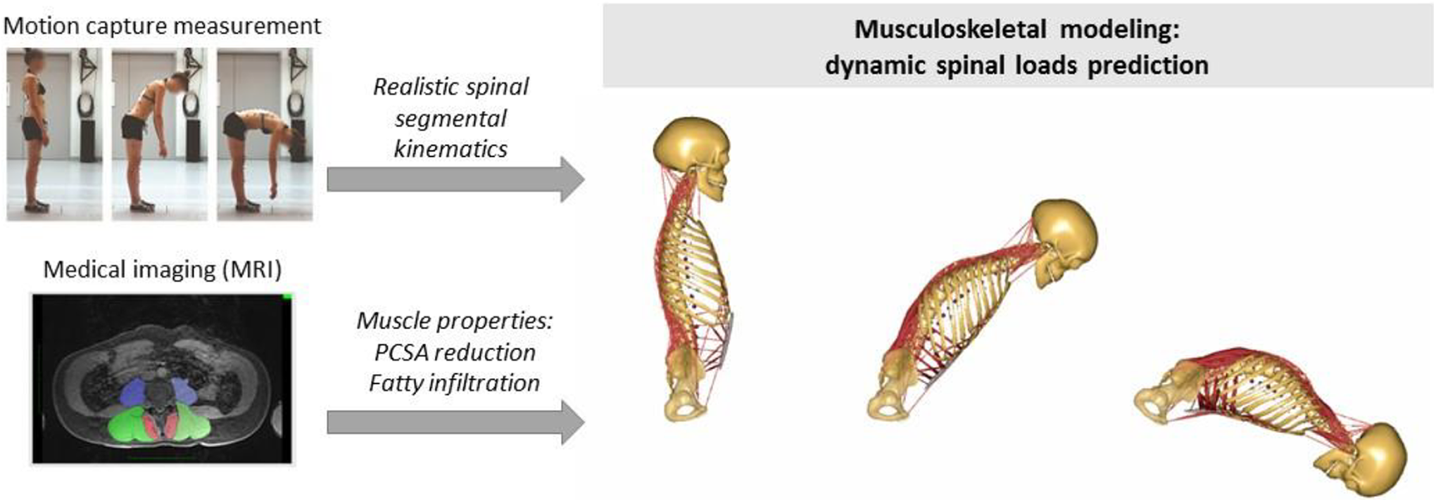
The concept of musculoskeletal modeling of the thoracolumbar spine combined with measurement-derived spine kinematics and paraspinal muscle properties obtained from previously reported imaging studies.

Maximum segmental compressive forces estimated for simulated stages of sarcopenia affecting erector spinae and multifidus muscles, normalized to default (unscaled muscle strength condition).
1. Hida T, Shimakata H, Sakai Y, et al. Sarcopenia and sarcopenic leg as potential risk factors for acute osteoporotic vertebral fracture among older women. Eur Spine J. 2016;25:3424-3431.
2. Mokhtarzadeh H, Anderson DE. The role of trunk musculature in osteoporotic vertebral fractures: implications for prediction, prevention, and management. Curr Osteoporos Rep. 2016;14:67-76.
3. Ignasiak D, Dendorfer S, Ferguson, SJ. Thoracolumbar spine model with articulated ribcage for the prediction of dynamic spinal loading. J Biomech. 2016;49:959-966.
4. Ignasiak D, Ferguson SJ, Arjamand N. A rigid thorax assumption affects model loading predictions at the upper but not lower lumbar levels. J Biomech. 2016;49:3074-3078.
5. Ignasiak D, Rueger E, Ferguson SJ. Thoracic spine kinematics in the young and elderly. Paper presented at: 21st Congress of the European Society of Biomechanics; July 5-8, 2015; Prague, Czech Republic.
6. Crawford RJ, Filli L, Elliott JM, et al. Age- and level-dependence of fatty infiltration in lumbar paravertebral muscles of healthy volunteers. Am J Neuroradiol. 2016;37:742-748.
7. Lang T, Streeper T, Cawthon P, Baldwin K, Taaffe DR, Harris TB. Sarcopenia: etiology, clinical consequences, intervention, and assessment. Osteoporos Int. 2010;21:543-559.
8. Kang CH, Shin MJ, Kim SM, Lee SH, Lee CS. MRI of paraspinal muscles in lumbar degenerative kyphosis patients and control patients with chronic low back pain. Clin Radiol. 2007;62:479-486.
This study has been supported by a research grant from AOSpine International, Switzerland (Project CPP FFOB_OC_14).
Introducing SpinePRO: Digital Clinical Data Collection for Spine Tango
T. R. Jansen1, H. Kohlhof1, R. Bornemann1, S. Hebel1, S. Koob1, D. C. Wirtz1, R. Pflugmacher1, and Y. Rommelspacher1
1Department of Orthopedics and Trauma Surgery, University Clinic Bonn, Germany
1. Melloh M, Staub L, Aghayev E, et al. The International Spine Registry SPINE TANGO: status quo and first results. Eur Spine J. 2006;17:1201-1209.
2. Roeder C, Chavanne A, Mannion AF, Grob D, Aebi M. SSE Spine Tango—content, workflow, set-up. Eur Spine J. 2005;14:920-924.
3. Goz V, Lakomkin N. Reliability of SRS-22 and ODI by phone: a step toward making PROs more accessible. Spine J. 2016;16:1047-1048.
4. Apple Inc. ResearchKit. https://developer.apple.com/researchkit/. Accessed September 20, 2018.
5. AngularJS. Home page. https://angularjs.org/. Accessed September 20, 2018.
The Expression of Transient Receptor Potential (TRP) Channels in Intervertebral Disc Cells Is Regulated by Pro-Inflammatory Cytokines
Olga Krupkova1, Joel Zvick1, Jürgen Klasen2, Stephen J. Ferguson1, Alfredo Franco-Obregon3, Karin Wuertz-Kozak1
1ETH Zurich, Switzerland

Gene expression of TRP channels in IVD cells treated with pro-inflammatory cytokines. IL-1β and TNF-α significantly induced the expression of TRPA1 and inhibited the expression of TRPC6. Graphs shows mean ± SEM (n = 5) evaluated by ANOVA with Tukey post hoc at P < .05, relative to untreated control.
1. Wu LJ, Sweet TB, Clapham DE. International Union of Basic and Clinical Pharmacology. LXXVI. Current progress in the mammalian TRP ion channel family. Pharmacol Rev. 2010;62:381-404.
2. Kaneko Y, Szallasi A. Transient receptor potential (TRP) channels: a clinical perspective. Br J Pharmacol. 2014;171:2474-2507.
3. Walter BA, Purmessur D, Moon A, et al. Reduced tissue osmolarity increases TRPV4 expression and pro-inflammatory cytokines in intervertebral disc cells. Eur Cells Materials. 2016;32:123-136.
4. Nilius B, Appendino G, Owsianik G. The transient receptor potential channel TRPA1: from gene to pathophysiology. Pflugers Arch. 2012;464:425-458.
5. Nummenmaa E, Hamalainen M, Moilanen LJ, et al. Transient receptor potential ankyrin 1 (TRPA1) is functionally expressed in primary human osteoarthritic chondrocytes. Arthritis Res Ther. 2016;18:185.
Biological Disc Replacement Using Tissue-Engineered Intervertebral Discs Combined With a Resorbable Stabilization System: A Proof of Concept Study in an Ex Vivo Beagle Model
Gernot Lang1,2, Jorge Mojica Santiago3, Rodrigo Navarro-Ramirez1, Ibrahim Hussain1, Roger Härtl1, and Lawrence J. Bonassar3,4
1Weill Cornell Brain and Spine Center, Weill Cornell Medicine, New York-Presbyterian Hospital, New York, NY, USA

(A) TE-IVD. (B) TE-IVD in situ. (C) TE-IVD with bioresorbable stabilization system. (D) Equilibrium modulus normalized to the intact motion segment. N = 12, Mean ± SD, *P < .05. (E) Instantaneous modulus normalized to the intact motion segment. N = 12, Mean ± SD, *P < .05.
Hypoplastic Vertebral Body and Recurrent Symptomatic Disc Herniation
Ralph Laeubli, MD1 and Nicolas Fragnière, MD2
1Head Spine Unit Hospitals FMI and Sportsclinic #1, Bern
Comparison of Gene Expression of Discs From Diffuse Idiopathic Skeletal Hyperostosis (DISH) and Healthy (Trauma) Patient
Rahel D. May1, Daniela A. Frauchiger1, Lorin M. Benneker2, and Benjamin Gantenbein1
1Tissue and Organ Mechanobiology, Institute for Surgical Technology and Biomechanics, University of Bern, Bern, Switzerland

TGFβ pathway transcriptome analysis. RNA transcripts that differed more than four times are indicated in re (up-regulated or in green (downregulated in DISH patient) relative to 16-years old trauma patient.
1. Resnick D, Guerra J Jr, Robinson CA, Vint VC. Association of diffuse idiopathic skeletal hyperostosis (DISH) and calcification and ossification of the posterior longitudinal ligament). AJR Am J Roentgenol. 1978;131:1049-1053.
2. Tekari A, May RD, Frauchiger DA, Chan SC, Benneker LM, Gantenbein B. The BMP2 variant L51P restores the osteogenic differentiation of human mesenchymal stromal cells in the presence of intervertebral disc cells. Eur Cell Mater. 2017;33:197-210.
The Influence of Primary Human Intervertebral Disc Cells on Primary Human Osteoblasts
Rahel D. May1, Adel Tekari1, Daniela A. Frauchiger1, Lorin M. Benneker2, Sandro Kohl2, and Benjamin Gantenbein1
1Tissue and Organ Mechanobiology, Institute for Surgical Technology and Biomechanics, University of Bern, Bern, Switzerland

Alizarin red (ALZR) staining and B. Quantification of ALZR staining of primary human osteoblasts, simulated with osteogenic medium and co-cultured with six or twelve, nucleus pulposus cell (NPC), annulus fibrosus cell (AFC) or cartilagenous endplate cell (CEPC) beads, respectively. Positive control groups were cultured with osteogenic medium (± insert and beads) and negative control group was cultured with αMEM only. Optical density of ALZR mean ± SEM. One-way ANOVA p-value *<0.03 ***<0.0001.
1. Watkins R 4th, Watkins R 3rd, Hanna R. Non-union rate with stand-alone lateral lumbar interbody fusion. Medicine (Baltimore). 2014;93:e275.
2. Chan SCW, Tekari A, Benneker LM, Heini PF, Gantenbein B. Osteogenic differentiation of bone marrow stromal cells is hindered by the presence of intervertebral disc cells. Arthritis Res Ther. 2015;18:29.
Cell Transplantation in Lumbar Spine Disc Degeneration Disease
H. J. Meisel1, C. Hohaus2, Y. Minkus1, and T. Ganey3
1Department of Neurosurgery, BG Klinikum Bergmannstrost, Halle/Saale, Germany
1. Ganey T, Libera J, Moos V, et al. Disc chondrocyte transplantation in a canine model: a treatment for degenerated or damaged intervertebral disc. Spine (Phila Pa 1976). 2003;28:2609-2620.
2. Ganey TM, Meisel HJ. A potential role for cell-based therapeutics in the treatment of intervertebral disc herniation. Eur Spine J. 2002;11(suppl 2):S206-S214.
Reduction of Pelvic Incidence and Patient-Reported Outcomes Following Surgical Correction and Stabilization of Lytic Spondylolisthesis
Michael Mokawem1 and Rajesh Shah1
1Department of Trauma and Orthopaedic Surgery, Hull and East Yorkshire Hospitals NHS Trust, Hull, UK
Bone Mineral Density of Vertebras at Instrumented Levels and Levels Above the Instrumentation Increases During the First Postoperative Year After the Lumbar Spine Fusion
Mikko A. Hakulinen1,2, Liisa Pekkanen3, Kati Kyrölä3, Marko H. Neva4, and Arja Häkkinen5,6
1Department of Applied Physics, University of Eastern Finland, Kuopio, Finland
Ferromagnetic Dissection and Discectomy in Scoliosis Surgery: Comparison With Electrical and Mechanical Tools
Alessandro Ramieri1, Giuseppe Costanzo1, and Cloe Curri1
1Orthopaedics, Don Gnocchi Foundation ONLUS, Milan and Rome Sapienza University, Rome, Italy
In Toto and Expanded Human Vertebral Bone Marrow Cells Cultured Under Normoxic and Hypoxic Conditions: A Novel Strategy for Spine Surgery
F. Salamanna1, S. Cepollaro2, G. Barbanti Brodano3, C. Griffoni3, A. Gasbarrini3, S. Bandiera3, S. Terzi3, R. Ghermandi3, S. Boriani3, and M. Fini1
1Laboratory of Preclinical and Surgical Studies, Rizzoli Orthopaedic Institute, Bologna, Italy
In Situ Photopolymerized Composite Hydrogels for Implants: Application to a Nucleus Pulposus Replacement
Andreas Schmocker1,2, Azadeh Khoushabi1,3, Daniela A. Frauchiger4, Benjamin Gantenbein4, Constantin Schizas5, Christophe Moser2, Pierre-Etienne Bourban3, Dominique P. Pioletti1
1Institute of Bioengineering, École Polytechnique Fédérale de Lausanne, Lausanne, Switzerland
Bitten by Translation: Are Veterinary Patient Dogs Good Models for Intervertebral Disc Repair Using Cell Therapy?
Alessandro Bertolo1, Frank Steffen2, and Jivko Stoyanov1
1Swiss Paraplegic Research, Nottwil, Switzerland
Success Stories and Challenges of Translational Studies
M. A. Tryfonidou, DVM, dipl ECVS1
1Department of Clinical Sciences of Companion Animals, Faculty of Veterinary Medicine, Utrecht University, The Netherlands
Successful translation of regenerative treatment strategies for degenerative disc disease demands a multidisciplinary approach. Results of in vitro and in vivo studies should be interpreted with a clinical directive and experiments need to be designed with the translation from bench to bed in mind. In-depth understanding of underlying degenerative and regenerative processes in animal models is facilitated by cutting-edge biomolecular techniques of “nonhuman species” and strong implementation of the 3Rs (reduction, refinement, replacement). Innovative technological platforms, including injectable drug delivery systems, enable local delivery and controlled release of medication for the purpose of pain management and tissue regeneration. On the course of collaborative projects, biomaterials that enable controlled release of anti-inflammatory medication and growth factors have been validated in a preclinical platform, that is, the canine model. The canine species is considered to be a suitable model to study the process of disc degeneration: like humans, dogs suffer from spontaneous disc degeneration and disease. The degenerative process involves similar macroscopic, histopathological, radiological, and biochemical changes as humans. To this end, we employed the Beagle as an experimental animal followed by successful first-in-dogs studies in canine patients suffering from back pain. Lessons learned during these preclinical studies will be discussed focusing on the 3Rs, fine tuning of the treatment strategies respecting the physiology of the IVDs (including needle size, injection volume, loading dose), and the additive value of imaging modalities for longitudinal follow-up.
Disc Regeneration Using MSC Transplanted via the Endplate Route
Gianluca Vadalà1, Fabrizio Russo1, Alessia Valentini2, Marco Bernardini2, Giulia De Benedictis2, Luca Denaro3, Rosaria Giordano4, and Vincenzo Denaro1
1Department of Orthopaedic Surgery, Università Campus Bio-Medico di Roma, Rome, Italy
The support of the Italian Ministry of Instruction, University and Research Grant (PRIN-200938NT8Z), the Young Investigator Research Grant of the Italian Ministry of Health (GR-2010-2 318 448), and the BIOSPINA Award of the Italian Society of Spine Surgery (SICV&GIS) are gratefully acknowledged.
1. Orozco L, Soler R, Morera C, et al. Intervertebral disc repair by autologous mesenchymal bone marrow cells: a pilot study. Transplantation. 2011;92:822-828.
2. Carragee EJ, Don AS, Hurwitz EL, Cuellar JM, Carrino JA, Herzog R. 2009 ISSLS Prize Winner: Does discography cause accelerated progression of degeneration changes in the lumbar disc: a ten-year matched cohort study. Spine (Phila Pa 1976). 2009;34:2338-2345.
3. Korecki CL, Costi JJ, Iatridis JC. Needle puncture injury affects intervertebral disc mechanics and biology in an organ culture model. Spine (Phila Pa 1976). 2008;33:235-241.
4. Vadala G, Sowa G, Hubert M, Gilbertson LG, Denaro V, Kang JD. Mesenchymal stem cells injection in degenerated intervertebral disc: cell leakage may induce osteophyte formation. J Tissue Eng Regen Med. 2012;6:348-355.
5. Vadala G, Russo F, Pattappa G, et al. A Nucleotomy model with intact annulus fibrosus to test intervertebral disc regeneration strategies. Tissue Eng Part C Methods. 2015;21:1117-1124.
6. Vadala G, Russo F, Pattappa G, et al. The transpedicular approach as an alternative route for intervertebral disc regeneration. Spine (Phila Pa 1976). 2013;38:E319-E324.
7. Vadalà G, Fabrizio R, De Strobel F, et. al. Novel stepwise model of intervertebral disc degeneration with intact annulus fibrosus to test regeneration strategies. J Orthop Res. 2018;36(9):2460-2468.
CD146-Positive Mesenchymal Stem Cells Possess a Superior Migration Potential Toward Induced Degenerative Intervertebral Discs
Sebastian Wangler1, Marianna Peroglio1, Zhen Li1, Ursula Menzel1, Lorin M. Benneker2, R. Geoff Richards1, Mauro Alini1, and Sibylle Grad1
1AO Research Institute Davos, Davos, Switzerland

Combined red and green fluorescent image of migrated MSCs in a sagittal section of bovine IVDs. Left: CD146− MSCs (blue); Right: CD146+ MSCs (red). The IVD region without endplates is marked with a yellow line. Scale bar = 1 mm.
1. Pattappa G, Peroglio M, Sakai D. CCL5/RANTES is a key chemoattractant released by degenerative intervertebral discs in organ culture. Eur Cell Mater. 2014;27:124-136.
2. Lee CH, Lee FY, Tarafder S, Kao K, Jun Y, Yang G, Mao JJ. Harnessing endogenous stem/progenitor cells for tendon regeneration. J Clin Invest. 2015;125(7):2690-2701.
Short- and Mid-term Effect of TNF-α Intradiscal Injection and Detrimental Dynamic Loading in Intervertebral Disc Organ Culture
Zhen Li1, Gernot Lang1,2, Yishan Liu1,2, Janna Geries1,2, Zhiyu Zhou1, David Kubosch2, Norbert Südkamp2, Mauro Alini1, and Sibylle Grad1
1AO Research Institute Davos, Davos, Switzerland

Phy: physiological loading (0.02-0.2 MPa; 0.2 Hz; 2 h/day) and high glucose (4.5 g/L) medium; Deg: degenerative loading (0.32-0.5 MPa; 5 Hz; 2 h/day) and low glucose (2 g/L) medium; TNF-α: TNF-α was injected into the nucleus pulposus (NP) tissue, 100 ng/IVD at day 1.
Conditioned medium was collected for nitric oxide (NO) and glycosaminoglycan (GAG) analysis. After 4 or 11 days, disc tissue was harvested and gene expression was analyzed using real-time RT-PCR. After 11 days, cell viability was evaluated by lactate dehydrogenase (LDH)/ethidium homodimer staining of disc cryo-sections. Graphpad software was used to perform the statistics. P < .05 was considered statistically significant.
Funded by the Foundation for the Promotion of Alternate and Complementary Methods to Reduce Animal Testing (SET). Zhiyu Zhou was funded by China Scholarship Council and Sino-Swiss Science and Technology Cooperation.