
Abstract
The present study aimed to investigate the effects of different disinfectants in preventing pin-site infection. A total of 32 healthy New Zealand white rabbits were selected as the research subjects. The rabbits were placed in an Ilizarov 3/4 circular external fixator for a period of time after feeding. They were then divided into four groups of eight animals each. After surgery, pin-site nursing was performed twice a day with a different disinfectant for each group (chlorhexidine gluconate alcohol disinfectant, Maokang iodine, 75% alcohol and physiological saline). Each pin site’s surrounding conditions were visually observed daily, and bacterial culture of the pin-site secretions was promptly performed. The changes of C-reactive protein (CRP) and white blood cell upon the first day, first week, second week, fourth week, and sixth week after the operation were compared, as were infection statuses and morphological changes in the surrounding tissue at the sixth week post operation. The differences in the CRP and white blood cell values and infection rates among the four groups were found to be statistically significant (P < 0.05), but the difference in infection levels among the four groups was not statistically significant (P>0.05). In addition, the difference in the pathologically inflammatory cells among the four groups at the sixth week post operation was statistically significant (P < 0.05).It was concluded that the effect of chlorhexidine gluconate alcohol disinfectant in preventing pin-site infection was superior compared to the other three disinfectants.
Keywords
Introduction
Ilizarov external fixation technology is widely used in the treatment of orthopedic diseases such as osteomyelitis, limb malformation and high-energy injury. Patients are generally treated with an external fixator for 6–18 months or longer. 1 Nevertheless, postoperative syndromes may manifest during the treatment process, of which pin-site infection has become one of the most common. 2 According to literature review reports,3–8 the infection rate of an external fixation pin-site ranges within 0.5%–100%. Pin-site infection not only loosens the fixed pin site but also affects functional exercise; in severe cases, osteomyelitis, nonunion, and treatment failure may also occur.9,10 Therefore, it is imperative to strengthen pin-site nursing in patients with an Ilizarov circular external fixator to prevent infection.
At present, domestic and foreign scholars have investigated the types, methods, and use frequencies of disinfectants in pin-site nursing, but there is no unified standard to date. Furthermore, there is a lack of relevant studies on the effect of disinfectants on the tissue morphology around pin sites. Thus, the present study investigated the effects of different disinfectants on pin-site infections as well as their morphological effects on the tissues surrounding the pin sites through animal experiments, providing a basis for choosing disinfectants during clinical pin-site nursing.
Materials and methods
Experimental animals and grouping
A total of 32 healthy New Zealand rabbits (5–6 months old and weighing 2.5–3.0 kg) were kept in the Bone and Soft Tissue Repair Laboratory of the Second Hospital of Shanxi Medical University for a period of time. This study was approved by Animal Use Committee of the Second Hospital of Shanxi Medical University.
Inclusion and exclusion criteria
Healthy New Zealand white rabbits of similar ages and genders were included, while rabbits with pre-operative skin ulcerations and fractures upon placement in the Ilizarov external fixator were excluded.
Methods
Way of insertion of the external fixator pins
Put the ring-shaped external fixator into the left knee; adjust the distance between the proximal and distal rings and the skin; insert a 1.5 mm Kirschner needle and a 2.0 mm threaded needle across the lower femur and upper tibia while avoiding important structures such as blood vessels, nerves, tendons, and so on; install a fixing nut; and adjust the joint hinge to be located at the center of rotation of the knee joint. These animals had not received any type of medication with antibiotics.
Pin-site nursing methods
(1) Nursing care was performed twice a day using different disinfectants on each group of animals as follows: group A, chlorhexidine gluconate alcohol skin disinfectant (Minnesota Mining and Manufacturing Medical Equipment (Shanghai) Co., Ltd.); group B, Maokang iodine (Maoming High-tech Industrial Development Zone, Maoming, Guangdong Province, China); group C, 75% alcohol (Taiyuan Qing Xu Kangjiu Pharmaceutical Excipients Company); group D, physiological saline (Baxter Healthcare, Shanghai, China). (2) No dressing was used to cover the surrounding area of each pin site postoperatively. A cotton swab was used to disinfect the pin sites and keep them clean and dry. If rabbit hairs were found growing around the pin sites, they were immediately removed using a shaving cutter and scissors. (3) Changes in the pin sites were visually observed daily during the nursing care. If any redness, swelling, or secretion was present at a pin site, a bacterial culture was immediately conducted for the secretions. If a pin site was suspected to be infected, the scab around the site was removed to maintain smooth drainage.
The pathological specimens were prepared within 6 weeks after the operation. A rabbit was taken from each group to be euthanized via excessive chloral hydrate, and the tissue around an uninfected pin site was collected.
Evaluation index
The infection level of each pin site was graded using the Checketts–Otterburn classification, in which grades 1–3 were considered minor infection and grades 4–6 major infection 11 (Table 1).
Checketts–Otterburn classification.

Pin-site problems and infections are classified broadly as minor or major based on Checketts–Otterburn classification. Minor infections can be managed on an outpatient basis, they respond to the appropriate treatment and external fixation can be continued. Major infections often require hospital admission, do not resolve with treatment, and usually involve more than one pin or group of pins. External fixation has to be abandoned.
C-reactive protein (CRP) and white blood cell (WBC) were measured at postoperative first day, postoperative first week, postoperative second week, postoperative fourth week, and postoperative sixth week for each group. For CRP, horseradish peroxidase (HRP)–labeled purified rabbit CRP antibody was used and absorbance of antibody–antigen–HRP conjugates was measured. For WBC values, the number of WBC were counted within a certain volume under the microscope after dilution and dissolving red blood cells.
Pathological evaluation indexes: Hematoxylin and eosin (H&E) staining was performed on the pin-site sections, and the number of inflammatory cells in the tissue surrounding each pin site was counted under a high-power microscope (400× light microscopy).
Statistical methods
The data were statistically analyzed using the SPSS 17.0 software, and the measurement data were analyzed by variance. Fisher’s exact probability method and a non-parameter test were used for the count data and rank data, respectively. A P value < 0.05 was considered statistically significant.
Results
Comparison of CRP values in the four groups of large white rabbits
After repeatedly measuring the variances in the general data of the four groups’ CRP values (Table 2), it was concluded that they were statistically significant (F = 11.707, P < 0.001), as were the CRP values at different time points (F = 204.3, P < 0.001). However, the interaction between the time and groups was not statistically significant (F = 1.821, P = 0.062), indicating that there were no differences in the CRP values among the groups with time.
Comparing CRP values of four groups (mg/mL).

The multiple comparison results revealed the following: Compared with group A, the CRP differences in groups B, C, and D were statistically significant (P = 0.003, P < 0.001, and P < 0.001, respectively). Compared with group B, there was no statistically significant difference in the CRP values between groups C and D (P = 0.061 and P = 0.067, respectively). There was no statistically significant difference in the CRP values between groups D and C (P = 0.961). However, the CRP value in group A was low compared to the other three groups.
The multiple comparison results revealed that the CRP values at postoperative first day, postoperative first week, postoperative second week, postoperative fourth week, and postoperative sixth week were statistically significant (P < 0.001). The CRP values at the first, second, fourth, and sixth weeks post operation were statistically significant compared with the values at day 1 post operation (P < 0.001). The CRP values at the second, fourth, and sixth weeks post operation were statistically significant (P = 0.045, P < 0.001, and P < 0.001, respectively) compared with the values at the first week post operation. The CRP values at the fourth and sixth weeks post operation were statistically significant (P = 0.041 and P = 0.001, respectively) compared with the values at the second week post operation. The CRP values at the sixth week post surgery were statistically significant (P = 0.006) compared with those at the fourth week post surgery. As shown in Figure 1, the CRP values of the four groups continued to increase over time.

The change trend of CRP values in each group.
Comparison of WBC among the groups
After repeatedly measuring the variances in the general data of the WBC values of the four groups (Table 3), it was concluded that they were statistically significant (F = 4.137, P = 0.015), as were the differences in the WBC values at different time points (F = 133.988, P < 0.001). However, the interaction between the time and groups was not statistically significant (F = 1.935, P = 0.078), indicating that the WBC values of the groups did not differ with time.
Comparing WBC values of four groups (×103/mm3).
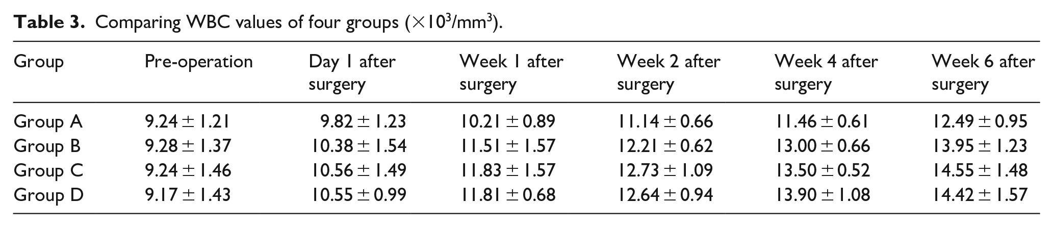
The multiple comparison of these results revealed that compared with group A, the differences in the WBC values in groups B, C, and D were statistically significant (P = 0.033, P = 0.005, and P = 0.005, respectively). Compared with group B, there was no statistically significant difference in the WBC values between groups C and D (P = 0.443 and P = 0.421). Compared with group C, the difference in the WBC values in group D was not significant (P = 0.970). Moreover, the WBC values in group A were low compared to the other three groups.
The multiple comparison results revealed that the differences in the WBC values at all time points were statistically significant (P < 0.001). As shown in Figure 2, the WBC values of the groups continued to increase over time.

The change trend of WBC values in each group.
Comparison of pin-site infections among the groups
Upon comparing the infection rates of pin-site in the four groups (Tables 4 and 5), it could be concluded that P = 0.033 using Fisher’s exact probability method and that the difference was statistically significant. The multiple comparison results revealed the following: Compared with group A, the differences in the infection rates among groups B, C, and D were statistically significant (P = 0.041). However, there were no statistically significant differences (P = 1.000) in the infection rates among groups B, C, and D. That is, the infection rate of group A was lower than that of the other three groups.
Comparing pin-site infection of four groups.

Comparing infection degree of four groups.

Comparison of pathological results among the groups
The changes in the tissues obtained from the uninfected pin sites in the four groups were observed via light microscopy from 6 weeks post surgery. It was observed that a small amount of inflammatory cells (mainly neutrophils) was present in the surrounding tissues of pin sites obtained from the rabbits in all groups, without any tissue hyperplasia or dead tissue (Figures 3). The comparison of inflammatory cells in the pathological sections of the four groups under a high-power microscope (400× light microscopy, Table 6) revealed that the difference in the number of inflammatory cells among the pathological sections was statistically significant (F = 65.414, P < 0.001).
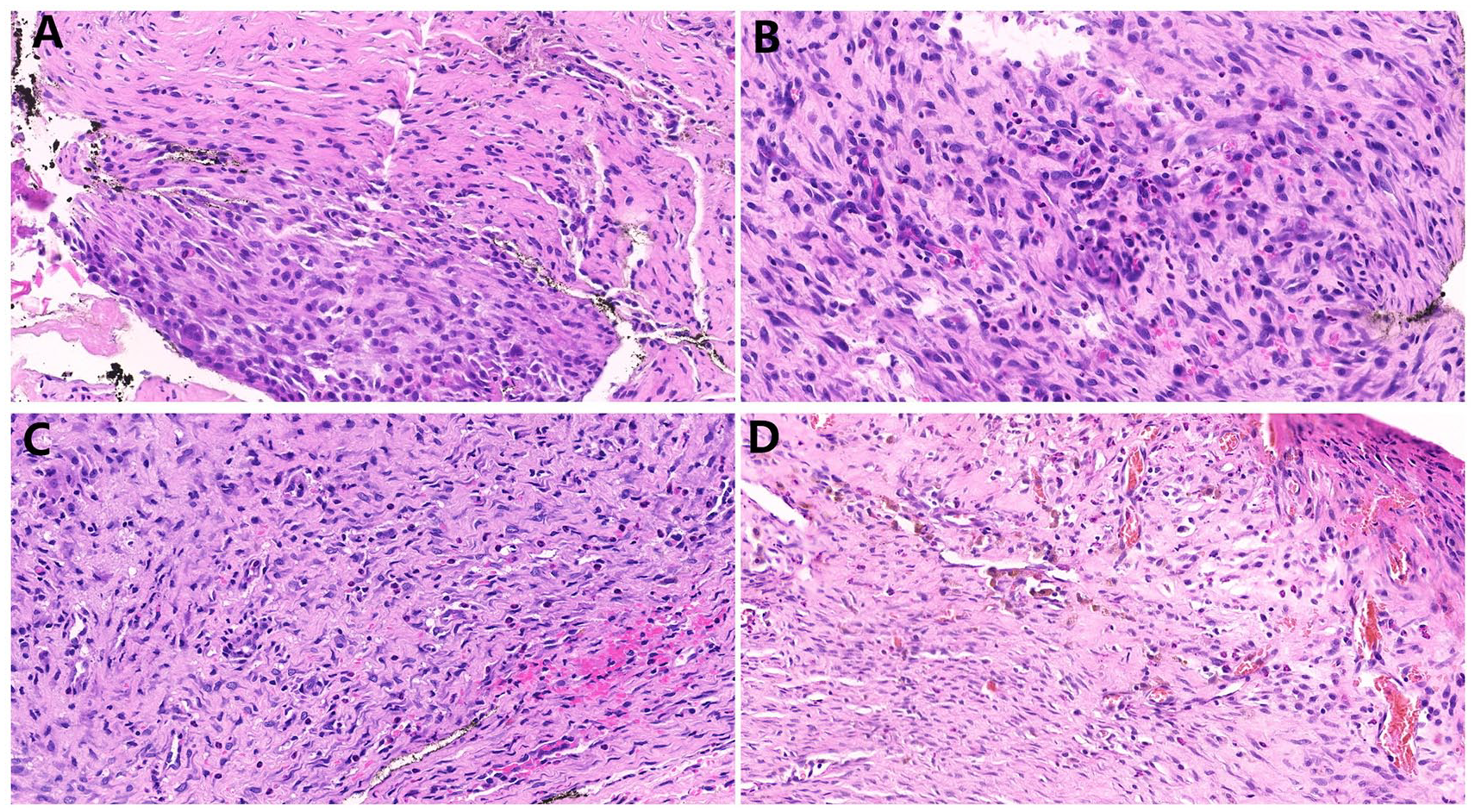
Pathological results of four groups of white rabbits under light microscopy from 6 weeks after surgery (HE staining, 10 × 20).
Comparing inflammatory cells in pathological sections of four groups.

Different superscript letters indicate statistically significant difference, while the same letter indicates no statistically significant difference.
Discussion
Effects of the different disinfectants
(1) The chlorhexidine gluconate alcohol skin disinfectant, which is mainly composed of 1.8%–2.2% (w/w) chlorhexidine gluconate and 63%–77% ethanol, is a long-acting disinfectant with a strong sterilization effect, few adverse reactions, and rapid action.12,13 At present, it is primarily used for clinical surgical site disinfection and the prevention of related catheter infection. Chlorhexidine gluconate is a cationic surfactant mainly used for disinfecting skin mucosa. The ethanol in glucose acid chloride has set the sterilization process with an effect of promotion and synergies. This not only rapidly prohibits bacterial breeding, mycobacterial and fungal spores, and lipotropic virus via short waiting time characteristics but also has a long-acting antibacterial effect. 14 For Staphylococcus aureus, Escherichia coli, and Candida albicans, it has a good killing effect.15–17 (2) The active iodine content of the main ingredient of Maokang iodine is 0.27 g/100 mL–0.33 g/100 mL, and the ethanol content is 50%–60%, which is considered as a medium and an effective disinfectant. It can coagulate and denature bacterial proteins and has a certain sterilization effect. When the effective iodine content is 25–100 mg/L, it can kill S. aureus, E. coli, and C. albicans. 18 However, the color of iodide is a bit dark, and skin pigmentation will occur after prolonged use, which is not conducive to observation. (3) The 75% alcohol can effectively kill bacteria and fungi but not bacterial spores. Furthermore, it has poor stability and is volatile and difficult to store. In addition, an improper concentration will lead to decreased efficacy of clinical disinfection and may cause allergic reactions. 19 A study conducted by Wang Xiaoli 20 revealed that the use of alcohol to sterilize the external fixation of pin-site could lead to dehydration and denaturation of the skin and subcutaneous soft tissues around the pin-site. If used for a long time, this will lead to enlargement of the pin-site and an increased chance of bacterial invasion, leading to pin-site infection. (4) Physiological saline, as a human body balance fluid, is non-sensitizing and irritating to skin mucosa. 21 It cleans pin sites during nursing care but does not kill bacteria; hence, it can be used to clean pin sites for only a short period of time.
In the present study, the pin-site infection was primarily mild, and the infection rate was significantly lower in group A compared to the other three groups. This was correlated with the strong sterilization effect, demonstrating its specific disinfectant applicability for clinical promotion.
Diagnostic indicators of inflammatory response (significance of inflammatory response indicators)
As a highly sensitive acute-phase reactive protein, CRP is a marker associated with tissue injury and inflammation that can accurately reflect the body’s infection status and is little affected by other factors. 22 The WBC count is neither sensitive nor specific to the prediction of infection, but it is the most commonly used indicator of infection in clinics, as an inflammatory response occurs in the body as the WBC increase. At present, the CRP and WBC counts are combined for the clinical diagnosis of infection. 23 Herein, the CRP and WBC values of the four groups increased at different levels; this increase was lowest in group A compared to the other three groups because chlorhexidine gluconate alcohol used in group A had a strong killing effect on bacteria. Thus, the inflammatory reaction in the body was relatively light.
At present, several studies 24 have combined interleukin 6 (IL-6) with the procalcitonin, CRP, and WBC counts when diagnosing inflammatory response. The relevant diagnostic indicators of inflammation should be improved in subsequent clinical studies.
Effects of disinfectants on tissues surrounding the pin site
In the present study, it was observed under a high-power microscope that a small amount of inflammatory cells (mainly neutrophils) appeared in the surrounding tissues of the pin sites. On one hand, the Kirschner pin site belongs to the foreign matter for the body when implanted and can easily stimulate the body’s immune response. On the other hand, the area surrounding the pin site may appear to have an inflammatory response after bacterial implantation. However, no fibrous tissue hyperplasia or necrotic tissue was found in the surrounding tissues in the four groups under a light microscope, and no erythema was observed in the skin surrounding the pin sites. Thus, little irritation was caused to the skin by the four disinfectants. In comparison, regarding the inflammatory cell numbers at high magnification, the value was lower in group A compared to the other three groups, indicating that the inflammatory response in that group was lightest and chlorhexidine gluconate alcohol has great effect against infections in the inoculation route.
Research limitations and predictions
In the present study, the effect of the disinfectants on the tissue morphology surrounding the pin site was added on the basis of commonly used clinical observation indexes, but there were some deficiencies. Clinically, the external fixator placement was relatively long. Moreover, due to the limitation of the experimental arrangement and specific operation time, the observation time was relatively short. Besides, subject used in this study was New Zealand white rabbits instead of human, and the effect on final results caused by the difference between these two species cannot be overlooked. Therefore, if the results of the present study are to be promoted in clinics, the crowd intervention time should be further extended with human beings selected as experimental subjects. And power analysis should be performed to determine the sample size before the experiment, which was missing in this study.
Brief summary
Pin-site infection is one of the most common postoperative syndromes after external fixator placement. Currently, although there are many kinds of nursing methods and disinfectants, regardless of which is available, there is a lack of solid evidence, and the curative effects are quite different. Therefore, the search for better and more evidence-based disinfection reagents and methods for preventing infection has become a top priority of clinical researchers.
Footnotes
Animal welfare
The present study followed international, national, and/or institutional guidelines for humane animal treatment and complied with relevant legislation.
Declaration of conflicting interests
The author(s) declared no potential conflicts of interest with respect to the research, authorship, and/or publication of this article.
Ethical approval
This study was approved by Animal Use Committee of The Second Hospital of Shanxi Medical University.
Funding
The author(s) disclosed receipt of the following financial support for the research, authorship, and/or publication of this article: Shanxi Provincial general Natural Fund Program (201701D121172).