
Abstract
This study investigates the expression of phosphatase and tensin homolog (PTEN) and Inositol polyphosphate 4-phosphatase type II (INPP4B) in children with acute myeloid leukemia. The levels of PTEN and INPP4B in bone marrow, from 95 acute myelogenous leukemia (AML) patients and 84 controls, respectively, were assessed by immunohistochemistry, quantitative polymerase chain reaction (qPCR), and Western blot. The prognosis was followed up and investigated and the correlation analysis was made. We found that the expression levels of PTEN and INPP4B were significantly lower in the AML group than those in the control group (P < 0.05). The survival time was lower in PTEN and INPP4B negative children relative to PTEN and INPP4B positive children (P < 0.05). In AML patients, INPP4B and PTEN expression was positively correlated (r = 0.552, P = 0.000). In conclusion, the levels of INPP4B and PTEN were reduced significantly and correlated positively in AML patients accompanying with abnormal karyotypes. The current investigation of INPP4B and PTEN could give new insight into targeted therapy for AML.
Introduction
Acute myelogenous leukemia (AML) mainly occurring in adults is a genetically and epigenetically heterogeneous disorder, which presents high morbidity and mortality all over the world, including in China. 1 At present, the etiological agent and pathogenesis of AML are not entirely clear. Only few AML cases can be accurately classified through traditional cellular morphological classification. Thus, it is very difficult to judge the disease condition and predict prognosis. The phosphatase and tensin homolog (PTEN) gene is a negative regulatory factor of PI3K/AKT signal channel activation processes and inhibits carcinogenic activity of PIK3 and facilitates cancer cell invasion or the possibility of transfer to achieve tumor inhibition. 2 Inositol polyphosphate 4-phosphatase type II (INPP4B) is a newly discovered lipid phosphatase and a potential tumor suppressor in the context of PTEN deficiency PMID: 26152921. This study aims to further elucidate the possible pathogenesis of AML by studying the expression of PTEN and INPP4B genes in children with myeloid leukemia and provide effective clinical reference values for the occurrence and prognosis of AML in the future.
Data and method
Ethical approval
The Institutional Ethics Committee of our hospitals approved the study, and written informed consent was obtained from all the participants.
Object of study
A retrospective study method was applied. In total, 59 child AML patients who were treated at the Leukemia Department of Baoding Children’s Hospital from February 2011 to May 2018 were chosen as the AML group. Inclusion criteria included the following: definitely diagnosed by bone marrow cytologic and immunological detection; complete clinical data; did not receive chemotherapy during the initial presentation; parents were informed and consented; Ethics Committee of Baoding Children’s Hospital approved the study; age 3–12. Exclusion criteria were as follows: suffered from other hematological system diseases; combined with a severe organ function disorder, including heart, lung, brain, liver, and kidney; lack of clinical data. In the same period, 84 children with nonhematological malignancies with a normal bone marrow examination were chosen as the control group, and the children were not infected.
Leukocyte count, sex, age, immunophenotype (CD34+ positive, CD117+ positive) and chromosome karyotype of all children were investigated and recorded. The total survival time and event-free survival time of children in the AML group were assessed by follow-up.
Immunohistochemistry
The two-step method was used to detect PTEN and INPP4B expression in bone marrow samples of the AML and control groups. All the specimens were fixed with 100 g/L neutral formalin, embedded with paraffin and cut into slices (4 μm) continuously. Each specimen was cut into three slices. One slice was used for hematoxylin and eosin (HE) staining, and immunohistochemical staining was performed using the other two slices. All the operating steps were performed in strict accordance with the reagent specifications. In the negative control, PBS replaced the primary antibody, and a gastric tissue specimen with known positive expression was used as the positive control. PTEN and INPP4B expression results were based on the revised Hercep Test scoring standard: 0 indicates no staining or <10% tumor cells have membrane staining; 1+ indicates >10% tumor cells have faint membrane staining or tumor cells only have partial membrane staining; 2+ indicates >10% tumor cells have weak-medium complete or substrate staining outside membrane; 3+ indicates >10% tumor cells have medium-strong complete or substrate staining outside membrane. In this experiment, 0 and 1 were regarded as negative; 2+ and 3+ were regarded as positive. Individual sections of each case were evaluated under the same conditions, and statistical comparisons of the degree of staining were performed on a QuantStudio 6 Flex polymerase chain reaction (PCR) system (Thermo Fisher, Waltham, MA, USA). Quantitative real-time PCR (qRT-PCR) primers for PTEN and INPP4B expression were commercially available from Applied Biosystems (Foster City, CA, USA). Glyceraldehyde 3-phosphate dehydrogenase (GAPDH) was used as an internal control for gene and miRNA quantitation, respectively. All independent PCR-based reactions were performed in triplicate. qPCR results were calculated by calibrator-normalized relative quantification with efficiency correction using LightCycler@480 Relative Quantification Software (Version 1.5) and are provided as normalized ratios. Full details of the experimental procedure are described in previous studies. 3
Western blotting
The whole cells were lysed in lysis buffer (Thermo Fisher), and protein concentrations were determined using a DC Protein Assay kit (Bio-Rad). Total protein lysates were fractionated on 4%–15% polyacrylamide gels and transferred onto nitrocellulose (Bio-Rad, Hercules, CA, USA). GAPDH was used as a housekeeping protein. The following primary antibodies were used: anti-PTEN (1:500 dilution; Bioss, Woburn, MA, USA), anti-INPP4B (1:800; Abcam), and anti-GAPDH (1:500; Sigma-Aldrich). After being washed with tris-buffered saline with Tween 20 (TBST) thrice, the membranes were incubated with horseradish peroxidase (HRP)-conjugated secondary Abs (Bio-Rad) for 1 h. All protein experiments were performed in triplicate. Separate blots were used for each independent experiment to avoid problems related to incomplete membrane stripping.
Statistical analysis
All data were analyzed by SPSS11.0 (Chicago, IL, USA), obtained from at least three independent experiments, and expressed as the mean ± standard deviation. Kruskal–Wallis H tests (nonparametric K independent sample test) were performed to evaluate the correlation between immunohistochemical markers within the different subgroups based on the origin. The correlation between PTEN and INPP4B expression was tested by Spearman’s correlation analysis.
Categorical data were analyzed by chi-square test, and the comparison of grading data was performed using rank sum test. A P value less than 0.05 was considered statistically significant.
Results
Comparison of general data
Comparisons of gender, age, and white blood cell count in both groups revealed no statistically significant differences. The proportion of CD34+ and CD117+ cells in the AML group was higher compared with that of the control group, and the proportion of karyotype abnormities was lower than that of the control group. The comparison was statistically significant (P < 0.05), as shown in Table 1.
Comparison of general data of children with different phenotypes.

AML: acute myelogenous leukemia.
Prognosis
In the AML group, the total survival time was (18.29 (+1.22) months, and the event-free survival time was (10.48 (+1.78) months. Compared with PTEN and INN4B negative subjects, the total survival time and event-free survival time of PTEN and INpp4B positive children were significantly increased (P < 0.05).
Immunostaining of PTEN in the AML and control groups
Positive expression rates of PTEN and INPP4B in bone marrow tissues of AML group was lower than that of the control group (40.1% vs 89.1%, 43.8% vs 86.2%, P < 0.05), as shown in Table 2 and Figure 1.
PTEN and INPP4B positive expression in bone marrow tissues of the AML and control groups.
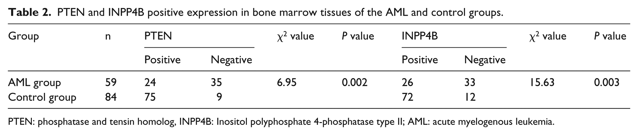
PTEN: phosphatase and tensin homolog, INPP4B: Inositol polyphosphate 4-phosphatase type II; AML: acute myelogenous leukemia.

Representative immunohistochemical staining for PTEN and INPP4B in bone marrow samples of the AML and control groups. (a) PTEN expression in AML group. (b) PTEN expression in control group. (c) INPP4B expression in AML group. (d) INPP4B expression in control group.
mRNA and protein expression of PTEN and INPP4B in the AML and control groups
This study found that the average PTEN and INPP4B expression levels in AML patients were significantly lower compared with that if the control group (P < 0.05) (Figure 2(a)). PTEN and INPP4B protein expression levels in the AML group were also significantly lower compared with that of the control group (P < 0.05) (Figure 2(b)).

The relative (a) mRNA and (b) protein levels of PTEN and INPP4B detected in the AML and control group, respectively.
Correlation between PTEN and INPP4B expression in AML patients
In this study, PTEN and INPP4B expression was correlated in AML patients, and PTEN and INPP4B expression was positively correlated (χ2 =34.17, r = 0.562, P = 0.000).
Discussion
In this study, we demonstrated that the expression levels of INPP4B and PTEN in the AML group was significantly lower than that in the control group. Such findings on PTEN and INPP4B expression in AML patients are basically consistent with studies in other tumors.4,5 Statistical analysis showed that INPP4B and PTEN expression was positively correlated in AML patients, suggesting that INPP4B and PTEN may participate together in the occurrence and development of AML. The common deletion of INPP4B and PTEN is one of the important factors involved in the development of breast cancer. 6 More interestingly, INPP4B protein loss was frequently found in tumors that also lacked PTEN. 7
The INPP4B gene harbors a series of special structures, such as a C2 lipid binding domain at the N-terminal that promotes the dephosphorylation and degradation of phosphatidylinositol-3,4 diphosphate, thereby inhibiting AKT activation, blocking PI3K/AKT signaling pathway transmission, and inhibiting tumor progression. 8 The PTEN gene, as the first cancer suppressor gene with bispecific phosphatase activity is widely expressed in normal tissues. 9 PTEN protein converts 3,4,5-phosphatidylinositol into 4,5-phosphatidylinositol, induces Caspase-9 protein expression, and mediates cancer cell apoptosis. 10 The absence of PTEN and INPP4B may lead to the accumulation of phosphatidylinositol-3,4,5-triphosphate and phosphatidylinositol-3,4-diphosphate catalyzed by PI3K, which maximize AKT activity, enhance cell activity, and lead to anchorage-independent growth of tumor cells.
To sum up, the levels of INPP4B and PTEN were reduced significantly and correlated positively in patients with AML in our study. This investigation of INPP4B and PTEN in AML could give new insight into targeted therapy for this aggressive blood cancer. Further experimentation is clearly needed; however, in light of these data, studies applying tissue-specific deletion of INPP4B and PTEN would shed light on the combined role of these two factors in controlling tumor progression and metastasis in other contexts.
Footnotes
Declaration of conflicting interests
The author(s) declared no potential conflicts of interest with respect to the research, authorship, and/or publication of this article.
Funding
The author(s) received no financial support for the research, authorship, and/or publication of this article.