
Abstract
One of the most devastating consequences of diabetes mellitus is a chronic condition, diabetic foot ulcer. Numerous investigations are being targeted to explore newer compounds for treatment of diabetic foot ulcer wounds in diabetic patients. Hesperidin (HSP), an isoflavone glycoside has been established to exhibit antidiabetic and antioxidant potential. In the current investigation, diabetes was induced in rats by administration by streptozotocin (STZ) intraperitoneally (50 mg/kg). Wound-healing capacity was estimated in hind paw of rats by artificially initiating wound injury on the paw dorsal surface. The injured animals were administered with incremental doses of HSP suspension orally (10, 20, 40, 60, and 80 mg/kg) and insulin subcutaneously (10 IU/kg). Parameters such as wound area were estimated every 2 days, and at the end of 20 days of study, biochemical estimations in serum and histopathological observations of the wound were made. HSP (60 and 80 mg/kg) revealed statistically significant (P < 0.05) improvement in wound dimension, glucose and insulin concentration, and glycated hemoglobin (HbA1C). Administration of HSP indicated significant (P < 0.05) modulation of mRNA associated with expression of vascular endothelial growth factor (VEGF), whereas the levels of tumor necrosis factor (TNF)-α and interleukin (IL)-6 levels were lowered compared to the control group of animals. Real-time quantitative polymerase chain reaction (RT-qPCR) indicated expression of vascular endothelial growth factor receptors 1 and 2 (VEGFR1 and VEGFR2) compared to glyceraldehyde 3-phosphate dehydrogenase (GAPDH). Histological observations indicated higher expression of VEGF in the groups receiving HSP, indicative of angiogenesis stimulation in the diabetic wound. The results advocate angiogenesis activity of HSP was enhanced owing to reduction in hyperglycemia and oxidative stress–induced damage, reduced expression of inflammatory mediators, and enhanced expression of growth-related factors, thereby promoting healing of diabetic foot ulcer.
Introduction
An alarming projection of diabetes mellitus (DM), a life-threatening intricate disease, in 2030 reveals its occurrence in around 366 million individuals globally.1–3 The individuals affected with DM will also indicate multi-organ complications related to eyes, kidneys, heart, and foot ulcer. 4 Around 40%–60% of DM victims are known to suffer from severe foot ulcer, which might further worsen into amputation of the affected organ. The healing of such diabetic ulcer is primarily determined by the extent of neuropathy and vasculopathy. 5 In majority of DM patients, neuropathy is associated with reduced or almost vanished sensation of pain in the lower extremities, which adds up to the development of larger wounds till being identified. Recovery from such detrimental condition is much difficult, as localized ischemia is coupled with number of metabolic deviations. 6 Statistical findings indicate prevalence of diabetic wound/foot ulcer in around 15% of diabetic patients, of which around 3% may be undesirably compelled to elimination of the lower limb. 7 Deviations in normal metabolic events may even worsen the surgical situation in patients with diabetes, making the amputation more complicated. 8
The process of wound healing is a finely coordinated and controlled process, regulated by numerous factors and is inclusive of—hemostasis, inflammation, granulation, and repair.9,10 Supporting players in the process of wound healing include blood cellular components, keratinocytes, various growth and proinflammatory mediators, and endothelial cells. The background mechanism of the wound-healing process has been vaguely clarified, but overall anyone of the above factors may modulate or delay the process of wound healing in diabetes. 11 Several studies have been documented that explain the delayed process of wound healing in diabetic condition. Reports claim that apoptosis induced by the presence of enhanced reactive oxygen species (ROS) may be a major contributor in the delayed wound repair. 12 Also, reduced expression or activity of the growth promoters—epidermal growth factor, insulin-like growth factor, transforming growth factor, vascular endothelial growth factor (VEGF) and platelet-derived growth factor—may contribute the situation. Reduction in the activity of inflammatory responses and reduced collagen levels in the vicinity of wound may lower the process of wound repairing. Reports provided by The Diabetes Control and Complications Trial reveal that restricted level of glucose in diabetic individuals could possibly reduce the neurologic and micro-vascular issues and thereby help to hasten the process of wound healing and reduction.
Clinical treatment of diabetic foot ulcer is an intricate process, as it involves a blend of other pathological implications such as hypo-perfusion, unusual angiogenesis and tissue repair, unusual hemodynamics, and localized neuronal damage. 13 These complications make the diabetic foot ulcer difficult to treat by single-treatment strategy. A few treatment tools target to remove the wound debris from the site of ulcer, use of antimicrobials, biological implants, corticosteroids, and proteolytic enzymes. However, the use of these strategies is limited due to narrow spectrum of clinical efficiency and precipitation of unwanted effects. 14 Research targets from few decades involve the use of growth factors such as epidermal growth factor, insulin-like growth factor, transforming growth factor, VEGF, and platelet-derived growth factor to accelerate the diabetic foot ulcer by cell regeneration and angiogenesis. 15 The use of such approaches has been limited due to non-uniformity in the process of wound healing and of course, the cost. Altogether, diabetic foot ulcer, a chronic condition needs to be clinically treated with number of therapeutic approaches for a long duration, might help to improve the quality of life of the patient, at the same time may increase the cost of treatment. 16 Hence, there is a serious need to explore new therapeutic agents that could effectively be able to treat such a multidimensional chronic disorder, with less side effects and cost effectivity.
Plant-derived compounds have received a widespread acceptability for the treatment of diabetic wounds and are evidenced by their strong clinical implications. Phytoconstituents belonging to the class of flavonoids, isoflavonoids, alkaloids, and triterpenes have been extensively investigated for their capacity to recover wounds in diabetic cases. Investigations carried out on some of these phytochemical constituents have strongly supported their significant therapeutic potential for treatment of diabetic foot ulcer on a clinical platform.17–19
Specific animal models that may have significant clinical utility are required for exploration of novel molecules with therapeutic potential. 20 Use of rats, induced with diabetes by injecting STZ has been a model of choice for studying diabetic foot ulcer. Reason being a model which mimics clinical and pathological symptoms and can reproducibly exhibit hyperglycemia, increased oxidative stress, localized reduction on oxygen, neuronal growth abnormalities, and impaired metabolic and immunological functionalities. 21
Hesperidin (HSP), an isoflavone glycoside molecule, can be isolated from citrus species, and is known for its various effects on biological systems.22,23 The effects of HSP have been known to exhibit cancer-preventive and cardiovascular-protective effects. These activities have been investigated and have been found to be linked with anti-inflammatory, free-radical scavenging, and subsequently the anti-oxidant effects. HSP has been known to potentially exhibit anti-cancer activity by its virtue to modulate target multiple cellular proteins, including caspase, B-cell lymphoma, and its associated X-protein, thereby induces apoptosis. 24 Moreover, HSP is known to attenuate cyclooxygenase-2, matrix metalloproteinase (MMP-2 and MMP-9), and thereby promote angiogenesis and stimulate metastasis. HSP has also recently been established for its activity against oxidative stress and production of anti-inflammatory cytokines in STZ-induced diabetic rat model. 25 Past explorations made on HSP, strongly endorse its potential to elicit possible activity against diabetic foot ulcer. The investigation presented, was targeted toward deeply exploring the wound-healing activity of HSP over rat model induced with diabetes using STZ. Apart from normal parameters to be evaluated in antidiabetic study over rat model, the studies were targeted toward investigation of biochemical parameters and understanding the mechanism of angiogenesis and anti-inflammatory potential after administration of HSP.
Materials and methods
Experimental animals
Adult male standard deviation (SD) rats (210–250 g) were procured from Central animal house of the University. The animals were stored in standard polypropylene cages (three rats per cage) and were exposed to optimized environmental conditions; 22°C–25°C, 55% ± 5% relative humidity and 12 h light and 12 h dark cycle. The animals were provided free access to standard chow diet (PicoLab Rodent Diet, PMI Nutrition International Inc, USA) and pre-filtered drinking water ad libitum. The experimental protocols approved by the Institutional Central Committee for Animals were carried out during day time, between 09:00 and 17:00 hours. HSP was purchased from Xian Yiyang Bioteh Co, China. STZ and thiopentone sodium were purchased from Sigma-Aldrich Co, USA.
Development and assessment of STZ-induced diabetes
DM was induced in experimental animals (n = 10) by intraperitoneally injecting a single dose of STZ (50 mg/kg in 0.1 M citrate buffer, pH 4.4).26,27 The rats from control group were administered with equivalent amount of the respective citrate buffer. The serum glucose concentration was analyzed in blood samples collected through retro-orbital plexus procedure using commercially available Hitachi 7072A Clinical Analyzer. All the experimental animals indicated serum glucose levels above 250 mg/dL and hence were considered for further investigation.
The diabetic animals were anesthetized using thiopentone sodium (40 mg/kg). The wound of standard dimension 2 mm × 5 mm was produced by removing skin layer from the dorsal surface of the right hind foot employing a non-opaque flexible template of plastic. The rats were shuffled into various groups of 10 animals each. The study groups involved for investigation were according to the experimental protocol approved by the Institutional Central Committee for Animals (Animal Ethical Order Key Laboratory for Molecular Diagnosis of Hubei Province/01/2017).
Experimental design
Each group involved in the experimental protocol had 10 SD rats. Total number of animals approved by the Institutional Central Committee for Animals were 90 SD rats.
Group I animals were non-diabetic and non-wounded (NDNW)
Group I: The rats involved in this group were not induced with diabetes and were not wounded. The rats received sterile water for injection (WFI) orally for 20 days.
Group II animals were non-diabetic and wounded (NDW)
Group II: The rats involved in this group (Sham group) were non-diabetic and were wounded. The rats received sterile WFI orally for 20 days.
Animal Groups from III to IX were diabetic rats and wounded (DW), as per the procedure indicated above.
Group III: The rats from this group were administered with sterile WFI orally for 20 days.
Group IV: The rats from this group were administered with aqueous suspension of HSP (10 mg/kg) orally for 20 days.
Group V: The rats from this group were administered with aqueous suspension of HSP (20 mg/kg) orally for 20 days.
Group VI: The rats from this group were administered with aqueous suspension of HSP (40 mg/kg) orally for 20 days.
Group VII: The rats from this group were administered with aqueous suspension of HSP (60 mg/kg) orally for 20 days.
Group VIII: The rats from this group were administered with aqueous suspension of HSP (80 mg/kg) orally for 20 days.
Group IX: The rats from this group were administered with insulin (10 IU/kg) subcutaneously.
Fresh HSP suspension was prepared everyday freshly in water for injection for administration to the animals of respective groups in doses 20, 40, 60, and 80 mg/kg beginning from the day, the wound was created. The wound area was measured periodically by an individual who was blind to the various treatment groups. Food and water consumption was analyzed periodically by keeping them in metabolic cages. After completion of 20 days of the study, the animals were anesthetized using ether, and the blood was collected by retro-orbital puncture and refrigerated for estimation of biochemical aspects.
Wound area measurement, wound contraction, and half-closure time
Wound area was manually measured after 1, 2, 4, 8, 12, 16, and 20 days by an observer unknown to the treatment groups. The percent wound closure (%WC), that is time during which the wound has been closed, was estimated using the following equation

A graph of %WC versus duration in days was plotted and statistically treated using GraphPad Prism software, and the value of half-closure time, that is, the time required to reduce the wound diameter to 50% (CT50), was calculated.
Quantification of glucose, insulin, and glycated HbA1c level determination
Glucose level was quantified using commercially available kits (Fine test, Hubei, China). The level of insulin was estimated using enzyme-linked immunosorbent assay (ELISA) kit following the technique reported by the supplier. Serum concentration of glycated HbA1c was determined using rat HbA1c ELISA kit (Fine test) according to the procedure provided by the manufacturer.
Estimation of biochemical parameters
Tissues collected from rat skin was disrupted and crushed evenly using a laboratory scale portable tissue homogenizer (Guangzhou Fan Bo Lun Co Ltd, China). The tissues were suspended in phosphate buffer pH 7.4 and subjected to homogenization at 4°C. The supernatant collected was subjected to cryocentrifugation at 5500g, and the resulting samples were analyzed for the determination of superoxide dismutase (SOD), reduced glutathione (GSH), hydroxyproline (HDP) content, lipid peroxidation and myeloperoxidase (MPO) using assay kits (Sigma-Aldrich).28,29
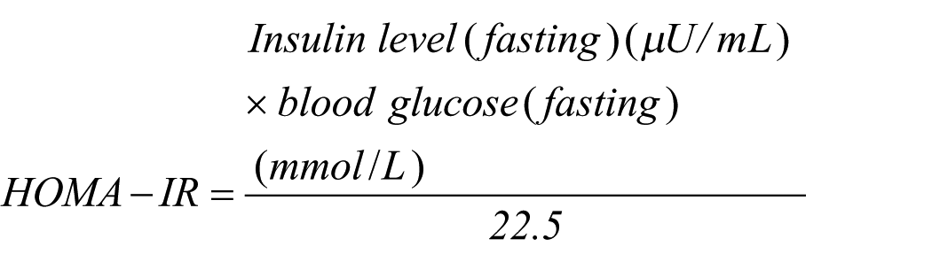
Homeostasis model of insulin resistance estimation
Resistance to insulin was determined by homeostasis model of insulin resistance (HOMA-IR) using following equation. HOMA-IR determination signifies importance of insulin resistance, considering standalone detection of glucose and insulin may not be sufficient to define the disorder30,31
TNF-α and IL-6 estimation
ELISA kits (Fine test) were used for the determination of serum proinflammatory cytokines levels of tumor necrosis factor (TNF)-α and interleukin (IL)-6. The ELISA kits were used as per the instructions enclosed by the manufacturer. The levels of these cytokines were analyzed using ultraviolet (UV) visible spectrophotometer at 450 nm. Unknown concentrations of TNF-α and IL-6 were determined by extrapolating the unknown absorbance’s of serum samples to the concentration axis on the standard curve.
Determination of VEGFR1 and VEGFR2 expression using RT-qPCR
Quantitative real-time polymerase chain reaction (RT-qPCR) technique was used for studying the expression of vascular endothelial growth factor receptors 1 and 2 (VEGFR1 and VEGFR2). RNA was isolated from plasma fractions using PureLink RNA kit (Thermo Fisher China Ltd, Shanghai, China), as per the instruction manual, and supported by the earlier studies. 32 The PCR template was generated by subjecting the total RNA for reverse transcription into complimentary DNA. RT-qPCR was carried out employing specific primer sequences: For VEGFR1—forward sequence 5′ TTTAAAAGGCACCCAGCACAT 3′ and the reverse sequence 5′ TTACTCACCATTTCAGGCAAAGAC 3′, for VEGFR2—forward sequence 5′ GGCCCAATAATCAGAGTGGCA 3′ and the reverse sequence 5′ TGTCATTTCCGATCACTTTTGGA 3′, and for glyceraldehyde 3-phosphate dehydrogenase (GAPDH)—forward sequence 5′ GGGAAACTGTGGCGTGAT 3′ and the reverse sequence 5′ AAAGGTGGAGGAGTGGGT 3′. To estimate the comparative expression of mRNA between various samples, the samples were normalized up to the level of GAPDH, and these values were expressed as percentage value of the control sample (vehicle blank).
Protein analysis by Western blotting
After breakdown of 1 g of wound tissues, by treatment with RIPA (radio immunoprecipitation assay) buffer and protease inhibitor, it was subjected to cryocentrifugation at 4°C for 10 min at 12,000g. The protein estimation was performed using Bradford kit (Beyotime, USA) and Western blotting. Membranes resulted were subjected to incubation with primary anti-β-actin antibodies (1:1000 ratio of dilution), and anti-VEGF (1:800 ratio of dilution) followed by mixing with horseradish peroxidase (HRP)-coupled secondary antibodies. Western blotting band detection system was employed for visualizing the separate protein bands and were further quantified using desitometry. Subsequently, membranes were dislocated and treated with β-actin-specific antibody. 33
Histopathological investigation for screening of VEGF
After the completion of treatment regimen with HSP, four animals from individual groups were randomly chosen and euthanized by administration of lethal dose of pentobarbital (800 mg/kg) intraperitoneally. Skin collected from the wound and the adjacent area was collected and treated with 4% paraformaldehyde solution and loaded in a block of solid paraffin. The preserved skin specimens were sliced into sections of 2 µm thickness. These fine sections were placed onto a slide and de-paraffinized and treated with VEGF antibody staining solution (1:100). The stain resulted in the conversion of VEGF into brown color granulated appearance of the endothelial capillaries. The specimens were visualized with the help of light microscope (Leitz Wetzlak 12 V, Germany) connected to Olympus CX44 camera, and the resulting images were gathered using processing software ImagePro plus 6.0.
Statistical treatment of data
The data was statistically treated with SPSS v17.0 software. Two-way analysis of variance (ANOVA) was applied to the data gathered for the interpretation of wound area and %WC and the data of biochemical parameters was treated using one-way ANOVA. Bonferroni and Tukey’s multiple range test were applied for post hoc analysis. The P values less than 0.05 were considered statistically momentous.
Results
HSP effect on glucose level and body mass
Animals from group III, comprising rats induced with diabetes and wounded but untreated, serum glucose levels were increased significantly (P < 0.5), whereas there was a significant drop in the body weight (P < 0.5), in comparison to animals from group I (non-diabetic non-wounded) and group II (non-diabetic and wounded). Treatment animal groups receiving HSP indicated significant drop in the elevated glucose levels (P < 0.05), coupled with remarkable improvement in the body weight (P < 0.05). These results were further compared to that indicated in group IX animals which were treated with insulin administration (10 IU/kg). As expected, insulin administration reduced the elevated serum glucose levels (P < 0.05), and the body mass was improved (P < 0.05). Among the animal groups receiving HSP, group VIII, receiving 80 mg/kg HSP, had indicated higher reduction in the serum glucose levels (P < 0.05) compared to group VII, receiving 60 mg/kg HSP (Table 1).
Impact of hesperidin treatment on various animal groups.
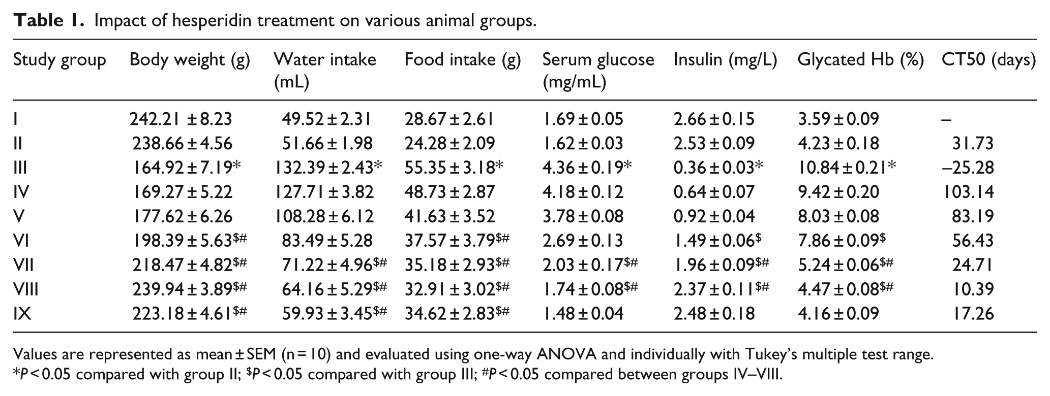
Values are represented as mean ± SEM (n = 10) and evaluated using one-way ANOVA and individually with Tukey’s multiple test range.
P < 0.05 compared with group II; $P < 0.05 compared with group III; #P < 0.05 compared between groups IV–VIII.
HSP effect on consumption of food and water in diabetic rats
In animals of group III, that is diabetic and wounded (DW), it was revealed that the consumption of food and water had enhanced significantly (P < 0.05) in comparison to non-diabetic groups I and II (NDNW and NDW). Administration of HSP in animals of groups IV–VIII (DW), indicated significant reduction (P < 0.05) in food and water consumption. Consecutively, in group VIII rats (DW with HSP dose 80 mg/kg orally), there was a remarkable reduction in water and food consumption, compared to the other rats administered with variable doses of HSP. Group IV, V, and VI rats, administered orally with HSP 10, 20, and 40 mg/kg, respectively, did not indicate marked reduction in food and water intake in comparison to rats from experimental groups VII and VIII (DW with HSP oral dose 60 and 80 mg/kg, respectively). Rats from group IX, administered with insulin (10 IU/kg), revealed significant reduction (P < 0.05) in symptoms of increased food and water intake in diabetic animals. The findings are indicated in Table 1.
HSP effect on serum insulin and glycated Hb levels
Serum insulin levels were found to be significantly (P < 0.05) lower, whereas glycated Hb concentration was significantly (P < 0.05) higher in rats of group III (DW), in comparison to rats of non-diabetic groups I and II (NDNW and NDW). Group VII and VIII rats (DW with HSP oral dose 60 and 80 mg/kg, respectively) depicted significant (P < 0.05) improvement in insulin levels, coupled with marked (P < 0.05) reduction in glycated Hb concentrations. Insulin administration in group IX (10 IU/kg) rats, indicated significant (P < 0.05) attenuation in the levels of insulin and glycated Hb. The observations are indicated in Table 1.
Influence of HSP on wound-related study parameters
HSP administration revealed wound healing in animals induced with diabetes. Group II animals (NDW) indicated wound healing, in comparison to group III (DW) animals, which were diabetic, wounded, and did not receive any treatment for wound healing, and the wound reduction rate was significantly (P < 0.05) lower in comparison to group I (NDW) animals. Administration of HSP indicated significant (P < 0.05) reduction in the wound area, together with enhanced wound contraction rate, compared to group III (DW) animals. Insulin delivered to animals in group IX, exhibited significant (P < 0.05) lowering of wound area, as well as increase in wound contraction rate. The wound half-closure time, that is CT50, was found to be lowest in animals administered with 80 mg/kg of HSP, as shown in Table 1.
Wound tissue analysis for estimation of SOD activity and GSH, HDP, MPO, and MDA levels
Statistically significant (P < 0.05) increase in the GSH levels were observed in animal groups treated with HSP, as indicated in Table 2. Significant (P < 0.05) lowering in the concentrations of GSH and HDP concentrations, and activity of SOD was visualized in group III (DW) animals in comparison to group I (NDNW) and II (NDW) rats. These levels were eventually redeemed with significant (P < 0.05) values with administration of HSP, compared with the groups not administered with HSP. Also, within the experimental animals receiving incremental dose of HSP (Groups IV–VIII), there was a statistically significant (P < 0.05) improvement in activity of SOD, GSH, and HDP levels, indicating dose-dependent relationship for wound healing in diabetic foot. Animal group IX, receiving insulin (10 IU/kg), did not reveal any significant modulation in the activity of SOD, as well as levels of GSH and HDP.
Impact of HSP treatment on levels of various biochemical parameters.
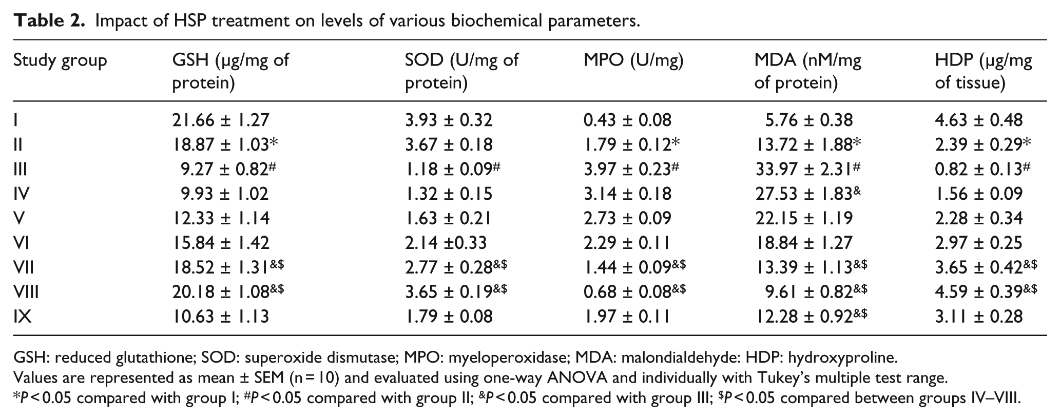
GSH: reduced glutathione; SOD: superoxide dismutase; MPO: myeloperoxidase; MDA: malondialdehyde: HDP: hydroxyproline.
Values are represented as mean ± SEM (n = 10) and evaluated using one-way ANOVA and individually with Tukey’s multiple test range.
P < 0.05 compared with group I; #P < 0.05 compared with group II; &P < 0.05 compared with group III; $P < 0.05 compared between groups IV–VIII.
Malondialdehyde (MDA) and MPO activity was significantly (P < 0.05) higher in diabetic rats from group I. Animal groups undergoing HSP administration indicated marked (P < 0.05) reduction in the levels of MDA and MPO in a dose-dependent manner, that is, marked lowering in group VIII (80 mg/kg of HSP) and comparatively reduced lowering in group IV (10 mg/kg of HSP).
Effect of HSP on HOMA-IR values
Diabetic animal groups indicated statistically significant (P < 0.05) increase in HOMA-IR value in comparison to the non-diabetic group (NDNW) of animals. The value of HOMA-IR was significantly (P < 0.05) improved in animal groups receiving HSP in a dose-dependent manner, compared to the diabetic group (Table 3).
HSP effect on insulin resistance index and levels of inflammatory mediators.

Values are represented as mean ± SEM (n = 10) and evaluated using one-way ANOVA and individually with Tukey’s multiple test range.
P < 0.05 compared with group II; $P < 0.05 compared with group III; #P < 0.05 compared between groups IV–VIII.
Influence of HSP on TNF-α and IL-6 levels
The effect of HSP on the levels of TNF-α and IL-6 has been indicated in Table 3. The levels of both TNF-α and IL-6 were found to be elevated significantly (P < 0.05) in group III (DW) not receiving any treatment for diabetes in comparison to group II (NDW). Animal groups (group IV–VIII) receiving incremental doses of HSP, indicated reduction in the levels of TNF-α and IL-6. Reduction in the TNF-α and IL-6 levels was comparatively significant (P < 0.05) in group VIII (80 mg/kg HSP) than in group IX on insulin therapy (10 IU/kg).
Western blotting reveals HSP induced VEGF expression
Analysis of Western blot reveals expression of VEGF protein compared to β-actin. Animal group administered with HSP (80 mg/kg) indicated maximum expression of VEGF. The Western blot results were further subjected to densitometry quantification, whereby VEGF levels were normalized by β-actin and the results were expressed as ratio of VEGF:β-actin, which indicates expression of VEGF comparably higher in the animal group receiving higher amount of HSP (Figure 1).

(a) Expression of VEGF expression using Western blot and quantified by desitometry, normalizing by β-actin levels. (b) Effect of HSP on VEGF:β-actin ratio in diabetic animals (*P < 0.05).
Histopathological exploration
Immunohistochemical treatment of wounds specimens with VEGF-specific antibodies indicated the presence of VEGF. Typical brown colored spots indicating focal of reaction between VEGF and its specific antibodies was seen under microscope. Increase in the number of brown spots, indicated higher VEGF content in the specimen (Figures 2 and 3).

Immunohistochemical treatment of wounds specimens with VEGF-specific antibodies indicating typical brown colored spots indicating reaction between VEGF and its specific antibodies seen under microscope.

Effect of percentage expression of VEGF in various animal groups after administration of HSP (*P < 0.05).
Impact of HSP on VEGFR1 and VEGFR2 expression
To study the angiogenesis capability of HSP, study of VEGFR1 and VEGFR2, well-established markers for angiogenesis, were explored using RT-qPCR. Relative VEGFR1 and VEGFR2 level was significantly (P < 0.05) higher in animal groups receiving HSP compared to group not receiving HSP treatment. Animal group receiving insulin (group IX) indicated reduced expression of VEGFR1 and VEGFR2, compared to those animal groups (groups VII and VIII) receiving HSP for 20 days (P < 0.05). This investigation reveals direct relationship between VEGFR1 and VEGFR2 expression and the increased dosing of HSP (Figure 4).

Effect of HSP administration in various animal groups on expression of mRNA in comparison to β-actin (*P < 0.05).
Discussion
Diabetes retards the process of wound healing, which is the co-ordination of several elements together to reduce the damage made to skin and tissues. All these elements work together to result into a complete wound closure and repair the damage tissues to achieve their original integrity. 34 This process of wound healing has been well established to be delayed owing to diabetes and is also known to result into significant morbidity. Retardation in the wound-healing process in individuals suffering from diabetes may be contributed by any one or more of the following factors—reduced insulin and growth hormone levels, improper flow of blood, disturbed cell membrane permeability, delayed tissue formation, reduction in collagen synthesis, enhanced ROS, and variability in apoptosis pattern. 35
Diabetic foot ulcer, is a condition normally associated with symptoms such as general reduction in strength and vitality and excessive hunger and thirst as a result of disturbed cellular metabolic functioning owing to lower levels of available glucose.36,37 Increased levels of glucose, is considered as one of the prominent factors that leads to retarded wound healing by lowering neutrophil-assisted chemotaxis attachment, followed by phagocytosis and killing of infectious. 29 Literature suggests that one of the clinical implications of diabetic foot ulcer may be increased level of glycated hemoglobin (HbA1C). 38 Advanced glycosylation end product levels in an extrapolated situation of long-term significant elevated levels of HbA1C. Cross-linking of advanced glycosylation end products with fibronectin is known to delay the process of wound healing in diabetes condition. 39 In the study reported hereby, HSP was found to markedly reduce the HbA1C levels at higher doses, which might be considered one of the factors that actively assists in healing diabetic foot ulcer.
Results indicate significant elevation of blood glucose coupled with reduced insulin levels in diabetic animals. Increased HOMA-IR values indicate that these animals have developed resistance to insulin. 31 This property in rats clearly resembles with that of metabolic features of the humans. Animals administered with HSP reveal lowered HOMA-IR values, which signify the beneficial impact of HSP in reducing the possible resistance to insulin, that is created in diabetic animal model.
Wound-healing steps are characterized by generation of granulation tissue, and coupled with regeneration of epithelium may further result into wound closure. 40 Administration of HSP in the experimental models accelerated the process of wound healing and reduced the CT50 value to almost one-third, compared to non-diabetic and wounded group of animals. The extent of wound contraction rate was significantly found to increase with HSP administration.
Flavonoids have been established to lower production of nitric oxide, leukotrienes, prostaglandins, cytokines, chemokines, and adhesion molecules, by modulating nitric oxide synthase and cyclo and lipoxygenase expression. Flavonoids are known to provide protective antioxidant effect by impeding the oxidative enzyme reaction chain. 41
SOD and GSH, free-radical scavenging anti-oxidants, are known to trigger cell defense system against the ROS. The cells in the vicinity of wounds may be more prone to oxidation-mediated injury, owing to reduction in the levels of SOD and GSH.42,43 Flavonoids have been well established as to reduce ROS and subsequently produce antioxidant effect. In the investigation reported hereby, HSP treatment significantly reclaimed the reduced levels of SOD and GSH at the site of diabetic wound. This may be as a result of enhanced relocation and production of keratinocytes and fibroblasts at the site of wound.
Elevated levels of lipid peroxidation (MDA) are primarily owing due to structural breakdown of unsaturated fatty acids present as lipid component in the cell membrane. Delayed healing of diabetic wound may be due to the inability of antioxidant enzymes to exhibit antioxidant effect at the injury site. Also, increased levels of MPO, signifies amassing of polymorphonuclear leukocytes. 10 Animals treated with HSP tend to exhibit marked reduction in the levels of MDA and MPO, revealing its possible role in antioxidant effect and subsequent acceleration in wound repair and closure.
Studies reported earlier indicate efficiency of HSP to lower diabetic complications including hyperglycemia and damage due to oxidative stress. 44 Importance of inflammatory process is of very importance in the process of wound healing. TNF-α and IL-6 have been authenticated as most crucial players in inflammatory response and correspondingly in neutrophil and macrophage recruitment to trigger the immune responses. This may alternatively be responsible for clearance of microorganisms from the site of wound. Siqueira et al. 41 revealed that the rate of wound healing in diabetes may be lowered due to elevated levels of TNF-α and IL-6, resulted by stimulation of apoptosis and reduced spread of fibroblasts. Reports reveal that HSP downregulates mRNA expression of cytokines such as TNF-α and IL-6. Reduction in the TNF-α and IL-6 after administration of HSP might therefore be responsible for increased rate of wound healing in diabetic animals.
Angiogenesis, an intricate several-step process, includes the conversion inherent blood vessels into of endothelial cells. The process of blood vessel development, organ maturation and cell revival, not only involves oxygen, but also requires various regulatory signaling instructions to the cells and tissues. 45 Any form of interruption in the angiogenesis process may further progress to variable diseases such as retinopathy, neuropathy, prolonged lung disorders, and arthritis. 46 Non-biologically differentiated microangiopathy during diabetes may lead to depletions of cellular growth requirements that progress to retarded wound healing. 47 VEGF may be among the various effective single growth treatment targets. Important factors required for effective wound healing include VEGF and collagen. Production of collagen is initiated in the area of wound after the injury to provide structural strength to the tissue network and enhances repair and wound healing in diabetes. 48 One of the significant precursors for synthesis of collagen is hydroxyproline, which is present at the site of tissue damage, and its presence is an indication of collagen biosynthesis. Diabetes is often characterized by reduced hydroxyproline, which subsequently reduces collagen and lowers tissue repair. 49 Administration of HSP in experimental animals indicated significant elevation in levels of hydroxyproline.
VEGF, a functionally diversified growth factor, is produced by thrombocytes, neutrophils, macrophages, and keratinocytes. It has specific roles to be performed on endothelial cells to carry out angiogenesis. The endothelial cells hold VEGFR1 and VEGFR2, which are responsible for the generation of blood vessels after being triggered by active VEGF. 50 Expression of mRNA related to VEGF has been reported to decline significantly at the wound site in diabetic rats and is thus considered as a fundamental cause of retardation of diabetic wound-healing process. 51 Literature reveals, expression of VEGF mRNA in diabetic induced rats triggers the process of angiogenesis and thereby accelerates the wound-healing process. 52 In the study reported hereby, HSP was found to stimulate the expression of VEGF mRNA and accelerate the process of angiogenesis, thereby accelerating the wound-healing process.
Conclusion
Results in the investigation envisaged advocate angiogenesis effect in diabetic wound induced in rats after treatment with HSP. Supporting aspects include reduced HbA1C and increase in HOMA-IR values. Wound healing was also measured using CT50 value and wound dimensions, thereby revealing efficacy of wound-healing potential of HSP. Reduction of inflammatory mediators such as TNF-α and IL-6 also marks acceleration in wound healing. Increase in level of hydroxyproline after HSP administration, marks tissue repair by increase in collagen in tissues. Increase in the expression of VEGF was identified by mRNA quantification, revealing angiogenesis with administration with HSP.
Footnotes
Acknowledgements
L.W. and T.H. contributed to this work equally.
Declaration of conflicting interests
The author(s) declared no potential conflicts of interest with respect to the research, authorship, and/or publication of this article.
Funding
This study was supported by Science and Technology Planning Project of Hubei Province, China (Grant No.2017CFC849) and the National Natural Science Foundation of China (Grant No.31400916).