
Abstract
Human exposure to radio-therapeutic doses of gamma rays can produce late effects, which negatively affect cancer patients’ quality of life, work prospects, and general health. This study was performed to explore the role of Piceatannol (PIC) in the process of “mitochondrial biogenesis” signaling pathway as possible management of disturbances induced in stressed animal model(s) either by gamma-irradiation (IR) or administration of reserpine (RES); as a mitochondrial complex-I inhibitor. PIC (10 mg/kg BW/day; orally) were given to rats for 7 days, after exposure to an acute dose of γ-radiation (6 Gy), or after a single reserpine injection (1 g/kg BW; sc). Compared to reserpine or γ-radiation, PIC has attenuated hepatic and renal mitochondrial oxidative stress denoted by the significant reduction in the content of lipid peroxides and NO with significant induction of SOD, CAT, GSH-PX, and GR activities. PIC has also significantly alleviated the increase of the inflammatory markers, TNF-α and IL-6 and apoptotic markers, cytochrome c, and caspase-3. The decrease of oxidative stress, inflammation, and apoptotic responses were linked to a significant amelioration in mitochondrial biogenesis demonstrated by the increased expression and proteins’ tissue contents of SIRT1/p38-AMPK, PGC-1α signaling pathway. The results are substantiated by the significant amelioration in mitochondrial function verified by the higher levels of ATP content, and complex I activity, besides the improvement of hepatic and renal functions. Additionally, histopathological examinations of hepatic and renal tissues showed that PIC has modulated tissue architecture after reserpine or gamma-radiation-induced tissue damage. Piceatannol improves mitochondrial functions by regulating the oxidant/antioxidant disequilibrium, the inflammatory and apoptotic responses, suggesting its possible use as adjuvant therapy in radio-therapeutic protocols to attenuate hepatic and renal injuries.
Introduction
Radiation therapy or radiotherapy is the use of ionizing radiation to destroy cancer cells and to control selected metastases. A large majority of cancer patients undergo radiation therapy as a part of cancer treatment including cancers of the breast, prostate, cervix, head and neck, lung, and brain, as well as sarcomas. 1 Side effects come from damage to healthy cells and tissues near the treatment area. Therapeutic abdominal irradiation for the treatment of bowel cancer is usually associated with higher rates of non-specific abdominal pain than those observed in radiation-naïve patients, years after treatment. 2 Goldberg and Lehnert 3 stated that avoidance of damage to non-cancerous tissues outside the target zone is a major priority in using ionizing radiation (IR). Radiation-induced oxidative stress involves a variety of deleterious cellular injuries including alterations in gene expression, augmented intracellular ROS production, mitochondrial damage, and apoptosis. Similarly, various reports linked xenobiotics-induced hepatic injury to a distinct type of factors including oxidative stress, inflammatory response, and cell death.
Mitochondrial complex-I, the largest and most complicated enzyme of the electron transport chain and the entry point for electrons to the respiratory chain, is considered the rate-limiting step in overall respiration, and thus play a crucial role in energy metabolism. 4 Mutations in its subunits lead to a wide range of inherited neuromuscular and metabolic disorders. 5 Reserpine is documented to inhibit complex-I, stimulate reactive oxygen/nitrogen species associated with inflammation, and mitochondrial dysfunction in various vital organs. 6
The cellular mitochondrial volume comprises about 4%–25% of the total cell volume; depending on the cell type and state, thus, the mitochondria represent a substantial target for cytoplasmic irradiation. 7 Mitochondrion plays a crucial role in the tissue bioenergetics status; therefore, its dysfunction is considered a prime trigger of diseases. 8 The peroxisome proliferator-activated receptor-gamma coactivator-1α (PGC-1α) is an important transcriptional co-activator that induces both mitochondrial metabolism and biogenesis at the molecular level. PGC-1α has been reported to possess a pivotal role in various diseases, where it showed an altered expression in many organs after chemo- and radiotherapy. 9 However, its role in radiation-induced mitochondrial dysfunction is still unclear. The activation of PGC-1α is controlled via Peroxisome proliferator-activated receptors (PPARs), AMP-activated protein kinase (AMPK), and Sirtuin1 (SIRT1). Briefly, AMPK phosphorylation promotes the expression and activation of PGC-1α; likewise, SIRT1 deacetylates and activates PGC-1α, regulates mitochondrial biogenesis. 10 Thus, agents activating the SIRT1-mediated PGC-1α signaling pathways might be of vital significance in regulating the antioxidants and mitochondrial functional states following the toxic destruction caused by certain exo or endotoxins. 11 An example of such agents is piceatannol (PIC), a polyphenol belonging to the stilbenes group; a resveratrol analog, existing naturally in plants. For example, both red and white grapes contain PIC at concentrations of 374 and 43 ng/g, respectively. 11 In addition, passion fruit and Vaccinium berries are rich in PIC.12,13 It was reported that PIC possesses a wide spectrum of biological activities including antidiabetic, neuroprotective, cardioprotective, anti-allergic, and anti-aging.14–16 It also protects retinal pigment epithelium cells against UV-B irradiation. 17 In light of the above-detailed information, this study aimed to modulate the radiation-mediated mitochondrial dysfunction as compared to that induced by the classical mitochondrial toxin; reserpine, with a focus on the potential effect of piceatannol against both stressors-induced liver and kidney injuries.
Materials and methods
Methods
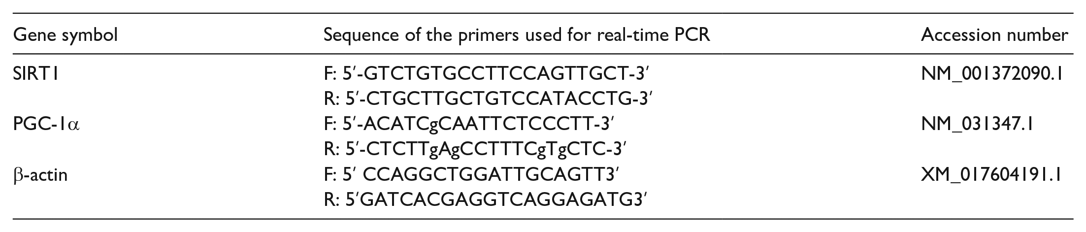
Protein levels were quantified by densitometric analysis of the autoradiograms using a scanning laser densitometer (Biomed Instruments). Results were determined after normalization for β-actin protein expression (as the housekeeping protein).
Histopathological examination
Samples of liver and kidney tissues were fixed in 10% formaldehyde solution and inserted in paraffin using standard methods. Sectioned tissues at 3-μm, were treated with hematoxylin-eosin (H&E) stain. Under a light microscope, the stained sections were inspected. 33 . The histopathological changes of hepatocytes and degree of periportal lymphocyte infiltration were recorded according to Ramos et al. 34 The histopathological findings of renal lesions were scored according to Zhang et al. 35
The hepatic lesions were scored according to the following criteria:

The renal lesions were scored according to the following criteria:

Statistical analyses
Comparisons among different groups were performed by one way analysis of variance (ANOVA). It is a parametric statistical analysis that compares between- and within-groups variance to measure differences between two or more groups. All statistical analysis were performed by using statistical package SPSS (Statistical Program for Social Science) (SPSS® Chicago, IL, USA) version 15.0 software program. Each value represents the mean ± standard deviation collected from eight animals in each group. The difference between means were considered significant at P < 0.05.
Results
Influence of PIC on hepatic and renal mitochondrial oxidative stress
As for RES and IR effects on the hepatic oxidative stress status, a notable increase in liver MDA content associated with an obvious inhibition in the mitochondrial activities of SOD, CAT, GSH-Px, and GR were observed in RES and IR groups in comparison to the control group. PIC treatment has significantly attenuated the RES and IR-induced rise in hepatic MDA contents and restored the hepatic mitochondrial activities of SOD, CAT, GSH-Px, and GR observed in PIC + RES and PIC + IR groups as compared to those of the RES and IR untreated groups (Table 1).
Effect of PIC on hepatic and renal mitochondrial redox status in rats exposed to RES or γ-radiation.

Data are presented as means ± SD (n = 8). Groups with different letters are significantly different, at P ⩽ 0.05. Groups with similar letters are not significantly different, at P ⩽ 0.05.
Regarding the effects of PIC against RES and IR-induced renal injury, nephrotoxicity biomarkers; serum urea and creatinine levels, were assessed. Results displayed in Table 1 revealed a marked elevation in serum urea and creatinine levels, accompanied by an elevation in renal MDA contents, together with the inhibition of renal mitochondrial SOD, CAT, GSH-Px, and GR activities in RES and IR groups as compared to the control group. On the other hand, PIC reversed those effects as it significantly reduced serum urea and creatinine levels as well as the renal MDA content. Moreover, PIC administration induced a significant elevation of renal mitochondrial SOD, CAT, GSH-Px, and GR activities in PIC + RES and PIC + IR groups, compared to RES and IR groups (Table 1).
Piceatannol effect on inflammatory markers and hepatic and renal functions
Serum liver enzymes activities; AST, ALT and ALP, were performed to determine the extent of liver injury induced by reserpine (RES) administration and ionizing radiation (IR) exposure. In Table 2, a significant increase in serum AST, ALT, and ALP activities in RES and IR groups; as compared to the control, is displayed. On the other hand, treatment of RES and IR rats with PIC significantly corrected the hepatic injury induced by each of the toxicants in serum AST and ALT activities as compared to the untreated RES and IR groups (Table 2). As displayed in Table 2, RES and IR groups showed a marked rise in serum levels of IL-6 and TNF-α, as compared to the control rats. Remarkably, administration of PIC to RES and IR groups significantly decreased serum IL-6 and TNF-α levels in PIC + RES and PIC + IR groups, compared to their corresponding control group.
Effect of PIC on inflammatory markers and hepatic and renal functions status in rats exposed to RES or γ-radiation.

Data are presented as means ± SD (n = 8). Groups with different letters are significantly different, at P ⩽ 0.05. Groups with similar letters are not significantly different, at P ⩽ 0.05.
Influence of PIC treatment on hepatic and renal mitochondrial changes in rats exposed to RES or γ-radiation
The activity of the mitochondrial Complex I enzyme and the ATP content were assessed as markers for both hepatic and renal mitochondrial functions. Each of reserpine injection and radiation exposure induced a significant decrease in hepatic and renal mitochondrial ATP contents and complex I activity, compared to untreated groups (Table 3). However, PIC-treated groups showed a significant increment in both the mitochondrial dehydrogenase (complex-1) activity and ATP content.
Effect of PIC on markers of hepatic and renal mitochondrial function in rats exposed to RES or γ-radiation.

Data are presented as means ± SD (n = 8). Groups with different letters are significantly different, at P ⩽ 0.05. Groups with similar letters are not significantly different, at P ⩽ 0.05.
Influence of PIC on hepatic and renal apoptotic markers
The mitochondrial dysfunction was also reflected in an obvious status of hepatic and renal cellular apoptosis. Data displayed in Table 4 indicated that each of RES and IR significantly increased the tissue contents of hepatic and renal cytochrome c and caspase-3 as compared to control group. Significant decrements were recorded in PIC + IR group (Table 4). In case of PIC + RES group, partial yet statistically significant improvements were observed, compared to RES group.
Effect of PIC on hepatic and renal apoptotic markers in rats exposed to RES or γ-radiation.
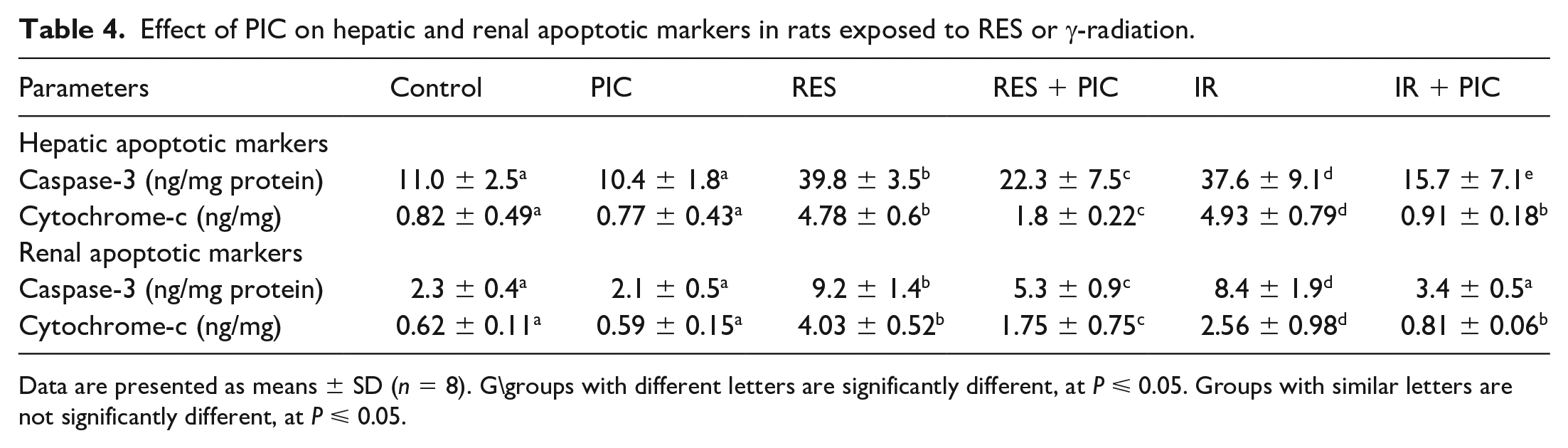
Data are presented as means ± SD (n = 8). G\groups with different letters are significantly different, at P ⩽ 0.05. Groups with similar letters are not significantly different, at P ⩽ 0.05.
PIC treatment upregulated the hepatocellular PGC-1α/p38 signaling molecules
As shown in Figure 1a to d, each of RES and IR induced a profound reduction in protein expression of hepatic and renal active p38 (p-p38Thr180/Tyr182) as indicated by the decrement in p-p38/t-p38 expression. This was accompanied by a significant decline in hepatic and renal PGC-1α gene expression and protein level as seen in RES and IR groups, compared to the control group. PIC treatment improved the RES and IR-induced disruption expression of hepatic and renal active p38 and PGC-1α proteins when compared to RES and IR untreated groups; respectively (Figure 2a–d).

Effect of whole body γ-irradiation (IR), reserpine (RES), and piceatannol (PIC) treatment on: rat hepatic and renal t-p-38/p-p38ratio protein expression (a and c), and the corresponding representative western blotting SDS-PAGE of hepatic and renal t-p38 and p-p-38 (40 kDa) normalized to β-actin (43 kDa) protein expression (b and d).

Effect of whole body γ-irradiation (IR), reserpine (RES), and piceatannol (PIC) treatment on: hepatic and renal PGC-1α mRNA fold change (a and b), hepatic and renal PGC-1α protein level (c and d).
On the molecular level, SIRT-1 mRNA and protein expression levels were markedly decreased in RES and IR treated groups, compared to the control group. Noteworthy, PIC administration induced an improvement in hepatic and renal SIRT-1 gene expression and protein levels, compared to RES or IR groups (Figure 3a–e).

Effect of whole body γ-irradiation (IR), reserpine (RES), and piceatannol (PIC) treatment on: rat hepatic and renal SIRT-1 mRNA fold change (a and b), hepatic and renal SIRT1 protein expression (c and d), and the corresponding representative western blotting SDS-PAGE of hepatic and renal SIRT-1 (110 kDa) normalized to β-actin (43 kDa) protein expressions (e).
Discussion
The current study investigated the alterations in mitochondrial biogenesis machinery induced by a single dose of radiation exposure, in comparison to that produced by the classical mitochondrial toxin; reserpine, intoxication via the induction of a state of oxidative stress along with subsequent changes of both liver and kidney mitochondrial functions. Other than oxidative stress, there are a variety of proposed mechanisms that may cause tissue injuries, such as the amplification of inflammatory reactions, the alternation in gene expression and cellular signaling pathways, and the induction of apoptosis. 36
In the present study, the significantly higher tissue contents of lipid peroxides and NO with significant inhibition of SOD, CAT, GPX, and GR activities indicated both radiation and reserpine-induced physiological toxicities in liver and kidney tissues. Such disruptions in the redox signaling status induced a consequent dysfunction in each liver and kidney as reflected by the observed changes in liver function biomarkers; ALT, AST, and ALP as well as those of renal function; serum urea, and creatinine as compared to control group. Comparable results were reported in the study of Moustafa et al. 37
A possible mechanism of those elevations is the RES-induced disruption of plasma membrane permeability hepatocytes leakage of these enzymes into serum. 38 On the same line, a recent study 39 reported that radiation-induced nephrotoxicity was triggered by ROS-mediated depression in Cyclin D1 expression in the renal nuclei and cells, thus inducing changes in the intracellular signaling with a consequent renal injury.
Following its systemic administration, reserpine is distributed to tissues based on their blood flow and easily penetrate cell membranes and then bind to the intracellular membrane components, particularly those rich in phospholipids, forming adducts inducing a nonstoichiometric inhibition of monoamine uptake, 40 with a consequent oxidative stress status due to the oxidation processes of those monoamines by monoamine oxidases. 41 Also, accumulation of tissue lipid peroxides induces an inflammatory status interfering with the mitochondrial functions in multiple organs including the liver and kidney.
In the current work, the overproduction of ROS; resulting from IR-induced tissue water radiolysis, and RES-induced oxidative mitochondrial injury, can react with macromolecules and other essential targets inducing pronounced autolysis in mitochondria 42 which results in mitochondria-mediated apoptosis of hepatocytes 43 as confirmed by the significant increase in hepatic and kidney cytochrome c and caspase-3 activity shown in the present results. Furthermore, the hepatotoxicity and nephrotoxicity in RES and IR treated rats were herein confirmed by histopathological findings (Figures 4 and 5).
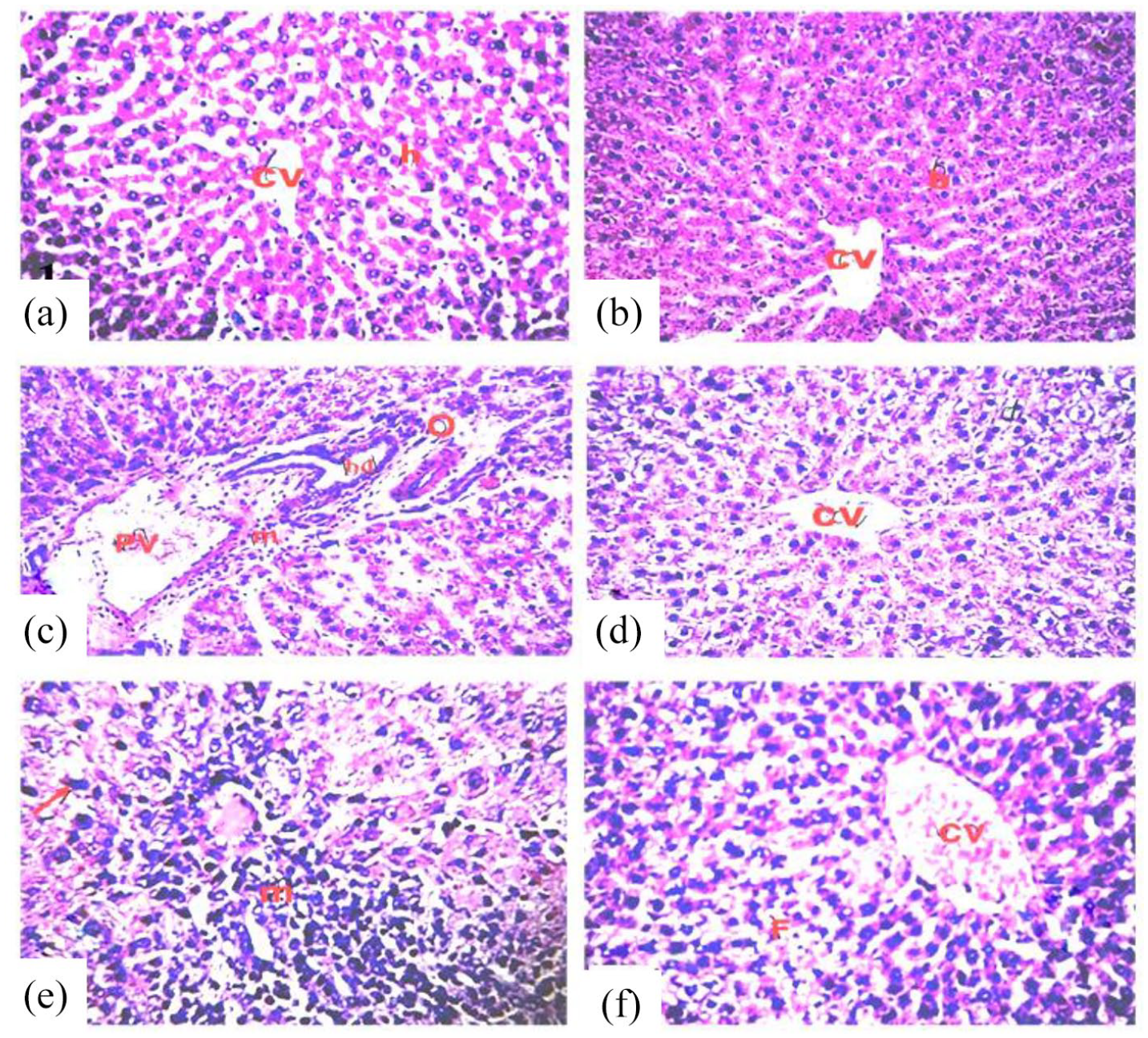
Photomicrographs of sections in the liver of rats in the different groups, stained with hematoxylin and eosin (H&E), magnification ×400. (a) Control group. (b) PIC group showing normal architecture of hepatic lobule (h), and normal central vein (CV) (score 0). (c) RES group showing congested vessels, dilated portal vein (PV) with infiltration of inflammatory cells (m) in portal area (score IV). (d) RES + PIC group showing improved hepatic lobule and portal area (score I). (e) IR group showing disorganization of hepatic cords and necrotic changes of hepatocytes with apoptotic hepatocytes (arrow) (score IV). (f) IR + PIC group showing some improvement in hepatic architecture and reduction in portal lymphocytic infiltration (score I).
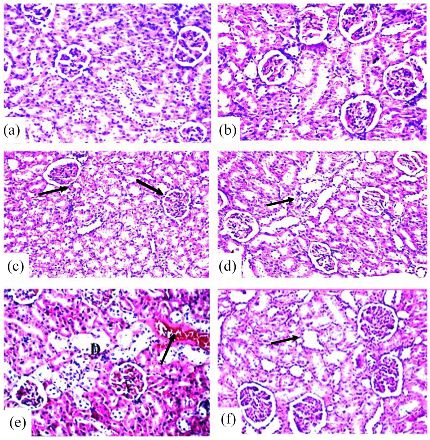
Photomicrographs of sections in the kidney of rats in the different groups stained with hematoxylin and eosin (H&E), magnification ×400. (a) Control group. (b) PIC group showing normal histological structure of renal parenchyma characterized by circumscribe glomeruli with normal structure of capillary tufts and Bowman’s capsule. The renal tubules of both proximal and distal convoluted tubules showed intact epithelial lining with empty lumen (score 0). (c) RES group showing tubular epithelial lining with granulo-vacuolar degeneration (arrows) and narrowing of tubular lumen. The epithelial lining of both proximal and distal convoluted tubules lining showed necrosis and apoptosis (score 4). (d) RES + PIC group showing improvement of the kidney architecture with some swelling of tubular epithelial lining (score 1). (e) IR group showing leukocytic infiltration, degeneration in lying epithelium (D) of some tubules with congestion of blood vessels (score 4). (f) IR + PIC group showed mild swelling of tubular epithelial lining without significant pathological alteration (score 1).
In agreement with the above, we have shown in this study that, reserpine significantly reduced the level of ATP and the activities of succinate-dependent Complex I, Sirt-1, PGC-1α, and p38 MAPK, indicating an impaired function in the mitochondrial respiratory chain in the liver and renal tissues. This could be attributed to an increased lipid accumulation inducing down-regulation of SIRT1, which in turn reduces the expression of β-oxidation genes in the liver and kidney. 44
The mitogen-activated protein kinases (MAPK) are involved in an activation loop consisting of ERK-1, ERK-2, JNKs, and the p38 kinase, a loop that is regulated by cellular environment-dependent mechanism affected by potentially injurious abiotic stress stimuli. 45 A recent investigation of Park et al. 46 stated that RES considerably induced MAPK, but inhibited p38 phosphorylation, which was noticed in the current data of hepatic and kidney tissues.
PIC is a potential candidate for the mitigation of hepatic and renal histological alterations in rats exposed to RES or γ-radiation
Altered levels of circulating cytokines might be non-invasive predictive biomarkers for radiotherapy-induced and clinical outcomes of cancer patients. 47 Various studies have reported that under inflammatory conditions the cytokines have a key role in changeable p38/PGC-1α axis levels. 48 In our study, RES and IR stimulated an inflammatory response reflected by the rise of TNF-α and IL-6 cytokines with reduced levels of p38/PGC-1α as compared to the corresponding control, suggesting the involvement of p38 AMPAK in hepatic and kidney injury. Thabet et al. 49 reported that PPAR-α/p38 inhibition in hepatocytes resulted in an increased cytokines activation, with a consequent rise in chemokine’s expression for the recruitment of inflammatory cells, leading to hepatocytes apoptosis. Mechanisms proposed; RES may induce hepatocytes injury via the integration of smooth endoplasmic reticulum proliferation, ribosomes, and mitochondrial damage. 38
PGC-1α plays a pivotal role in mitochondrial biogenesis in skeletal muscle 50 and liver. 51 Its deletion leads to reduced mitochondrial content. 52 PGC-1α functions as an activating transcription factor and is involved in the coordinated regulation of nuclear and mitochondrial genes such as nuclear respiratory factors 1 and 2 and mitochondrial transcription factor-A. 53
In the current study, piceatannol treatment was investigated for its regulatory and inducing effects on Sirt-1/PGC-1α involved in mitochondrial biogenesis and thus affecting its integrity and functions. The results revealed that piceatannol reduced lipid peroxidation and NO contents and increased GSH-PX, GR, SOD, and CAT activity in the liver and kidney. These effects of piceatannol could be attributed to its structural similarity with resveratrol and pterostilbene whose comparable effects have been reported in various studies. For example, resveratrol and pterostilbene were reported to augment the antioxidant status in diabetic animals.54,55 Similarly, the antioxidant effects of pterostilbene were attributed to its unique structure with one hydroxylic group and two methoxylic groups that scavenge ROS 56 ; which are implicated in the initiation and pathogenesis of several diseases.
The results revealed also that piceatannol was able to attenuate the inflammatory and apoptotic responses against IR and RES toxicities in the liver and kidney. Piceatannol diminished the increase of the inflammatory markers, TNF-α and IL-6 and the apoptotic markers, cytochrome c and caspase-3. Furthermore, piceatannol boosted the expressions and proteins’ tissue contents of p-p38, SIRT1, PGC-1α. In this context, pterostilbene structurally similar to piceatannol was found to protect rats against diabetic myocardial oxidative stress and apoptosis via increased phosphorylation of adenosine monophosphate-activated protein kinase (AMPK). 57 Moreover, pterostilbene has been reported to decrease cardiac oxidative stress through stimulation of AMPK/nuclear factor erythroid 2-related factor-2 (Nrf2)/heme-oxygenase (HO-1) pathway, 58 and to possess neuroprotective properties against β-amyloid-induced inflammation through the inhibition of NLRP3/caspase-1 inflammasome pathway in the brain. 59
PGC-1α is the “main key” of mitochondrial biogenesis that stimulates the mitochondrial gene expression and mitochondrial DNA replication. 10 In the current study the results revealed that PIC treatment indorses deacetylation of PGC-1α and target gene expression, with increased protein levels of PGC-1α, which the SIRT1-dependent pathway activation such as a resveratrol analog on SIRT1 mRNA activation in the kidneys of diabetic nephropathy rats. 53
The increased PGC-1α expression elicited by piceatannol; herein, was associated with enhanced activity of the antioxidant enzymes. In agreement with this observation, Rius-Pérez et al., 60 Valera-Alberni and Canto 59 reported that PGC-1α is involved in the induction of ROS detoxifying enzymes. Thus, we speculated that the ability of piceatannol to suppress ROS improved antioxidant and reduced inflammatory and apoptotic responses might function through the SIRT1/p38-AMPK signaling pathway.
In rats treated with PIC after exposure to IR and RES, the decrease of oxidative stress, inflammatory, and apoptotic markers was accompanied by an amelioration in mitochondrial biogenesis. The higher levels of ATP content, and complex I activity indicates improved mitochondrial function. The modulator role of PIC in liver and kidney injury was confirmed by the improvement of liver and kidney functions and histopathological examinations. Because of its outstanding antioxidant function, piceatannol (PIC) is a beneficial compound. PIC derived its antioxidant property from the ethylene double bonds and phenolic hydroxyl groups. 61 Li et al. 62 found that PIC showed strong antioxidant activity in scavenging hydroxyl radicals, due to its four phenolic hydroxyl groups on the benzene ring. Setoguchi et al. 63 have demonstrated that piceatannol revealed higher metabolic stability than resveratrol since, under their experiment; the pharmacokinetic curve for piceatannol was 2.1 times higher than that for resveratrol.
The results obtained in the current study go in line with the previous findings of who reported that strawberry fruits are a rich source of phytochemicals activates AMP-activated protein kinase (AMPK) signaling pathway, involved in mitochondrial biogenesis and antioxidant defenses.64–66 In addition, Wahdan et al. 67 demonstrated that, PIC at a dose of 10 mg/kg was effective to cure kidney injury and the results obtained corroborate also that PIC (10 mg/kg BW) treatment of rats exposed to Cadmium-induced toxicity revealed inhibition of oxidative stress via upregulation in mRNA- and protein expression levels of Nrf2 pathway. 18 In this study, we provide a new mechanism for PIC-mediated protection by demonstrating that PIC could protect against gamma radiation-induced oxidative stress, mitochondrial dysfunction and apoptosis via decreasing the tissue contents of lipid peroxides, NO and inducing SOD, CAT, GPX, and GR activities, together with the regulation of SIRT1/p38-AMPK, PGC-1α signaling pathway. Yet, our research has some limitations; for instance, the assessment of mitochondrial permeability transition, the assessment of complex II and inflammatory cytokines tissues contents, biochemical and transmission electron microscopy (TEM) of mitochondrial morphology would have been supportive parameters to describe the mechanisms of action of PIC in more details.
Conclusion
It could be concluded that piceatannol improve mitochondrial biogenesis and functions by regulating the oxidant/antioxidant disequilibrium, the inflammatory, and apoptotic responses, suggesting that the protective action of piceatannol might be a step forward in the development of adjuvant therapy for patients undergoing radiotherapy.
Footnotes
Acknowledgements
The authors would thank Prof. Dr. Ahmad Othman (Professor of Pathology, Pathology Department, Faculty of Veterinary Medicine, Cairo University) for the histopathological preparation, examinations, and reviewed of hepatic and renal tissue sections in this work.
Declaration of conflicting interests
The author(s) declared no potential conflicts of interest with respect to the research, authorship, and/or publication of this article.
Ethics approval
Ethical approval for this study was obtained from *the Ethical Committee of the National Centre for Radiation Research and Technology (NCRRT), Cairo, Egypt (Ref. No.: 195/7/2020)*.
Funding
The author(s) received no financial support for the research, authorship, and/or publication of this article.
Animal welfare
The present study followed international, national, and/or institutional guidelines for humane animal treatment and complied with relevant legislation.