
Abstract
We aimed to analyze the action of berberine on the neuropathic pain and neuroglia activation in experimental diabetes mellitus (DM) model. Diabetes in mice was induced by intraperitoneal injection of streptozotocin (STZ) followed by the administration of berberine. Mechanical allodynia and thermal hyperalgesia and activations of microglia and astrocytes were evaluated. The levels of pro-inflammatory cytokines and protein expressions of inflammatory proteins were assessed by enzyme-linked immunosorbent assay (ELISA) and western blot, respectively. Our results revealed the anti-nociceptive effects of berberine in DM mice, supported by the improved mechanical threshold and thermal latency. In addition, berberine suppressed the activations of microglia and astrocytes in the spinal cords of diabetic mice. Berberine inhibited the expression of pro-inflammatory cytokines including tumor necrosis factor (TNF)-α, interleukin-6 (IL-6), and interleukin-1β (IL-1β), along with inflammatory proteins including iNOS and COX-2. Berberine suppressed neuropathic pain in STZ-induced diabetic mice, and this effect is related to the reduction on the neuroglia activation and inflammation associated with DM.
Introduction
Diabetes causes various secondary complications in different organs. Previous studies have suggested that cognitive dysfunction and type 2 diabetes are interrelated and type 2 diabetes increases risk of developing cognitive impairment. 1 In addition to the common angiopathic complications, approximately 50% of patients with diabetes mellitus (DM) will develop polyneuropathy. 2 The manifestations of polyneuropathy include positive symptoms including dysesthesia and tingling/itching, as well as negative symptoms including muscle weakness, numbness, and balance loss. Notably, up to 25% of patients with diabetic neuropathy also develop neuropathic pain. Painful diabetic neuropathy is associated with increased distress and poor life quality in comparison with the general healthy population.
Berberine, an alkaloid extracted from Chinese herbs, has been extensively utilized in Oriental Medicine. Berberine possesses a great variety of pharmacological and biological activities, including anti-inflammatory, anti-oxidant, and anti-tumor effects. Furthermore, it was reported that berberine has potent neuroprotective effects against brain ischemia, Alzheimer’s disease, and brain tumor. Specifically, research suggested that blockades on potassium currents by berberine contributed to its protective action against ischemic brain damage. 3 Berberine suppressed surgery-induced neuroinflammation, preventing the development of postoperative cognitive dysfunction. 4 Although the full mechanisms of the neuroprotection of berberine remains unclear, an inflammatory response occurs alongside astrocyte and microglial activation in the hippocampus of diabetic mice. Herein, we aimed to analyze the action of berberine on the neuropathic pain and neuroglia activation caused by experimental DM in mice.
Materials and methods
Animals
All animal experimental protocols were approved by the Ethics Committee of Jinan Second People’s Hospital, and all procedures were performed in accordance with the Guide for the Care and Use of Laboratory Animals from the National Research Council (8th edition). Male C57BL/6 mice were purchased from SLAC (Shanghai, China). Animals were housed 4–5 per cage in temperature (22°C ± 1°C) controlled animal facility with 12 h light and dark cycles. Mouse food and water is available ad libitum. Animals were randomly assigned to four experimental groups as described in the following.
Establishment of diabetic mouse model and experimental groups
Diabetes was induced by intraperitoneal injection of 200 mg/kg streptozotocin (STZ; Sigma-Aldrich, St. Louis, MO, USA) dissolved in 0.1 M citrate buffer, pH 4.5. The control mice received citrate buffer at equal volume. The blood glucose levels in the tail vein blood were measured to confirm diabetes. The mice with a glucose level above 220 mg/dL were chosen for the subsequent study.
Starting at 2 weeks after STZ/vehicle injection, control and diabetic mice were randomly assigned in four groups (n = 13): Group 1, control + vehicle: non-diabetic control mice received a subcutaneous (s.c.) injection of vehicle; Group 2, control + berberine, non-diabetic control mice received a s.c. injection of berberine (dissolved in 0.9% saline; Sigma-Aldrich); Group 3, DMs + vehicle, diabetic control received s.c. injections of vehicle; and Groups 4, DMs + berberine, diabetic mice which received berberine s.c. injections.
Mechanical allodynia and thermal hyperalgesia testing
Quantitative sensory reflex testing of mechanical allodynia and thermal hyperalgesia was performed on different groups of animals. The baseline values were measured prior to the injection of berberine. Mechanical thresholds were measured using a Dynamic Plantar Aesthesiometer (Shanghai Yuyan Instruments Co., Ltd., Shanghai, China). Briefly, mice were placed in acrylic boxes. A metal filament releases a linearly increasing force ramp to the plantar surface of the hind paw of the mouse. The force which elicited the paw withdrawal was recorded as mechanical threshold (g).
Thermal hyperalgesia was evaluated by the Hargreaves’ thermal stimulator (Shanghai Yuyan Instruments Co., Ltd.). The device releases a quantified heat to the plantar surface of the hind paw, and the paw withdrawal latency was recorded in seconds.
Immunohistological analysis
For the microglia activation analysis, the spinal cords of mice were removed, post-fixed with 4% paraformaldehyde, and embedded with paraffin; 5-µm-thick sections of lumbar segment of the spinal cord were stained with hematoxylin and eosin (H&E) and immunohistochemistry to visualize microglia activation using antibody against Iba-1 (Abcam, Cambridge, MA, USA).
For the astrocyte activation assessment, spinal cord sections were processed for antigen retrieval with citrate buffer (pH 6.0) and blocked with 10% goat serum for 20 min at 37°C. Rabbit polyclonal anti-glial fibrillary acidic protein (GFAP; Abcam) was kept overnight at 4°C in humidity chambers. Fluorescence images of these sections were captured at 400× magnification. Quantification of the relative average optical densities of Iba-1 and GFAP was performed using Image-Pro Plus software (Media Cybernetics, Rockville, MD, USA). At least five randomly selected sections were analyzed.
Measurement of cytokine levels
The spinal cord and dorsal root ganglia (DRG) of mice were removed and homogenized in pre-chilled phosphate-buffered saline (1× PBS) containing a protease inhibitor. The samples were centrifuged at 10,000×g for 15 min. The cytokine levels in the supernatant were measured using mouse tumor necrosis factor-α (TNF-α), interleukin-6 (IL-6), and interleukin-1β (IL-1β) using Quantikine enzyme-linked immunosorbent assay (ELISA) kits (R&D Systems, Minneapolis, MN, USA) according to the manufacturer’s instructions. The absorbance was red using a microplate reader at 450 nm (Molecular Devices, San Jose, CA, USA).
Western blot analysis
The total protein concentration in the supernatant of spinal cord and DRG extraction was estimated by the Bradford method. The proteins were applied on sodium dodecyl sulfate–polyacrylamide gel electrophoresis and transferred onto polyvinylidene fluoride membrane. After blocking with 5% non-fat milk in tris-buffered saline and Tween-20, the membranes were incubated with primary antibodies against iNOS (1:1000 dilution; Santa Cruz Biotechnology, Dallas, Texas), COX-2 (1:500 dilution; Abcam), and β-actin (1:2000 dilution; Sigma-Aldrich). The membranes were developed with Pierce ECL Western Blotting Substrate (Pierce, Waltham, MA) and the density of bands were measured by Amersham Imager 600 (GE Healthcare, Buckinghamshire, UK).
Statistical analysis
Statistical analysis in the study was performed by SPSS (version 20.0; SPSS Inc., Chicago, IL, USA). Data are expressed as mean ± standard error of mean (SEM). Differences among groups were assessed by one-way analysis of variance (ANOVA). P < 0.05 was considered as statistically significant.
Results
Anti-nociceptive effects of berberine to diabetic mice
Using STZ-induced diabetic mouse model, we evaluated the effects of berberine on the diabetic neuropathic pain. Two weeks after STZ injection, control or diabetic mice were treated with berberine. As shown in Figure S1(a), we can see the mechanical threshold in the DMs + vehicle group, in which diabetic control mice received vehicle, is significantly lower than the groups of control + vehicle (P < 0.01), indicating the successful establishment of the diabetic models. At days 28 and 42, the berberine treatment significantly improved the mechanical threshold when compared to the diabetic control mouse group (P < 0.01). Notably, the berberine itself did not show significant effects on the sensory reflex testing of mechanical allodynia, suggesting the relative drug safety of berberine at the effective dose used in this study. Similarly, thermal hyperalgesia testing showed that berberine treatment improved the thermal latency of the diabetic mice at days 28 and 42 (Figure S1(b), both P < 0.01), indicating the anti-nociceptive effects of berberine.
Berberine inhibited spinal microglia and astrocyte activation in the diabetic mice
To determine whether the administration of berberine inhibited microglia activation in the diabetic mice, we evaluated the protein expression of Iba-1 in the spinal cords by immunohistochemical staining. As shown in Figure 1(a) and (b), the relative Iba-1-positive area in the diabetic control mice is significantly higher than the non-diabetic ones in the control + vehicle and control + berberine groups (P < 0.01). In contrast, the treatment of berberine significantly inhibited Iba-1-positive area in the diabetic mice (P < 0.05), indicating the suppression of berberine on the microglia activation.
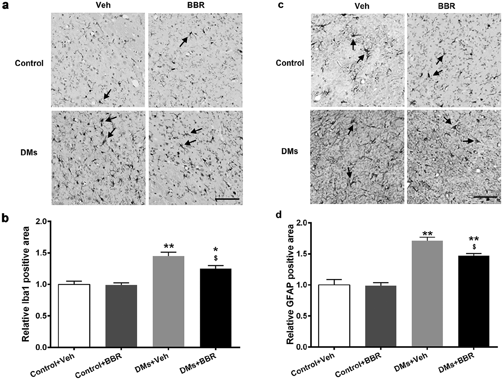
The effects of berberine treatment on Iba-1-positive microglia and GFAP-positive astrocytes in the spinal cords of DMs mice. (a) Iba-1-positive microglia were detected by immunohistochemistry and relative Iba-1-positive area (b) was quantified. Black arrows indicate Iba-1-positive microglia. (c) GFAP-positive astrocytes were detected by immunohistochemistry and (d) relative GFAP-positive area was quantified. Black arrows indicate GFAP-positive astrocytes. Data are presented as mean ± SEM (n = 6 for each group).
As shown in Figure 1(c) and (d), the relative GFAP-positive area reflected by the GFAP fluorescence density was significantly elevated in the diabetic mice (P < 0.01), indicating the spinal astrocyte activation in the diabetes. As expected, the administration of berberine significantly decreased the astrocyte activation (P < 0.05) in comparison with the diabetic control mice. However, there was no significant difference observed between the berberine and vehicle control mice.
Berberine reduced the levels of cytokines in the spinal cord and DRG
To further examine the spinal inflammatory response after berberine treatment, the expressions of inflammatory mediators were measured by ELISA. As shown in Figure 2(a)–(f), ELISA analysis showed that the levels of TNF-α, IL-6, and IL-1β were significantly increased in the spinal cord and DRG in diabetic mice than the non-diabetic mice (P < 0.01). Significantly, treatment with berberine inhibited the increased production of all tested cytokines in the spinal cord and DRG in the diabetic mice (P < 0.05). However, there was no significant difference between control and berberine-injected non-diabetic mice.

Effects of berberine on the increased pro-inflammatory cytokine levels in spinal cord and DRG of DMs mice. The levels of (a and d) TNF-α, (b and e) IL-6, and(c and f) IL-1β from spinal cord and DRG were determined by ELISA. Data are presented as mean ± SEM (n = 6 for each group).
Berberine downregulated the expression of iNOS and COX-2 in the spinal cord and DRG
As seen in Figure 3, when compared with the non-diabetic mice, the relative protein expressions of iNOS and COX-2 in the diabetic mice were significantly upregulated in both of the spinal cord (Figure 3(a), (c), and (d)) and DRG (Figure 3(b), (e), and (f); P < 0.01). The administration of berberine in the diabetic mice significantly inhibited the expression of iNOS and COX-2 in the spinal cord and DRG (P < 0.05). These results showed the inhibitory effects of berberine on the expression of inflammatory protein in diabetic mice.

Effects of berberine on inflammatory protein levels in spinal cord and DRG of DMs mice. Protein expressions of iNOS and COX-2 in (a) DRG and (b) spinal cord were detected by western blot and (c, d, e, and f) relative expressions were analyzed. Data are presented as mean ± SEM (n = 6 for each group).
Discussion
The role of microglial in the neuropathic pain is limited. The microglia inhibitor, although alleviated the development of neuropathic pain, frequently failed to alleviate existing neuropathic pain. Astrocytes are one type of glial cells in the central neural system. In addition to microglia, more and more evidence has revealed that spinal astrocytes also take part in the pathology neuropathic pain, indicated by the altered morphology and function. Previous study has shown that inhibition of astrocytic activation by fluorocitrate, the astrocyte inhibitor, led to an analgesic effect in paclitaxel-evoked neuropathic pain. 5 Consistent to the microglia activation, we also observed significant elevation of astrocyte numbers in the diabetic mice than non-diabetic ones. Moreover, berberine treatment significantly lowered the numbers of astrocytes compared to the vehicle-treated diabetic mice, suggesting the suppression of berberine on the activation of astrocytes in the DM mice.
To this end, we understood that berberine possessed the neuroprotective effects to alleviate diabetes-associated neuropathic pain, and this protection is related to the activation of microglia and astrocytes. Studies deciphering the correlation between inflammation and diabetes have offered plenty of data implicating the role of inflammation toward the development of insulin resistance and pathogenesis of type 2 diabetes. 6 Inflammatory markers such as TNF-α, IL-1β, and IL-6 are increased in the tissues and serum of patients with type 2 diabetes. 7 More studies have shown that these pro-inflammatory cytokines may be involved in the development and maintenance of neuropathic pain. 8 It has been shown that minocycline diminished the levels of IL-6 and IL-18 cytokines at the spinal cord level in neuropathic pain and this effect was correlated with an inhibition of microglial cell activation and a reduction in the neuropathy symptoms.9,10 In this study, we investigated the protein levels of TNF-α, IL-1β, and IL-6 in the homogenate of spinal cord and DRG tissues, and our results showed significantly elevated levels of these pro-inflammatory cytokines in the diabetic mice compared to the non-diabetic control mice. Moreover, the administration of berberine decreased the levels of all of the cytokines, indicating the anti-inflammatory effects of berberine.
It has been reported that the induction of diabetes in rat experimental model increased the expression level of iNOS in vascular tissue, 11 while the deletion of the iNOS gene prevents the impairment of endothelial vasodilatation in carotid arteries of mice caused by diabetes and preserves cerebral arteriolar vasomotor function in diabetic mice. 12 Diabetes also interferes with the activity of vascular COX, the key enzyme inducing inflammatory pain. 13 In this study, berberine treatment also decreased the levels of inflammatory proteins, including iNOS and COX in the diabetic mice. Given the important roles of iNOS and COX in the pathogenesis of diabetes, berberine could serve as a potential anti-diabetic component owing to its neuroleptic and anti-inflammatory properties.
Supplemental Material
Supplementary_Materials_2 – Supplemental material for Berberine reduces neuroglia activation and inflammation in streptozotocin-induced diabetic mice
Supplemental material, Supplementary_Materials_2 for Berberine reduces neuroglia activation and inflammation in streptozotocin-induced diabetic mice by Mei Liu, Linlin Gao and Na Zhang in International Journal of Immunopathology and Pharmacology
Footnotes
Declaration of conflicting interests
The author(s) declared no potential conflicts of interest with respect to the research, authorship, and/or publication of this article.
Funding
The author(s) received no financial support for the research, authorship, and/or publication of this article.
Supplemental material
Supplemental material for this article is available online.
References
Supplementary Material
Please find the following supplemental material available below.
For Open Access articles published under a Creative Commons License, all supplemental material carries the same license as the article it is associated with.
For non-Open Access articles published, all supplemental material carries a non-exclusive license, and permission requests for re-use of supplemental material or any part of supplemental material shall be sent directly to the copyright owner as specified in the copyright notice associated with the article.