
Abstract
Cardiac metastases are the most frequent cardiac tumors. They can cause dysrhythmia, myocardial dysfunction, pericardial effusion, and heart failure. In decreasing order, the major primary malignancies associated with cardiac metastases are pleural mesothelioma, lung adenocarcinoma, undifferentiated carcinomas, lung squamous cell carcinoma, and breast carcinoma. Cardiac metastasis of sarcomas is uncommon, and only a limited number of cases have been found in literature. We report the case of an incidentally discovered cardiac metastasis when assessing the extension of a thigh mass in a 45-year-old man.
Introduction
Cardiac metastases are the most frequent cardiac tumors. 1 They can cause dysrhythmia, myocardial dysfunction, pericardial effusion, and heart failure. 2 Their exact incidence is not exactly known, 1 but it has increased in the last decades because of the improvement of diagnostic tools. 3 In decreasing order, the major primary malignancies associated with cardiac metastases are pleural mesothelioma, lung adenocarcinoma, undifferentiated carcinomas, lung squamous cell carcinoma, and breast carcinoma. 1 Cardiac metastasis of sarcomas is uncommon, and a limited number of cases have been found in literature. 4
We report the case of an incidentally discovered cardiac metastasis when assessing the extension of a thigh mass in a 45-year-old man.
Case report
A 45-year-old man with no medical history presented with a slow growing mass in his right thigh evolving over the past year associated with an altered general condition and weight loss. He was addressed to our department for local and distant extension assessment of his thigh mass.
Thigh MRI revealed a voluminous mass of the right thigh (Fig 1(a)–(b)), occupying almost the entire quadricipital space. This mass presents a low signal on T1WI, an intermediate heterogenous signal on T2WI, and an irregular peripheral enhancement. The mass invades the upper 1/3 of the femur and the various chiefs of the quadriceps muscle. The thigh mass was biopsied and revealed a soft tissue sarcoma (Fig 2). (a) STIR-weighted coronal image showing the voluminous thigh mass with irregular borders and heterogeneous high signal. (b) Enhanced T1WI coronal image showing peripheral enhancement of the voluminous thigh mass with central necrosis. Histological appearance at medium magnification (×20) showing a largely necrotic, pleomorphic, and hyperchromatic sarcomatous proliferation. The immunohistochemical results are consistent with a pleomorphic rhabdomyosarcoma grade 3 according to the FNCLCC classification.

To evaluate the presence of distant metastasis, a CT scan of the chest, abdomen, and pelvis was performed, revealing a predominantly cystic myocardial mass that enhanced after contrast injection. The mass, measuring 57 × 34 mm, appeared to be attached to the left ventricle and was consistent with a cardiac metastasis (Fig 3). CT also revealed the presence of other secondary localizations in the lungs, liver, lymph nodes, spleen, bone, and right paravertebral muscles with intramedullary invasion (Fig 4(a)–(b)). It should be noted that the secondary lesions found in the liver, lungs, and lymph nodes also had a cystic component. Axial enhanced CT image displaying a cystic myocardial mass, which enhances after contrast injection and appears to be attached to the left ventricle, consistent with a cardiac metastasis. (a) Axial CT image showing hepatic metastasis and muscular paravertebral metastasis. (b) Axial CT image with lung window and maximal intensity projection (MIP) showing multiple pulmonary metastasis.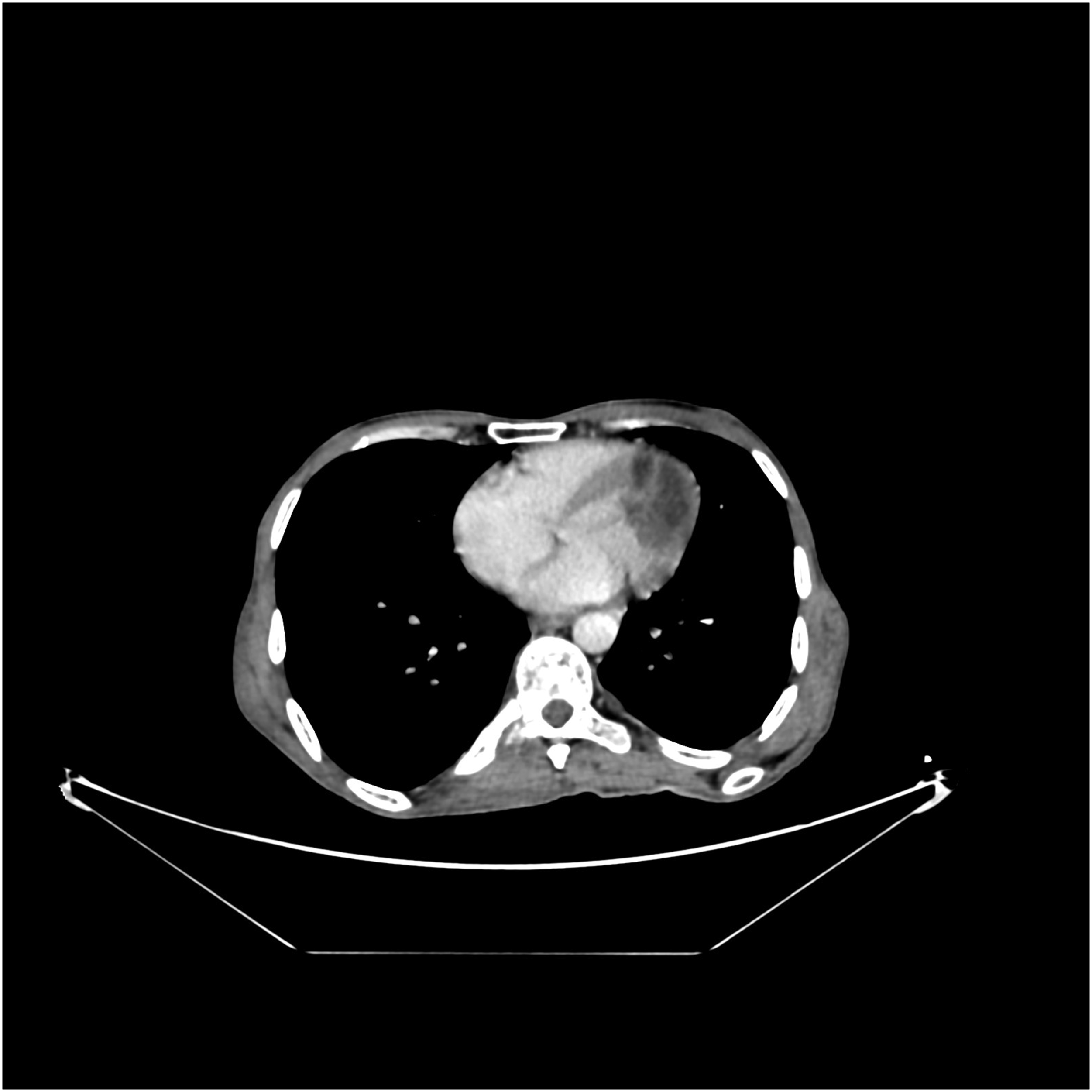

Discussion
The incidence of cardiac metastasis in patients with known primary tumors is 9%. 5 This incidence has increased due to the increased ability to detect disease of the myocardium in vivo and the prolonged survival of cancer patients. 5 The most common primary neoplasms to metastasize to the heart are melanoma, carcinoma (lung, breast, esophagus, and rarely colorectal), and hematologic malignancies (leukemia and lymphoma). 6 Cardiac metastasis of a soft tissue sarcoma is very rare, with only a limited number of cases published in literature.3,4,7
According to Abbas et al., in their 12-year series reporting primary and metastatic cardiac sarcomas at a German heart series, only 4 cases were found to have sarcoma metastatic to the heart, three of which were soft tissue sarcoma. 8
According to Burazor et al., in their large retrospective study evaluating metastatic cardiac tumors in two medical centers, only two patients were found to have a sarcoma cardiac metastasis, both were from a uterine sarcoma. 6
In most case reports and series found in literature, most patients with soft tissue sarcoma cardiac metastasis had other organ metastasis either concurrent to or preceding cardiac metastasis.3,4,8 In our patient, we found lung, liver, lymph nodes, spleen, bone, and right paravertebral muscles metastasis at the diagnosis.
We also observed on those series that heart metastasis was not present on initial staging.3,4,8 The mean duration between primary soft tissue sarcoma diagnosis and heart metastasis varied greatly among series, ranging from 44.5 months to 109 months.3,8 In our case, the cardiac metastasis was discovered at initial staging.
Most patients of cardiac metastasis disease are asymptomatic. When present, the symptoms are often nonspecific, and their severity depends on the size and the location of the metastasis. They can mimic myocardial ischemia, heart failure, or cardiac injury related to chemotherapy or radiation therapy. 5 In our case, there were no clinical cardiac manifestations.
Echocardiography is the first-line imaging modality to be realized in patients with known malignancies presenting with cardiac symptoms. 5 It helps to assess pericardial effusion and intra- or pericardial masses and their impact on cardiac function. 5 CT can also be used to assess cardiac metastatic disease. Its speed and high spatial resolution combined with cardiac gating can help localize lesions and evaluate anatomic relationships with cardiac structures. It also helps to assess mediastinal lymph nodes. Its main limitation is poor tissue characterization. 5 Cardiac MRI is the best imaging modality to evaluate cardiac metastatic disease; its high tissue characterization can easily distinguish between an infiltrating tumor and intracardiac thrombus. The main imaging characteristics of cardiac metastasis on MRI are the following: Low signal intensity on T1-weighted imaging, high signal intensity on T2-weighted imaging, and contrast enhancement after gadolinium injection. 5 Larger masses often show heterogeneous enhancement, primarily due to tumor necrosis resulting from tissue hypoxia. 9 In our case, the cystic component of the cardiac metastasis is likely the result of tissue necrosis.
In conclusion, cardiac metastases have a very poor prognosis and they can be rapidly fatal. 1 Their management consists mainly of treating the primary tumor or palliative care. 1 Surgical treatment can be considered when important symptoms of obstruction outweigh the mortality risk of operating and the benefit of medical therapy alone. 1
Footnotes
Declaration of conflicting interests
The author(s) declared no potential conflicts of interest with respect to the research, authorship, and/or publication of this article.
Funding
The author(s) received no financial support for the research, authorship, and/or publication of this article.