
Abstract
Background
For many common malignancies, including breast cancer, evaluation for metastatic disease using multiphase computed tomography (CT) has fallen out of favor and been replaced by studies performed only in the portal venous phase. However, differences in tumor vascularity could produce differences in appearance on post-contrast imaging.
Purpose
To assess non-contrast phase and portal venous phase computed tomography in detection and measurement of hepatic metastases from breast carcinoma.
Materials and Methods
A total of 75 CT scans from 52 breast cancer patients were independently assessed by three body imagers for lesion presence, number and size. Readers randomly assessed portal venous phase or combined phase images at one session with cross-over reads performed four to six weeks later.
Results
In the 58% of cases where index lesions measured larger on combined phase, the mean difference in lesion size was 5.7 mm. In this group, combined phase reads demonstrated an 8.4 mm increase in sum of largest diameters, and a mean percentage sum of largest diameters increase of 19% compared to portal venous phase-only reads.
Conclusion
Addition of non-contrast phase images results in increased index lesion size in most patients with hepatic metastases from breast cancer. If only the portal venous phase is utilized, there is potential for incorrectly diagnosing disease progression on follow-up due to underestimation of lesion size.
Introduction
Breast cancer is the most commonly diagnosed malignancy in women in the United States. 1 It also represents one of the most common sources of metastatic disease, including hepatic metastases. 2 The accurate detection and characterization of hepatic metastatic disease in this patient population is important for both initial staging and assessment of response to treatment.
With the widespread use of contrast-enhanced multi-detector computed tomography (MDCT), it is easy to obtain images at multiple phases after intravenous contrast administration during a single examination. However, as the vascularity of metastases of different primary tumors can affect their appearance on post-contrast imaging, conclusions about the optimal phase in which to image metastases of a particular primary tumor will not necessarily apply to metastases of another.
The most common sources of hepatic metastases, namely lung and colon cancer, are hypovascular and therefore hypoattenuating during the portal venous phase (PVP). It has been confirmed that hepatic metastases from these primary malignancies are best visualized at the PVP. 3 These hypovascular lesions image differently in comparison to primary neoplasms which produce hypervascular hepatic metastases, including thyroid, neuroendocrine, renal cell carcinoma, and melanoma. There has been a reported significant increase in the number of metastatic lesions detected with the combination of non-contrast phase (NCP) and PVP images, relative to PVP images alone in this patient population. 4 , 5 However, multiple studies comparing non-enhanced to PVP imaging have demonstrated that, while there are lesions detected on non-contrast imaging that are not seen in the PVP, the overall number of lesions identified on PVP images alone is significantly greater than on non-enhanced images alone. 6 These findings have also been seen specifically in the setting of hepatic metastases from breast cancer. 7 More importantly, it was shown in this study that per-patient sensitivity and specificity for the presence of metastatic disease are not improved by the addition of non-enhanced images relative to PVP images alone. This finding has also been seen with other hypervascular metastases. 8 These results led to the conclusion that non-enhanced images confer minimal benefit in the diagnosis of the majority of hepatic metastases from breast cancer and may be unnecessary.
Since the advent of Response Evaluation Criteria in Solid Tumors (RECIST) in 2000 and their revision in 2008, assessment of metastatic disease and subsequent treatment decisions has become more dependent on accurate determinations of the numbers and sizes of individual lesions, 9 , 10 rather than the simple presence or absence of disease. As a result, imaging decisions based on previous studies showing non-inferiority of PVP imaging alone may need to be reevaluated. Further, the focus on the sizes of individual lesions lends new relevance to previous observations of size variability of metastases based on the phase in which they are imaged. PVP imaging alone frequently underestimates the sizes of hepatic metastases of multiple tumor types, as the peripheral regions of metastases can be iso-attenuating with normal hepatic parenchyma on portal venous-phase imaging. 4 , 11 Such falsely low measurements may in turn lead to false-positive findings of progression post treatment, as these same peripheral regions necrose, causing their apparent sizes to increase on PVP images. 12
The purpose of this study was to assess the utility of NCP and PVP helical CT in detection and measurement of hepatic metastases from breast carcinoma.
Material and Methods
A total of 214 female patients over the age of 18 with breast cancer who had CT imaging between 1 April 2015 and 1 September 2016 were identified through a search of the electronic medical record. Of these, 72 patients were excluded due to not having both NCP and PVP images of the abdomen. The remaining 142 patients had CT images performed as part of a clinically requested oncology protocol which included both NCP and PVP images. CT protocol was as follows: MDCTs were performed with and without weight-based contrast dosing (Omnipaque 350 Iohexol, GE Healthcare), fixed 65-s PVP delay, and 3 mm slice thickness. Multiple CT platforms were used to acquire the images, including single-energy machines from Siemens and GE, as well as dual-energy scanners from Siemens. Coronal and sagittal images with 3 mm slice thickness were reconstructed from the axial data set. Multiple CT platforms were used to acquire the images.
Of the 142 patients with combined phase (CP) images including both PVP and NCP, 81 had normal liver parenchyma. True negative status in these patients was confirmed by radiology and clinical follow-up for at least 12 months. A randomly selected subset of 11 of 81 patients without hepatic lesions was included in the final data set for the evaluation of sensitivity and specificity. Of the 61 patients with hepatic lesions, 22 patients were excluded due to lack of confirmation of benign or malignant disease. Metastatic disease was confirmed using imaging and review of the medical record. Confirmation was achieved by biopsy, positron emission tomography (PET), magnetic resonance imaging (MRI), progression on CT, or response to chemotherapy. Twenty patients with benign hepatic lesions were also included. A total of 75 CTs in 52 patients were included in the study (Fig. 1).

Inclusion criteria for breast cancer patients in this study.
Images were independently assessed by three fellowship-trained body imagers for lesion presence, number, and size. Readers randomly assessed PVP or CP images at one session with cross-over reads performed four to six weeks later to prevent measurements from one study from influencing measurements on the other. Up to two index lesions were identified by each reader for PVP and CP scans with up to three additional non-index lesions being measured, if present. Measurements were performed using digital calipers on an Agfa Impax PACS system. A single measurement of greatest diameter of each selected lesion was performed by each reader on axial images. For each patient, the sum of largest diameters (SLD) for the index lesions was calculated. Each reader was also asked to document their preference for the best phase for detection and measurement of each index lesion, either PVP or NCP. A binomial test was used for the evaluation of these data.
To complete a lesion-based analysis, data from all three readers was pooled for 97 individual metastases from 44 individual CT studies. Differences in diameter between NCP and CP images were measured using a two-tailed repeated measures t-test. All analyses were conducted using statistical software (SPSS, IBM).
Radiation dose data were recorded for each CT, including volume CT dose index (CTDI vol) and dose length product (DLP). Estimated effective radiation dose was calculated using a conversion factor of 0.015 mSv/(mGy⋅cm). 13
Results
Pathology or imaging-confirmed hepatic metastases were identified in 21 patients. The pooled per-patient sensitivity and specificity for the detection of metastases was 91/67% and 94/71% on PVP and CP reads, respectively.
In 58% of cases, index lesion size measured larger on CP. When all patients with metastatic disease were included, mean index lesion size on CP was slightly larger in comparison to PVP, although this difference was not statistically significant (1.93 cm vs. 1.81 cm, p=0.13). However, in the subset of patients where index lesion size measured larger on CP, the mean index lesion size measured an average of 5.7 mm (range 1–28 mm) greater than on the PVP alone. In these same patients, CP reads demonstrated 8.4 mm (SD 0.77 mm) increase in sum of largest diameters (SLD), and a mean percentage SLD increase of 19% compared to PVP-only reads (Table 1).
Sum of largest diameter for individual readers.
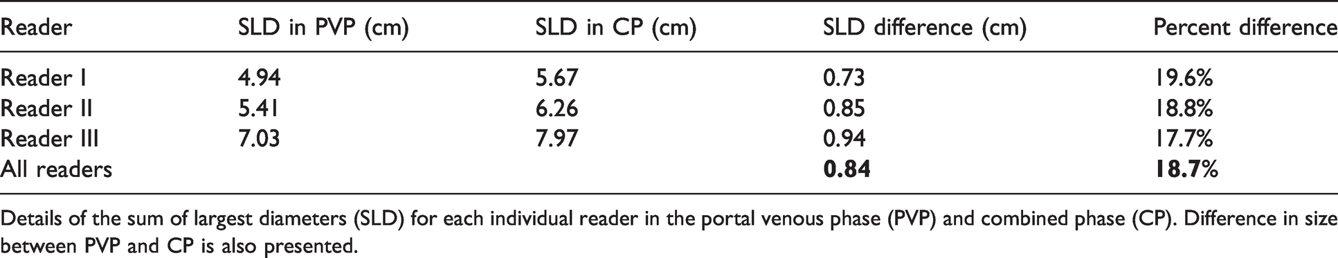
Details of the sum of largest diameters (SLD) for each individual reader in the portal venous phase (PVP) and combined phase (CP). Difference in size between PVP and CP is also presented.
When a per-lesion analysis of 97 pooled metastases was performed, the measurements of lesion size were significantly larger on NCP images (mean 2.94 cm, SD = 2.21 cm) than on the PVP (mean 2.78 cm, SD = 2.16 cm), p < .001.
Readers overwhelmingly preferred the PVP method to the NCP method for index lesion selection and chose PVP 78% of the time (222 of 285 lesions), p < .001.
Mean CTDI vol and DLP of NCP was 9.31 mGy and 257.6 mGy⋅cm, respectively, with an estimated dose of 3.86 mSv for NCP scans. Mean CTDI vol and DLP of PVP was 14.41 mGy and 648.9 mGy⋅cm, respectively, with and estimated effective dose of 9.74 mSv.
Discussion
Our study found that the inclusion of NCP images in addition to the PVP results in larger lesion size measurement in the majority of breast cancer patients (Fig. 2), which could have an impact on response assessment on follow-up imaging.

CT images obtained in the non-contrast phase (NCP; a) and portal venous phase (PVP; b) demonstrate significant differences in measurement of hepatic metastasis due to iso-enhancing viable tumor at the periphery of the lesion.
It is generally held that multi-phase imaging improves lesion detection and quantification in hypervascular metastases. 4 , 5 However, in recent years, radiation risk and cost concerns have prioritized single-phase post-contrast imaging over multiphase imaging. While breast cancer is generally considered in the differential of hypervascular hepatic lesions, 14 many have argued against the routine use of additional phases in this patient population. 7 These prior studies have demonstrated no difference in the sensitivity or specificity for the detection of metastatic disease in breast cancer patients on a per-patient basis when NCP images are included in the imaging protocol. Our study found similar results, with a minimal increase in per-patient sensitivity and specificity. However, unlike many other hypervascular liver lesions, such as hepatocellular carcinoma and carcinoid tumor, breast cancer metastases are usually not treated with surgical resection or local therapy. Therefore, while there are breast cancer metastases that are seen better or only on NCP images (Figs. 3 and 4), these individual lesions are rarely the only sites of metastasis. As a result, additional phases are generally not considered necessary if the only clinical question is the presence or absence of disease. While sensitivity and specificity may not be significantly different for this group of patients, as breast cancer can be at least partially iso-attenuating during the PVP, it stands to reason that the addition of non-contrast images could result in more accurate assessment of lesion size. We confirmed a statistically significant increase in metastatic lesion size and SLD in a majority of our breast cancer population. To our knowledge, our study is the first to confirm this finding in breast cancer patients, although other studies have suggested that NCP is the most accurate assessment of lesion volume. 15

(a) Non-contrast phase (NCP) and (b) portal venous phase (PVP) images demonstrating a right hepatic metastasis that is only seen on the NCP image (arrow).

NCP (a) and PVP (b) images of patient with breast cancer. While innumerable hepatic metastatic lesions are seen on the NCP (arrows), no metastases are visualized on the PVP portion of the examination.
The development of RECIST guidelines has dramatically changed the impact of imaging on oncology patients. Rather than simply detecting the presence or absence of metastatic disease, the clinical management of patients relies on accurate measurement and follow-up of individual lesions. Changes in number and size of individual metastases can affect treatment decisions and, as such, the ability of a given imaging modality to accurately characterize these changes is becoming more of a priority. The most common indications for oncologic imaging the abdomen are lung and colon cancer, both of which are generally hypovascular malignancies. 16 In this subset of patients, the addition of NCP may not add significant value as the true size of the lesions are likely accurately represented on PVP images. However, in hypervascular metastases such as breast cancer, PVP images can underestimate the true lesion size. 15 Our study found that CP reads demonstrated 8.4 mm increase in SLD, and a mean percentage SLD increase of 19% compared to PVP-only reads. This difference in size likely represents iso-attenuating areas of viable tumor on the PVP images. After treatment, these areas could become necrotic and hypoattenuating on the PVP images, which would result in an artificial increase in lesion size if only PVP images are compared. This could incorrectly classify the patient as having progressive disease, when, in fact, the patient was responding well to their current therapy. This is a clinical phenomenon we have witnessed at our institution and has been termed pseudoprogression (Fig. 5). This is distinct from pseudoprogression described with other primary tumors, particularly in the context of immunotherapy, where lesion size actually increases after initial treatment before decreasing on subsequent studies. 17 In contrast to our findings, this change in lesion size with immunotherapy appears to be real and seen on multiple sequences and modalities, whereas in our breast cancer population the lesions are not truly changing in size. Rather, treatment is altering the appearance of peripheral iso-attenuating tumor that was already present.

Patient with known breast cancer metastasis. Dual-phase CTs including non-contrast (a and c) and portal venous phase (b and d). CTs were performed before (a and b) and after (c and d) treatment with chemotherapy. Lesion size on portal venous phase image after treatment appears larger in comparison to the prior examination. However, after reviewing the non-contrasted images, the lesion appears very similar in size to the pretreatment examination. Lesion size changed by greater than 20% which when combined with additional lesions would meet RECIST criteria for disease progression. However, this lesion was stable on the subsequent examination, indicating that this change in measurement was likely due to treatment change, and not disease progression.
It is worth noting that in our patient population, the CP reads resulted in an average increase of greater than 5 mm and approaching 20% of the SLD, which would fulfill RECIST criteria for progression. Given that lesion size measured greater on NCP than PVP images in 58% of patients, a significant number of these patients could be affected.
Radiation dose was increased slightly with the addition of the NCP images, with an average increased effective dose of 3.86 mSv. While this is an important consideration, in patients with known metastatic disease, the ability to avoid mischaracterizing stable disease as treatment failure may outweigh the marginal risks of additional radiation exposure. With newer scanners capable of producing quality images at much lower radiation doses, it is possible to perform a multi-phase technique with minimally increased dose to the patient.
Our study has several limitations. First, patients were included regardless of whether or not they had been treated with systemic chemotherapy or selective estrogen receptor modulators (SERM), such as Tamoxifen, which could affect individual lesion vascularity. While this could alter the results, it is also possible that inclusion of all women could disguise an even larger effect on treatment-naïve patients. Second, the sample for this preliminary study was relatively small which makes it difficult to extrapolate our conclusions to a larger population.
Future directions for this research include an evaluation of the percentage of patients and lesions that result in apparent progression by RECIST criteria on subsequent imaging after treatment. It would also be interesting to correlate change in lesion size based on the type of treatment and the biology of the tumor itself, with the hypothesis that anti-angiogenesis treatments would have a greater impact. Lastly, we have started to investigate if the use of dual-energy CT (DECT) with the creation of virtual non-contrast (VNC) images could provide similar information without additional radiation dose.
In conclusion, a majority of breast cancer patients demonstrated an increase in index lesion size and SLD with the inclusion of NCP images. If only PVP imaging is utilized, there is potential for incorrectly classifying treatment response as disease progression due to underestimation of true lesion size at PVP-only imaging. We believe that this area needs further research, emphasizing the possible uses of DECT and VNC images. There may also be the possibility of targeting multiphase protocols to specific breast cancer treatment regimens.
Footnotes
Declaration of Conflicting Interests
The author(s) declared no potential conflicts of interest with respect to the research, authorship, and/or publication of this article.
Funding
The author(s) received no financial support for the research, authorship, and/or publication of this article.