
Abstract
Background
Crystal X-ray interferometer-based phase-contrast X-ray computed tomography (C-PCCT) enables the depiction of internal structures of biological tissue without contrast agents.
Purpose
To determine the advantage of this technique in visualizing detailed morphological structures of a rare spontaneous brain tumor in an aged rat.
Material and Methods
An aged rat’s spontaneous brain tumor was imaged by C-PCCT without contrast agent. Three-dimensional (3D) images of the tumor microvasculature were reconstructed and compared with pathological pictures.
Results
C-PCCT depicted the tumor’s various pathological features clearly, e.g. its cell density and vasculature, and blood clots caused by hemorrhaging and/or hematomas. The obtained images resembled pathological pictures with a magnification of ×20 and were used to reconstruct 3D images of the tumor vascularity up to approximately 26 µm in diameter.
Conclusion
Since C-PCCT is able to depict various pathological conditions, it might be useful for cancer research.
Keywords
Introduction
The overall incidence of brain tumors, including primary or metastatic malignancies of the central nervous system, is increasing worldwide. Since naturally occurring brain tumors are very rarely used for cancer research, brain tumor models are generated by implanting tumor cells into the brains of mice in most cancer studies. In our laboratory, a rare spontaneous brain tumor was found by chance in a 2-year-old male Wistar rat. In a previous carcinogenicity study involving 11,705 Wistar rats, the incidence of spontaneous nervous system tumors was found to be 2.04%, and most of them occurred in rats age more than 1.5 years (1). Thus, reports about naturally occurring brain tumors in aged rats are very rare.
Various non-invasive imaging techniques have been used to assess the pathophysiology of animal brain tumor models. In vivo micro-magnetic resonance imaging depicts brain tumor angiogenesis of small-animal, but its spatial resolution is limited around 60 µm even with microscopic magnetic resonance angiography (4.7T) using contrast agents (2). Conventional X-ray absorption imaging is often used to assess brain tumors in small animals during biological research (3). However, due to the small absorption differences between biological soft tissues, conventional X-ray absorption imaging techniques cannot depict the internal structures of soft tissue tumors in detail without the use of contrast agents. On the other hand, phase-contrast X-ray technique, in which the phase-shift caused by the sample is utilized to provide image contrast, is approximately 1000 times more sensitive than the conventional absorption X-ray technique (4). Many phase-contrast X-ray imaging techniques such as an interferometry with a crystal X-ray interferometer, grating X-ray interferometer, diffraction enhanced imaging (DEI), and a propagation-based (in-line phase-contrast) method, have been developed. Crystal X-ray interferometric technique detects the phase shift directly, while the other methods detect first or second special gradient of the phase shift. Actually, in comparative experiments among absorption, DEI, grating and crystal X-ray interferometry, the sensitivity was higher, approximately 230 times in crystal X-ray interferometer, approximately 40 times in DEI, and approximately 13 times in grating X-ray interferometer compared to absorption image with monochromatic X-ray of 17.8 keV (5,6). Therefore, crystal X-ray interferometer-based phase-contrast X-ray CT (C-PCCT) enables to depict the internal structures of biological structures, such as amyloid deposits within the brain (7) and cancerous lesions (8,9), in detail. In addition, C-PCCT exhibited higher image quality and contrast than MRI for formalin-fixed cancer lesion (10).
In the present study, the detailed morphological structures of a rare brain tumor that arose spontaneously in an aged rat were clearly depicted using C-PCCT, and the subsequent three-dimensional (3D) image processing revealed the tumor’s pathological characteristics.
Material and Methods
Animal preparation
A 2-year-old Wistar male rat (body weight, 500 g) exhibited sudden ataxia of the lower limbs. Thus, the rat was promptly anesthetized via the intraperitoneal injection of sodium pentobarbital (50 mg/kg body weight), and the apex of the left ventricle was surgically cannulated. The rat’s blood was then replaced with physiological saline solution containing heparin in order to eliminate blood coagulation artifacts within the rat’s blood vessels. An abnormal brain mass containing hemorrhages was seen by chance, so the brain was extracted quickly and fixed using 10% formalin. Non-destructive 3D observation of the sample using C-PCCT was performed before pathological preparation. The experimental protocol was approved by the President of Kitasato University through the judgment of the Animal Care and Use Committee of Kitasato University (approval no. 0402).
Phase-contrast X-ray CT imaging system
A two-crystal X-ray interferometer-based phase-contrast X-ray imaging system was used in this study (Fig. 1) (11). An X-ray CCD camera with a 1300 × 1000 pixel sensor (pixel size, 13 × 13 µm2) and a field of view of 16 × 13 mm2 was used to detect interference patterns. The spatial resolution determined by Gaussian fit was estimated as about 26 µm. The experiment was performed at the vertical wiggler beamline 14 C of the Photon Factory, Tsukuba, Japan.
Schematic picture of the crystal X-ray interferometer-based phase-contrast X-ray CT imaging system.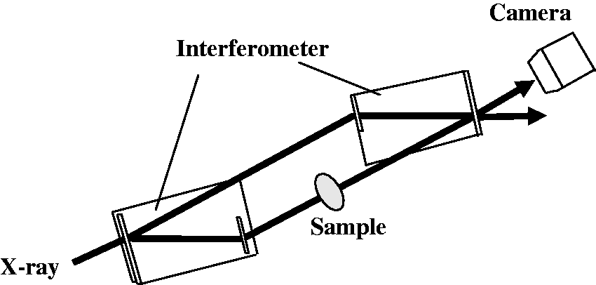
During the C-PCCT imaging, the specimen was placed in a 3-cm thick sample cell filled with formalin to prevent the drying of sample. The sample cell was then inserted into the beam path between the mirror and analyzer of the interferometer. The X-ray energy was set at 35 keV. The exposure time was 5 s for each interference pattern, and 250 projections were delivered over 180°. A phase map depicting the spatial distribution of the phase shifts caused by the sample was obtained using a three-step fringe scan. Sectional images were reconstructed by using a filtered back-projection algorithm.
Quantitative image analysis
During the analysis of the C-PCCT images, mass density


3D visualization of tumor vessels
Numerical analysis of 3D image data was performed using 3D volume-rending software (Real INTAGE; KGT Inc., Tokyo, Japan). A gray value of 30% was chosen as the threshold for extracting the tumor vascular network.
Histopathological analysis
After the C-PCCT imaging, the brain was embedded in paraffin. Then, 3-µm thick sections were cut in the coronal plane. Hematoxylin-eosin (H&E) staining and immunostaining for a neuronal marker (synaptophysin) were carried out to examine the tumor’s histopathological structures. The stained sections were imaged using an optical microscope (Olympus FSX100; Olympus, Tokyo, Japan).
Results
Coronal images of the rat’s brain obtained using C-PCCT are shown together with the corresponding pathological sections in Fig. 2a and b. C-PCCT demonstrated excellent tissue conspicuity, and hence, was able to reveal the detailed anatomical structures of the rat’s brain, including the cortex, lateral ventricles, caudate putamen, hippocampus, corpus callosum, thalamus, and the tumor, which was located in the brain stem. The tumor was easy to differentiate from the surrounding normal cerebral tissue. A pathological examination based on H&E staining confirmed that the tumor location and cerebral anatomy corresponded to those observed on C-PCCT. Specifically, on C-PCCT, the tumor appeared round, and oval high density structures were seen within it. In addition, the tumor also contained small low density duct-like structures, mainly in its inner portion (Fig. 2c). A pathological examination revealed that the high density and small low density structures corresponded to hematoma-derived blood clots/dilated vessels, and hypervascular small vessels that had been perfused with physiological saline, respectively. Marked dilation of the lateral ventricle was also observed. In addition, it was found that a region of the normal cerebral tissue surrounding the tumor, which exhibited a relatively low density, had been affected by edema. Absolute density obtained from C-PCCT images were showed in Table 1.
Coronal section of the rat’s brain containing the tumor. (a) Crystal X-ray interferometer-based phase-contrast CT image. (b) Corresponding histological section (H&E stain): a, cortex; b, lateral ventricle; c, caudate putamen; d, hippocampus; e, corpus callosum; f, thalamus; g, tumor. (c) Coronal section of the tumor obtained with crystal X-ray interferometer-based phase-contrast X-ray CT: 1, blood clot-filled blood vessel; 2, inner tumor region; 3, outer tumor region; 4, tumor-surrounding tissue; 5, normal cerebral tissue. Absolute density values obtained from crystal X-ray interferometer-based phase-contrast X-ray CT.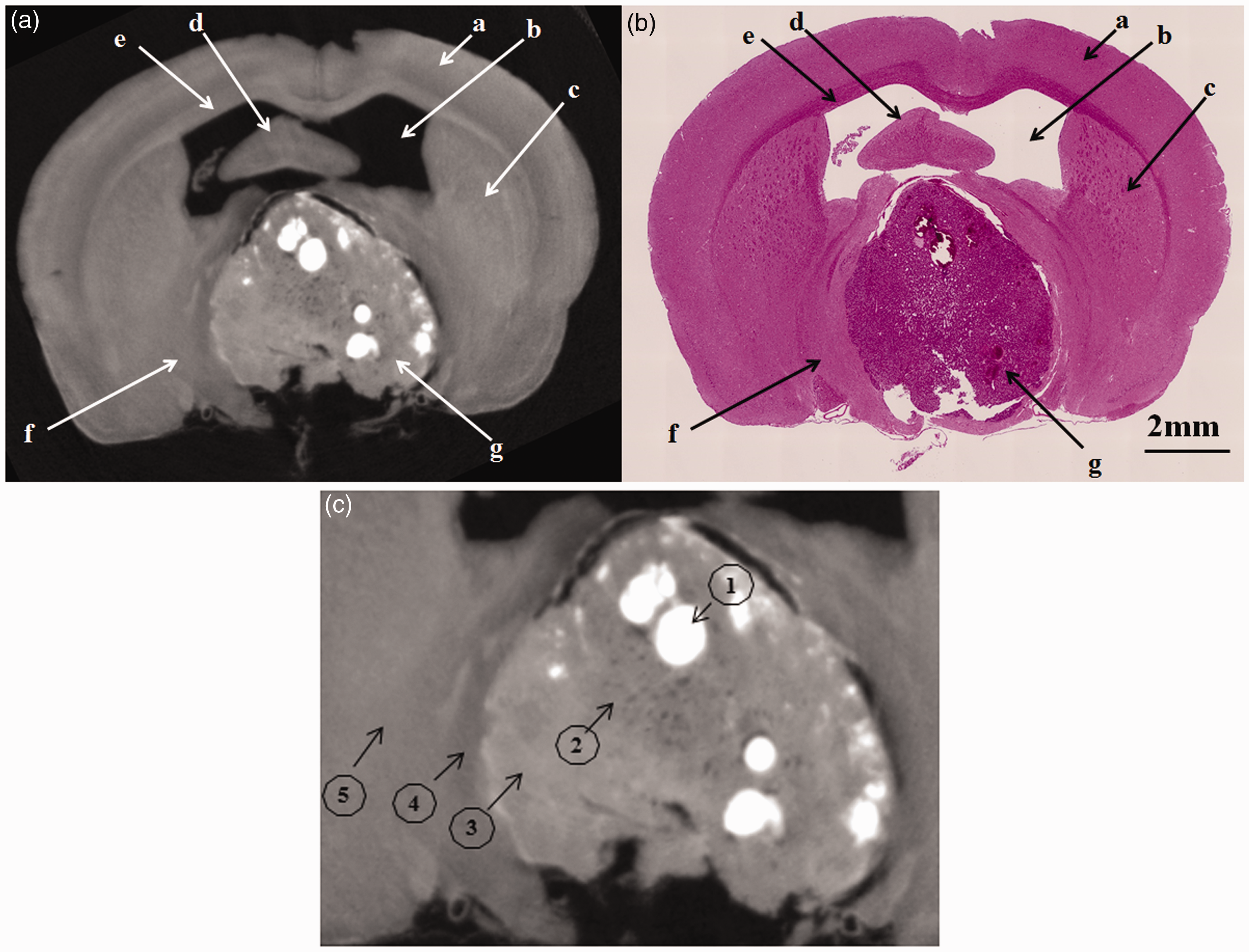

3D images of the tumor microvessels were reconstructed from the C-PCCT images, as shown in Fig. 3a and b. The 3D images made it possible to view the tumor microvasculature from different angles (Fig. 3c), and the features of these blood vessels were depicted clearly, e.g. tortuous, saccular (arrow), and dilated blood vessels, which exhibited haphazard patterns of interconnection, were observed. Small branch vessels of up to ∼26 µm in diameter were clearly visible in the reconstructed images.
Three-dimensional reconstruction of the tumor in the rat’s brain based on crystal X-ray interferometer-based phase contrast CT images. (a) Overview of the brain and tumor microvasculature. (b) Magnified image of the square region shown in (a). (c) The tumor microvasculature viewed from different angles. The characteristics of the tumor microvasculature were well depicted, e.g. tortuous, saccular (arrow), and dilated blood vessels were observed, which exhibited haphazard patterns of interconnection.
In the pathological examination, the tumor displayed high cellularity and hypervascularity. In addition, the tumor cells were round to elongated and possessed hyperchromatic nuclei that exhibited a salt and pepper chromatin pattern and an indistinct cytoplasm. Some of the tumor cells formed Homer-Wright rosettes (H&E stain). Immunostaining demonstrated that the tumor cells were positive for the neuronal marker synaptophysin (Fig. 4a and b).
Pathological section of the tumor. The tumor exhibited high cellularity and vascularity. The tumor cells were round to elongated and possessed hyperchromatic nuclei displaying a salt and pepper chromatin pattern (black arrow) and indistinct cytoplasm. Some of the tumor cells formed Homer-Wright rosettes (yellow arrow). (a) H&E stain. Immunostaining demonstrated that the tumor cells were positive for the neuronal marker synaptophysin (b).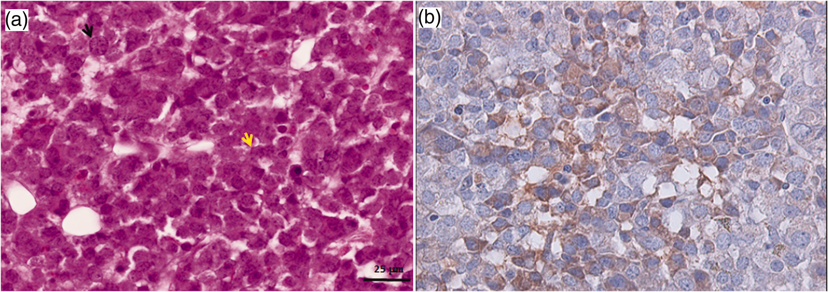
Discussion
A hypervascular tumor that arose spontaneously in an aged rat’s brain, was imaged clearly by C-PCCT within formalin solution. The resultant images resembled pathological pictures (magnification ×20), and depicted the tumor’s pathological features well. The microvasculature of the rat’s brain could be identified well because the formalin solution, with almost the same density of physiological saline, causes high image contrast between formalin within the vessel and the surrounding soft tissues (12). Furthermore, 3D tumor microvasculature of up to ∼26 µm in diameter was reconstructed because the image was obtained CT configuration. Tumor vessels were tortuous, dilated, and were connected to each other in a random manner. Grating X-ray phase-contrast imaging was obtained lung metastasis microvasculature of mice at dry stage (13). However, the tissue drying with shrunken and deformity caused unnatural geometry.
In C-PCCT, the gray level of an image corresponds to its absolute density, and the present imaging system has a density resolution of approximately 0.5 mg/cm3 (5). The absolute densities of the outer parts of the tumor, which contained dense cellular components, and the hypervascular inner region of the tumor were 1.060 g/cm3 and 1.046 g/cm3, respectively. Thus, minute density differences caused by tumor cell characteristics and the presence/absence of blood vessels can be distinguished on C-PCCT images without contrast agents. In the present study, blood clots due to hemorrhaging/hematomas exhibited high density, as reported previously (10). Moreover, the absolute density of the tumor-surrounding tissue affected by edema was less dense than the normal cerebral tissue. The dilatation of the left ventricle resulted in hydrocephalus was probably caused by the tumor obstructing the flow of cerebrospinal fluid.
This tumor was pathologically diagnosed as a primary brain tumor of the central nervous system, probably a medulloblastoma. This is the most common type of embryonic brain tumor affecting humans, but such tumors are rarely seen in rats (prevalence, 0.018% in 5480 Wistar rats). In rats, medulloblastoma tend to be seen in younger animals; however, they have also been reported to occur in animals of aged more than 100 weeks in necropsy studies (1).
Intratumoral vessel growth is a hallmark of tumor development, and the inhibition of vessel development is a valid target for anticancer therapy (14). Thus, the ability to visualize intratumoral vessels might be useful for preclinical anticancer research.
The present C-PCCT imaging system is limited to obtaining in vivo imaging because of the long image acquisition time. Currently, we are planning to use a new X-ray CCD camera system to perform the high speed data acquisition within 10 min/CT image.
In conclusion, C-PCCT makes it possible to depict the minute pathological structures such as intratumor and its vascular architecture up to approximately 26 µm without the use of contrast agents. Thus, it might be a useful tool for cancer research.
Footnotes
Declaration of conflicting interests
The authors declared no potential conflicts of interest with respect to the research, authorship, and/or publication of this article.
Funding
This research was carried out under the approval (proposal nos. 2009S006, 2012G044, 2013G584) of the committee of the High Energy Accelerator Research Organization and was supported by a grant from Kitasato University School of Allied Health Sciences (Grant-in-Aid for Project, no. 2013-6659).