
Abstract
Case summary
A 4-year-old male neutered domestic shorthair cat was presented to the Oregon State University cardiology service for suspected pericardial effusion. Cardiac tamponade was documented and pericardiocentesis yielded purulent fluid with cytologic results supportive of bacterial pericarditis. The microbial population consisted of Pasteurella multocida, Actinomyces canis, Fusobacterium and Bacteroides species. Conservative management was elected consisting of intravenous antibiotic therapy with ampicillin sodium/sulbactam sodium and metronidazole for 48 h followed by 4 weeks of oral antibiotics. Re-examination 3 months after the initial incident indicated no recurrence of effusion and the cat remained free of clinical signs 2 years after presentation.
Relevance and novel information
Bacterial pericarditis is a rare cause of pericardial effusion in cats. Growth of P multocida, A canis, Fusobacterium and Bacteroides species has not previously been documented in feline septic pericarditis. Conservative management with broad-spectrum antibiotics may be considered when further diagnostic imaging or exploratory surgery to search for a primary nidus of infection is not feasible or elected.
Introduction
Pericardial disease in cats is relatively uncommon, with a reported prevalence ranging from 1.0-2.3% in post-mortem studies.1,2 The most common causes of pericardial effusion in cats results from congestive heart failure secondary to cardiomyopathic disease and neoplasia. 1 Other less frequently cited etiologies include trauma, disseminated intravascular coagulation, uremic pericarditis, peritoneopericardial diaphragmatic hernia, feline infectious peritonitis, coagulopathy, hypoalbuminemia and infective pericarditis. Infective pericarditis includes viral, bacterial, fungal or parasitic colonization of the pericardium, and is rarely reported in cats.1–4 This case report describes a cat with bacterial pericarditis with previously undocumented microorganisms and a favorable clinical response to conservative medical management.
Case description
A 4-year-old male neutered domestic shorthair cat presented to the primary veterinarian after being found recumbent and dyspneic at home. Mild pyrexia (39.5

Right lateral radiograph. There is a generalized increase in cardiac silhouette size with dorsal deviation of the thoracic trachea. The cardiac silhouette spans nearly four intercostal spaces and has a globoid shape. The increased opacity of the cranial mediastinum is most consistent with fat infiltration
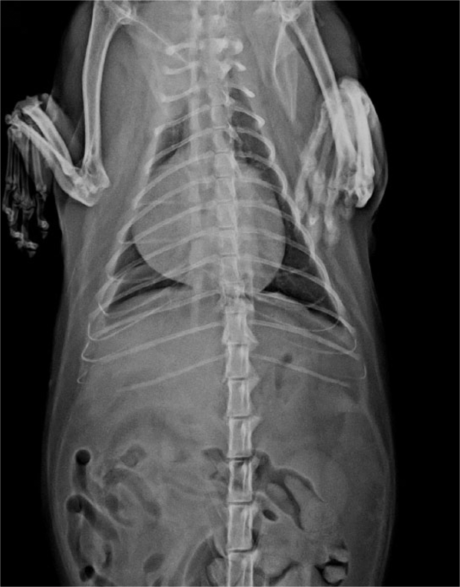
Ventrodorsal radiograph. There is a generalized increase in cardiac silhouette size with a globoid shape. A small volume of pleural effusion is also present. The cat is positioned obliquely
Upon presentation to OSU, the cat was depressed but responsive. It was hypothermic (36.7

Right parasternal short axis view. A large volume of anechoic pericardial effusion is present. Diastolic collapse of the right atrium and ventricle was noted, consistent with cardiac tamponade. The lumens of the right ventricle (RV) and left ventricle (LV) are labeled. The pericardial effusion is diffusely present around the ventricles (*)
The signs of cardiogenic shock resolved following pericardiocentesis. The echocardiographic examination was completed with no evidence of structural heart disease or neoplastic masses. Cardiac tamponade was abolished and a small amount of residual pericardial and pleural effusions remained after pericardiocentesis. Other diagnostics performed included complete blood count (CBC), chemistry panel, urinalysis, feline immunodeficiency virus (FIV) and feline leukemia virus (FeLV) antigen testing (IDEXX Laboratories), blood pressure measurement and ECG. The cat’s blood pressure was 130 mmHg (Doppler method) and its ECG indicated sinus rhythm with a left anterior fascicular block-like pattern. The relevant laboratory test abnormalities are listed in Table 1; abnormalities were consistent with sepsis (eg, transitional leukogram, mild hyperbilirubinemia). The FIV/FeLV test was negative. The urine sample was obtained several hours after fluid therapy, indicating a urine specific gravity of 1.018 without bilirubin, glucose, protein, cells or bacteria present.
Laboratory data

WBC = white blood cell
Diagnostic and therapeutic options were discussed with the cat’s owners, including the recommendation to pursue computed tomography (CT) to search for a local thoracic or distant source of infection. Exploratory surgery with pericardiectomy via thoracotomy or thoracoscopy was also discussed. Owing to financial limitations, the owners elected to pursue conservative management with hospitalization and empiric parenteral antibiotic therapy. Ampicillin sodium/sulbactam sodium (30/kg mg IV q8h; Aurobindo Pharma), metronidazole (15 mg/kg IV q12h; Claris Lifesciences) and intravenous fluid therapy (Lactated Ringer’s solution) were initiated. The cat’s 5% dehydration deficit was replaced over 6 h, followed by a maintenance fluid rate of 20 ml/h.
Over the next 48 h, the cat stabilized and serial recheck echocardiograms indicated diminishing residual pericardial effusion. A CBC and chemistry panel were repeated 2 days after presentation; the relevant results are presented in Table 1. After 2 days of hospitalization, the cat was transitioned to oral medications and discharged with instructions to receive amoxicillin/clavulanic acid (20 mg/kg PO q8h; Pfizer) and metronidazole (10 mg/kg PO q12h; Watson Pharma Private) for 4 weeks. At the time of discharge, the cat had no overt pericardial effusion and trace pleural effusion. The pleural effusion was likely either residual pleural fluid from impaired diastolic filling of the right heart during cardiac tamponade, or the result of pericardial effusion leakage following percardiocentesis. The final aerobic culture results yielded heavy growth of both Pasteurella multocida and Actinomyces canis. The anaerobic culture indicated heavy growth of A canis, Fusobacterium and Bacteroides species. Aerobic antimicrobial susceptibility testing suggested P multocida was sensitive to ampicillin though resistant to clindamycin and tobramycin. Susceptibility testing was not performed for the other bacterial isolates.
The cat presented to OSU for re-evaluation 1 week after discharge and was reported to have normal appetite, energy and demeanor at home. The physical examination was unremarkable. A recheck CBC indicated normalization of the white blood cell count (8970/µl; SI 8.97 × 10⁹/l) with an unremarkable leukogram. Furthermore, the chemistry panel indicated complete resolution of the previous electrolyte disturbances, as well as normalization of creatine kinase and alanine aminotransferase. There was no echocardiographic evidence of pericardial or pleural effusion. Continuation of the antibiotic regimen was recommended and recheck examination was advised following completion of the course. The cat was re-evaluated by echocardiography 3 months after its initial presentation, and no recurrent effusion was noted. The cat remains free of clinical signs 2 years after presentation.
Discussion
Feline bacterial pericarditis has been sparsely reported in the veterinary literature. In a large-scale retrospective study of pericardial effusion in 146 cats, no cases of septic pericarditis were identified. 5 Several mechanisms can result in septic pericarditis, including pericarditis secondary to localized spread of infection (eg, pneumonia, suppurative mediastinal lymphadenitis), hematogenous infection, extension of endocarditis/myocarditis, or direct inoculation resulting from penetrating wounds, migrating foreign material or surgery. 6 The associated clinical signs are often related to hemodynamic compromise from pericardial effusion or related to systemic infection (eg, pyrexia, weight loss).
Of the published case reports, some have shown a documented predisposing infection, whereas the inciting etiology was not conclusively identified in others. A cat in one report developed bacterial pericarditis in close temporal proximity to a dental prophylaxis with a common oropharyngeal microorganism, Peptostreptococcus. 3 The mechanism of infection was theorized to be bacteremia following the dental procedure. The cat was treated with amoxicillin–clavulanic acid for approximately 3 weeks and a full recovery was reported 6 weeks after completion of the antibiotic course. Another recent case report described bacterial pericarditis in an intact female cat with pyometra and hematogenous spread of Escherichia coli. That cat responded well to a 4 week course of enrofloxacin and metronidazole following peri-cardiectomy, and remained clinically stable 1 year later. 7 Conversely, another published report of feline bacterial pericarditis identified infection with a mixed population of Enterobacteriaceae species and coagulase-negative Staphylococcus species without an obvious predisposing cause. 4 An anaerobic culture was not performed, and the cat responded favorably to an 8 week course of broad-spectrum antimicrobial therapy. Owing to the multiple organisms involved, direct inoculation via a penetrating wound or foreign material was thought to be more likely than septicemic inoculation of the pericardial sac.
The case reported here did not have a readily apparent systemic or focal infection, or any immune-modulating systemic diseases; however, an exhaustive search was not pursued. There was no history of recent dentistry or substantial evidence of dental disease. The polymicrobial nature of this infection with mixed aerobic–anaerobic isolates is suggestive of a penetrating bite wound or foreign body. Bite wounds are generally associated with the oral flora of the biting animal rather than the skin flora of the victim. 8 The most common aerobic isolate in cat bites is P multocida, and anaerobic isolates commonly include Fusobacterium and Bacteroides species.8,9 Though the cat reported here was exclusively housed indoors, another cat was present in the household and may have incited the infection through an unwitnessed altercation. Less likely, this infection may have resulted from atraumatic hematagenous spread of bacteria and alternate sources of infection (eg, odontogenic, esophageal, pleuropulmonary) were not systematically excluded.
While the incidence and causes of septic pericarditis vary across species, the diagnostic approach and therapeutic goals remain the same. Identification of the specific pathogen(s), related antibiotic susceptibility and source of the infection are paramount. Cytology and culture of the pericardial fluid is crucial in the diagnosis of septic cases, unlike most other causes of pericardial effusion. In addition, blood cultures are advised in humans as they are positive in 40–70% of cases. 6 Aerobic and anaerobic susceptibility testing is also recommended, although most laboratories do not routinely perform susceptibility testing on anaerobic isolates. As anaerobic pericarditis is associated with odontogenic infection, esophageal disease, pleuropulmonary infection, abdominal infection and pelvic infection in humans, a thorough search for adjacent and distant infections is warranted. 10 Both magnetic resonance imaging and CT are ideal modalities for imaging multiple body cavities.
Owing to the low incidence and small number of published case reports, the ideal therapy for septic pericarditis in cats is unclear. Results of susceptibility testing facilitate antibiotic selection, although, if unavailable, broad-spectrum coverage is recommended based on the wide range of bacterial isolates reported. The standard of care in affected humans involves aggressive medical management, including an indwelling pericardial catheter for drainage and targeted antibiotic therapy. In dogs affected by bacterial pericarditis, a combination of exploratory surgery, pericardectomy and antimicrobial therapy is advocated.11,12 Eradication of the source of the infection is imperative and surgical exploration is indicated in cases that have failed medical management or have suspected abscessation identified on diagnostic imaging. Consequently, advanced imaging, exploratory surgery and pericardectomy could be advised in affected cats, although, interestingly, complete recovery has been reported with antibiotics alone in previous cases and the cat reported here.3,4
The development of constriction is an important potential sequela to septic and non-septic pericarditis, and should be assessed on patient follow-up. Constriction is theorized to occur secondarily to the influx of inflammatory cells into the pericardial space, ultimately leading to proliferation of fibrotic connective tissue and neovascularization. 6 These changes can result in a loss of pericardial elasticity and thus limit diastolic filling. In a prospective human study, 9/500 (1.8%) patients developed constriction over a median follow-up time of 72 months. While overall constriction is a rare complication of human viral or idiopathic pericarditis (<0.5%), the risk of constriction was highest in the bacterial pericarditis group (33%). 13 The prevalence of constriction in cats is unknown, although echocardiographic evidence of constrictive physiology was identified in one cat with septic pericarditis. 7 Constrictive pericarditis is difficult to definitively diagnose, but echocardiographic findings of a thickened pericardium and respiratory variation of >25% in mitral inflow velocities can be highly suggestive. 14 In the case presented here, no subjective or clinical evidence of constrictive pericarditis was identified during the limited echocardiograms during recheck examinations; however, concern exists for its development nonetheless.
Conclusions
Bacterial pericarditis is a rare but potentially curable cause of pericardial effusion in cats. The cat reported here fully recovered following conservative therapy consisting of pericardiocentesis and broad-spectrum antibiotic treatment. Larger retrospective or prospective studies could help further characterize treatment strategies and prognosis in affected cats.
Footnotes
Acknowledgements
The authors thank Dr Gray for the provision of the thoracic radiographs.
Supplementary material
Video of the right parasternal long axis cine loop showing a large volume pericardial effusion. The effusion is mostly anechoic with hyperechoic flecks throughout; severe underfilling of cardiac chambers is noted.
Funding
The authors received no financial support for the research, authorship and/or publication of this article.
Conflict of interest
The authors declared no potential of interest with respect to the research, authorship and/or publication of this article.
References
Supplementary Material
Please find the following supplemental material available below.
For Open Access articles published under a Creative Commons License, all supplemental material carries the same license as the article it is associated with.
For non-Open Access articles published, all supplemental material carries a non-exclusive license, and permission requests for re-use of supplemental material or any part of supplemental material shall be sent directly to the copyright owner as specified in the copyright notice associated with the article.