
Abstract

Case report
We present the case of a 73-year-old male who presented with left-sided hearing loss on a background of controlled hypertension and right hand osteoarthritis. Under microscopic exploration, a solid mass located in the attic of the left ear could be demonstrated. Pure tone audiogram showed a mean conductive hearing loss of 40 dB on the left ear and mild presbyacusis on the right (Figure 1). A cone beam computed tomography was requested demonstrating a lesion, which appeared to be attached to and partially enveloping the handle of the malleus (Figure 2).
Pre-operative (a) and post-operative (b) pure tone audiometry (PTA). Closure of the conductive GAP on the left ear can be appreciated. Cone bean CT demonstrating the presence of a bone mass attached to and enveloping the handle of the malleus and in contact with the scutum anteriorly. (a) Axial cut. (b, c and d) Coronal cuts.

Due to the working diagnosis of conductive hearing loss caused by an external ear canal exostosis extending into the middle ear, the patient was listed for exploration of left ear, removal of bony exostosis and tympanoplasty. Consent for surgery and publication is routinely obtained and kept in the clinical notes. At surgery, through an endaural approach, a tympanomeatal flap was lifted showing a lesion in the left ear canal composed of a soft deposit of bone-like material, which crumbled readily. We followed this into the middle ear through the tympanic membrane and into the area around the head of the malleus. The lesion was removed grossly and sent for histopathological analysis (Figure 3), delaying any further management until a definitive diagnosis could be achieved due to the unexpected consistency and possibility of a malignant tumour. The ossicular chain demonstrated normal movement.
Specimen removed measuring 1 cm.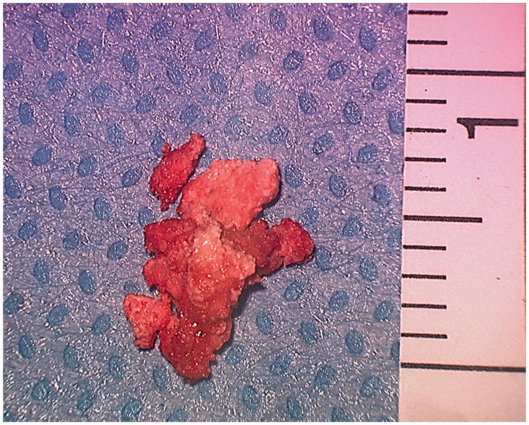
Two weeks after the surgery, the ear canal packing was removed and a complete restoration of the conductive hearing loss was demonstrated (Figure 1). The histopathological report was consistent with calcium pyrophosphate dehydrate deposition disease (pseudogout), and consequently, the patient was referred to the rheumatology department.
Discussion
Calcium pyrophosphate dihydrate crystal deposition in articular tissues, mainly in fibrocartilage and hyaline cartilage, causes an arthropathy. The European League Against Rheumatism divides this pathology depending on the clinical presentations into four groups: (1) asymptomatic calcium pyrophosphate dihydrate; (2) osteoarthritis with calcium pyrophosphate dihydrate; (3) acute calcium pyrophosphate crystal arthritis; (4) chronic calcium pyrophosphate inflammatory crystal arthritis, 1 that can be (a) degenerative arthritis in younger patients with deposition in the meniscal cartilage or (b) arthropathy in older patients where crystals are found within the cartilage along with extensive areas of sclerotic bone articular surface and (c) a rather uncommon tumoural form that clinically and radiologically mimics neoplasia involving the temporomandibular joint. 2 The aim of this article is to describe the unusual and unexpected isolated otologic presentation of this disease that could be added to the previous classification.
The differential diagnosis of external ear canal bone tumours include exostosis, osteoma, bone dysplasia, bone dystrophy, chondroma, osteochondroma, sarcoma and chondrosarcoma. 3 In addition, all tumours and pseudotumours of the temporomandibular joint can invade the external auditory canal.3–6 In the study of the expansive processes of the ear, computed tomography and magnetic resonance imaging are very useful, orientating the diagnosis according to the location, signal, density and characteristics of the lesion, and an estimation of its extension can be achieved. Only with the definitive histopathology, however, is the diagnosis certain, as the radiographic findings are non-specific and difficult to recognise in middle ear joints. 7 In the present case, the computed tomography demonstrated an isolated lesion between the middle and external ear, suggesting an osteoma as the main cause of the hearing loss. No further symptoms were present. During the surgery, with the possibility of a malignancy and the knowledge that an isolated surgical treatment would not be suitable if this was the case, the lesion was largely debulked to restore the normal movement of the ossicular chain and tympanic membrane.
In the incudomaleolar joint, calcifying arthropathies may occur due to the saddle-shaped synovial joint capsule and a meniscus that separates both bones. For the study of crystal deposition disorders, synovial fluid analysis helps in characterising the morphological features of the clinically relevant crystals, but in tumour manifestation, clinically relevant investigations include imaging, cytology, biopsy and biochemical analysis for a definitive diagnosis.2,8,9
The unusual diagnosis of calcium pyrophosphate dihydrate deposition affecting the ear as an extension of a lesion from the temporomandibular joint is rare, but it is the most described otological presentation.5,10 The isolated involvement of the middle ear has only been described once before and is thus of interest. 7 Consideration of an otological tumour as the presentation of chronic calcium pyrophosphate inflammatory crystal arthritis 2 should be added to the pseudogout diagnostic classification and also to the differential of an expansive lesion in the ear. Finally, a combined treatment of surgery to restore hearing and medical rheumatologic management seems the most reasonable therapeutic option.
Key message
Consider pseudogout with deposition of calcium pyrophosphate dihydrate crystals as an unexpected isolated lesion in the middle ear.
Consent for publication
The consent for surgery and publication was obtained prior to the surgery and kept in the medical notes.
Availability of data and materials
The data and materials presented in this case record are available at St George’s NHS Foundation Trust of London on both computer system and ENT clinical notes of the institution.
Supplemental Material
sj-pdf-1-shr-10.1177_2054270419894818 - Supplemental material for A description of unilateral conductive hearing loss from pseudogout: a case report and review of the literature
Supplemental material, sj-pdf-1-shr-10.1177_2054270419894818 for A description of unilateral conductive hearing loss from pseudogout: a case report and review of the literature by Manuel Gomez Serrano, Natalie Anne Watson and David Selvadurai in JRSM Open
Footnotes
Declarations
References
Supplementary Material
Please find the following supplemental material available below.
For Open Access articles published under a Creative Commons License, all supplemental material carries the same license as the article it is associated with.
For non-Open Access articles published, all supplemental material carries a non-exclusive license, and permission requests for re-use of supplemental material or any part of supplemental material shall be sent directly to the copyright owner as specified in the copyright notice associated with the article.