
Abstract
Lesson
The first case of bilateral distal tibiofibular joint fusions for osteochondromas is reported with excellent long-term outcomes.
Case report
A 26-year-old male presented with large osteochondromas arising from the distal tibiae.
There was a strong family history of hereditary multiple osteochondromas. Exostoses had been removed from his left scapula, right femur and twice from the left proximal tibia. His first surgery was at 10 years old.
He worked in university management. Pain began five years earlier with no history of injury. The left ankle was more severely affected. Both in intensity and duration, the pain was becoming increasingly severe affecting his walking distances. Sometimes he had difficulty weight bearing. The history of osteochondromas, multiple surgeries and a perception of poor balance had limited his sports.
Plain X-rays (Figure 1) revealed osteochondromas arising from the distal tibiae. The left lesion was larger. Both were deforming and thinning the fibula confirmed on CT scan (Figure 2). There were no features of arthritis or malignant change.
AP weight-bearing X-rays of both ankles demonstrating deforming distal tibial osteochondromas. CT scan. Axial CT scan showing thinning and deformity of fibulae. Worse on the left side.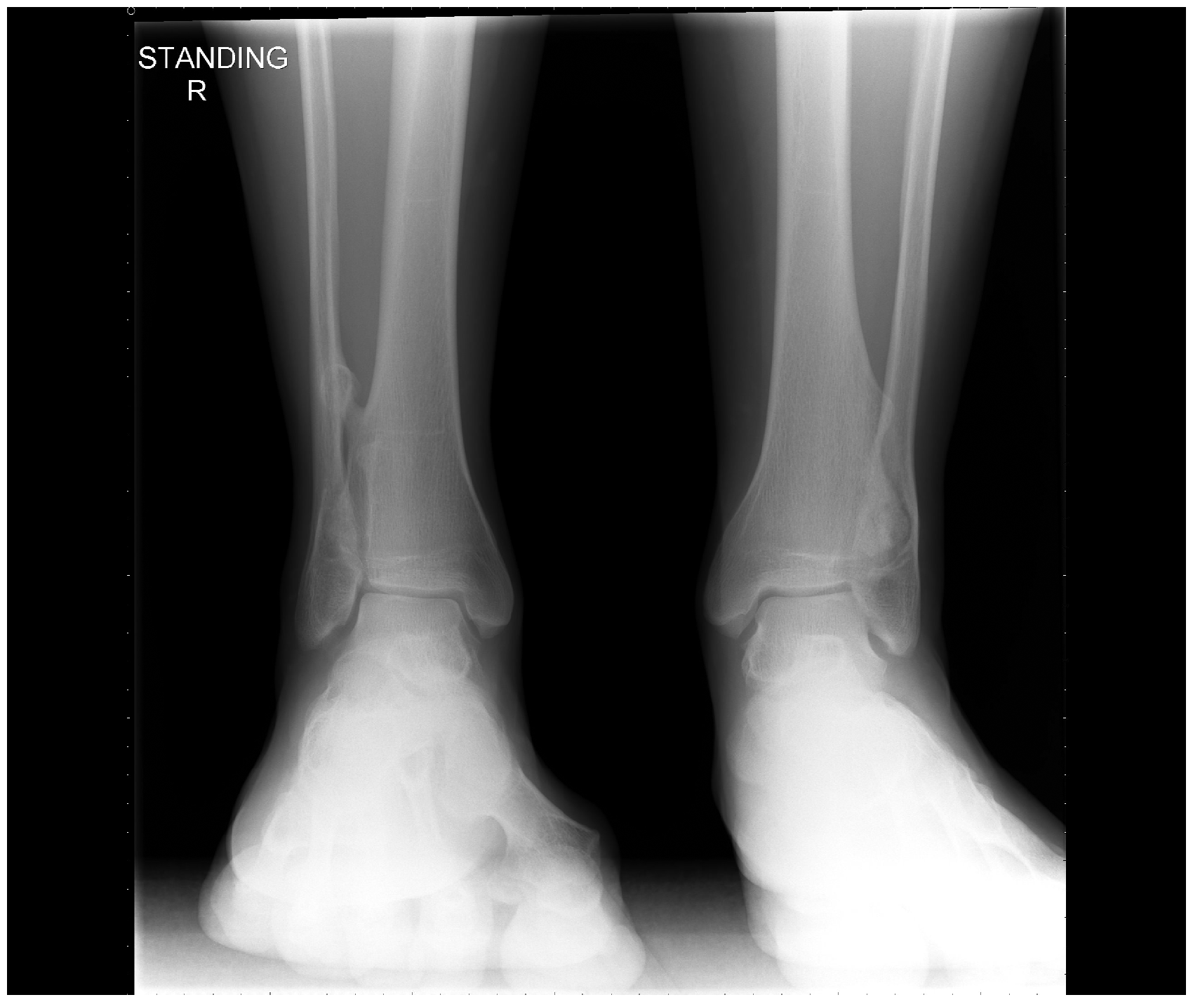

Simple resection for the left side was adjudged to risk the thinned fibula fracturing or leave the syndesmosis and ankle unstable with risk of continuing pain and later arthritis. The patient was advised to undergo a fusion of the distal tibiofibular syndesmosis.
In 2010, the left distal tibiofibular joint was arthrodesed via an anterolateral approach. The superficial peroneal nerve was protected, osteochondroma removed and tibial and fibular surfaces decorticated. Iliac graft was utilised with a fibular plate and syndesmotic screws to stabilise the construct. He was immobilised in a below knee cast for six weeks followed by six weeks protected weight bearing in a removable boot.
Delighted with pain relief and retained mobility, the patient stated a preference for the same procedure on the right side. The second surgery was performed six months later with a similar outcome.
The patient was reviewed 77 (left) and 71 months (right) post-surgery. He had made an excellent recovery and was pain free. Walking distances were unlimited and he was comfortable on uneven ground. He had no restrictions with footwear and never required analgesia. He swims, attends the gym and participates in kayaking. There was occasional stiffness of the ankles in winter months. There was no discernible limp and an excellent range of ankle movement. Radiologically both joints were fused with no metalwork issues (Figure 3).
AP weight-bearing X-rays of both ankles ((a) right and (b) left) at over six years’ follow-up after distal tibiofibular fusions.
Discussion
Osteochondromas are benign tumours found incidentally in 2% of patients. 1 They account for 40% of benign bone tumours 2 but are uncommon around the ankle joint. Seventy-three foot and ankle benign and malignant bone tumours in a Musculoskeletal Tumour Unit reported over a 20-year period found three osteochondromas. 3
Lesion growth follows normal skeletal development and ceases to enlarge post-osseous maturity. Later growth can indicate malignant change in ≤1% of cases. Ankle osteochondromas usually present in teenage years causing pain and deformity secondary to physeal disturbance.3–5 Resection of symptomatic lesions is usually recommended through either anterolateral, posterolateral or fibular osteotomy approaches.4–7
The largest series reported describes 19 tumours over a 16-year period. 4 Twenty-one per cent recurred and complications included nerve injury, infection and growth arrest. Three adults had resections with only one symptom free and not requiring further surgery.
Distal tibiofibular fusions are rare with the most common indications including:
Salvage following syndesmotic injury with a chronic, symptomatic diastasis following Weber B and C and ankle fractures or, more rarely, isolated chronic syndesmotic injuries with instability. During certain total ankle replacements (e.g. Agility Total Ankle Replacement)
Outcomes from these infrequently performed procedures are difficult to ascertain.
Espinosa et al.’s 8 stated that ‘no studies have reported the outcome after syndesmotic arthrodesis’ and that they were limiting and should be regarded as salvage procedures.
Peña and Coetzee 9 reported no outcomes of the procedure in the English literature. He described it as a salvage operation for syndesmotic incongruency following metalwork removal and over six months of symptoms and that it was not compatible with active athletic life.
Both papers described operative techniques but no outcomes.
Olson et al. 10 published the largest outcome series of distal tibiotalar fusions with 10 cases. Historical doubts were noted about the procedure including loss of normal fibula motion during gait causing pain, stiffness and ankle arthritis development. All had chronic syndesmotic instability following rotational ankle fractures. All were offered a tibiofibular fusion elsewhere. Each united with 100% patient satisfaction. Average AOFAS scores improved from an average 37 to 87 at minimum two-year follow-up. There were no reports of ankle joint arthritis.
Van Dijk 11 described nine patients with osteochondromas who all reported good to excellent results and return to sport even at the highest level following fusion.
The Agility Total Ankle Replacement requires a syndesmotic fusion as the tibial component design resurfaces the distal tibial surface and both malleoli. It requires a stable contact area between the tibia and fibula. Non-unions are relatively common − 34 to 49% reported by various authors.12–14
The favoured fusion approach is open anterolateral as described here. Graft can be autograft, allograft or demineralised bone matrix. Improved fusion rates have been reported by the addition of autologous platelet-derived growth factors and a recent report described performing the procedure arthroscopically.
Syndesmotic fusion fixation has evolved from a single 3.5 mm screw to two and, later, either larger 4.5 mm screws or a fibular plate.
Olson et al. 10 emphasised that, for fusions for chronic instability, other procedures are often required including fibular osteotomy for mal- or non-union, Achilles lengthening and medial gutter debridement.
Distal tibiofibular arthrodesis for osteochondromas in adults has only been described twice previously.15,16 Both were treated with screws only. One arose in the tibia and the other the fibula. Both fused and returned to sports without pain. The rationale for selecting a fusion over resection was pain levels, lesion size, distortion and fibula thinning 15 and tibiotalar involvement. 16
This case presentation is only the third patient to undergo a distal tibiofibular fusion for a painful, deforming osteochondroma and the first to undergo bilateral fusion. At over a six-year follow-up, the patient has excellent pain relief, function and return to sport.
Footnotes
Declarations
Acknowledgements
The patient
Provenance
Not commissioned; peer-reviewed by Jonathan Wright