
Abstract
Human orf should be considered based on a typical presentation with erythematous papule/nodule to avoid unnecessary over-treatment.
Case report
A previously healthy woman aged 48 years presented with a bloodshot papulo-bullous cutaneous lesion on the extensor surface of her right index finger (Figure 1(a)). Two weeks before she had first noticed a fissure developing into a small singular indolent pink papule that enlarged with a progressive accompanying erythema within the next four days. She did not remember any trauma. The self-maintained local application of antiseptics did not improve recovery. She had no fever, lymphedema, pain nor tenderness. On questioning, the patient reported that she had fed the lambs from a neighbouring peasant, but she did not remember them to have been ill. Based on the clinical presentation and the patient’s history concerning contact with small ruminants, we assumed the diagnosis of orf disease. The lesion was sparingly debrided and fractions of the crusts’ border were sent for histological examination and for parapoxvirus real-time polymerase-chain reaction (rt-PCR) testing according to Nitsche et al.
1
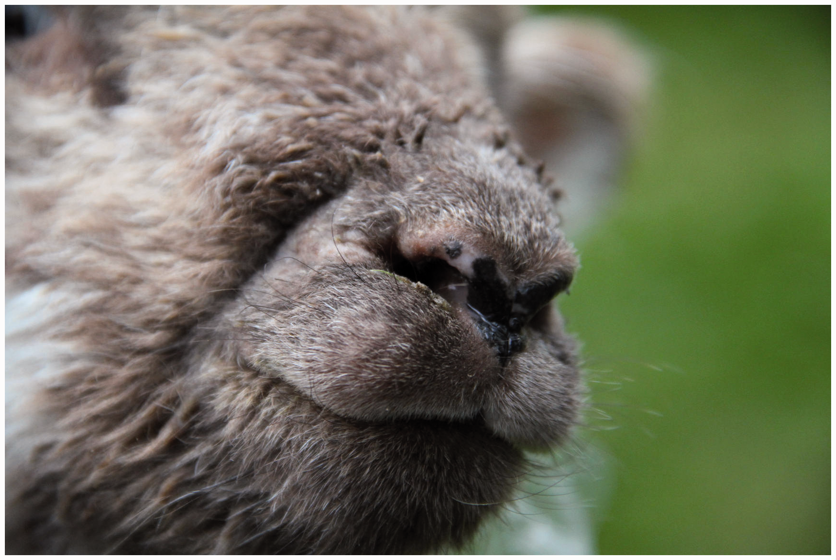
Histological examination revealed inflammatory cellular infiltrates and typical eosinophilic intracytoplasmatic inclusion bodies in epithelial cells (Figure 1(b)). Parapoxvirus infection was confirmed by rt-PCR. Further evaluation of the sheep herd revealed a lamb demonstrating a typical mucous eschar (Figure 2). Crusts taken from the lamb’s lesion were positive for parapoxvirus by rt-PCR demonstrating the epidemiological link. No specific treatment was initiated. Three weeks after the initial evaluation the patient’s lesion healed without sequelae.
(a) Clinical presentation with papulo-bullous lesion on the digit and (b) histological examination showing vacuolisation of squamous epidermal cells and intracytoplasmatic eosinophilic inclusion bodies (H&E × 200). Sore/scabby mouth disease with the typical mucosal eschar.
Discussion
Orf is caused by the orf virus, a member of the genus parapoxvirus in the family Poxviridae. Like most members of the poxviridae family, it has a tropism to epidermal cells. 2 Orf virus infection is endemic in many sheep and goat herds worldwide, where the disease is more commonly known as ecthyma contagiosum, contagious pustular dermatitis, ‘sore mouth’ or ‘scabby mouth’ disease. The animal’s lesions are typically located in the mucosal area of lips, muzzle, nostrils and eyes and may occur on udder and teats.
Transmission to humans occurs through direct contact in conjunction with a skin lesion, seldom through contact with contaminated meat or objects.2,3 Human orf infection has become rather uncommon due to increasing hygiene and, possibly, the availability of veterinary vaccines. It is most frequently seen in professionally exposed persons such as farmers, butchers, sheep shearers and veterinarians.2–5 Clusters have been described in shepherds’ communities or after the feast of sacrifice in Muslim countries.4,5 Recently, nosocomial human-to-human transmission in patients on a burn unit has been described. 6 The actual prevalence of human orf infection is presumably underestimated because orf is self-limiting and recognised by people at risk who do not seek medical care. 3
Given the transmission mode human orf generally manifests at sites of direct contact after an incubation period of two to six days. The lesions are typically seen on the hand, occasionally on other body regions. 2
The lesions usually progress through five distinct clinical and histopathological stages. 2 In the first, ‘maculopapular’ stage an erythematous papule develops from an initial macule. A red centre surrounded with a white middle ring and a red halo characterises the second ‘target lesion’ stage. The third ‘nodular’ stage shows an erythematous weeping nodule. It is followed by the fourth papillomateous ‘regenerative’ stage where the lesion dries. The fifth ‘regression’ stage is characterised by dry crust. The initial lesion generally heals spontaneously without scarring within four to six weeks. 2 Histologically, the parapoxvirus infection is characterised by the appearance of hyperkeratosis and pathognomonic eosinophilic inclusion bodies in the cytoplasm of vacuolated epidermal cells in the upper epidermis. 7
Constitutional symptoms such as fever, malaise and lymphadenopathy may accompany the clinical presentation but are uncommon. 2 In immune-compromised patients, the cellular immune dysfunction alters the classic clinical presentation. This can result in giant orf, multiple lesions, a protracted course of illness, or failure of spontaneous resolution.8–10
Orf is mainly a clinical diagnosis based on the history of relevant exposure and the typical clinical appearance of the lesion. 2 The diagnosis may be confirmed by histopathological examination, fluorescent antibody tests, electron microscopy, PCR and identification of specific viral nucleic acid sequences.2,4 Differential diagnosis includes, depending on the disease stage, other members of the genus parapoxvirus causing milker’s nodule and similar disease, fish-tank granuloma (Mycobacterium marinum), cutaneous anthrax, fungal infections, pyogenic granuloma and keratoacanthoma.2,4
The treatment of choice in healthy patients is to await the spontaneous resolution of the lesion. It is recommended to keep the lesion clean with local antiseptic to avoid secondary bacterial infection. 2 Treatment in immunocompromised hosts can be problematic. Topical imiquimod or cidofovir may be indicated in these patients.8,10 Giant hand lesions that do not respond to medical therapy may even require complete excision and skin grafting. 2 In the case of bacterial superinfection presenting with purulent secretion, lymphadenitis and onset of systemic symptoms systemic antibiotics may be indicated. 2
In conclusion, ecthyma contagiosum – orf – is an endemic zoonotic infection. Humans at risk may be infected by direct contact with diseased sheep or goats, in conjunction with skin lesions. Diagnosis is based on the patients’ history of relevant exposure and characteristic clinical lesions. Human orf is usually self-limiting and no specific treatment is indicated; complications are mainly due to over-treatment, in particular surgical debridement.