
Abstract
Liposomes and liposome-derived nanovesicles such as archaeosomes and virosomes have become important carrier systems in vaccine development and the interest for liposome-based vaccines has markedly increased. A key advantage of liposomes, archaeosomes and virosomes in general, and liposome-based vaccine delivery systems in particular, is their versatility and plasticity. Liposome composition and preparation can be chosen to achieve desired features such as selection of lipid, charge, size, size distribution, entrapment and location of antigens or adjuvants. Depending on the chemical properties, water-soluble antigens (proteins, peptides, nucleic acids, carbohydrates, haptens) are entrapped within the aqueous inner space of liposomes, whereas lipophilic compounds (lipopeptides, antigens, adjuvants, linker molecules) are intercalated into the lipid bilayer and antigens or adjuvants can be attached to the liposome surface either by adsorption or stable chemical linking. Coformulations containing different types of antigens or adjuvants can be combined with the parameters mentioned to tailor liposomal vaccines for individual applications. Special emphasis is given in this review to cationic adjuvant liposome vaccine formulations. Examples of vaccines made with CAF01, an adjuvant composed of the synthetic immune-stimulating mycobacterial cordfactor glycolipid trehalose dibehenate as immunomodulator and the cationic membrane forming molecule dimethyl dioctadecylammonium are presented. Other vaccines such as cationic liposome–DNA complexes (CLDCs) and other adjuvants like muramyl dipeptide, monophosphoryl lipid A and listeriolysin O are mentioned as well. The field of liposomes and liposome-based vaccines is vast. Therefore, this review concentrates on recent and relevant studies emphasizing current reports dealing with the most studied antigens and adjuvants, and pertinent examples of vaccines. Studies on liposome-based veterinary vaccines and experimental therapeutic cancer vaccines are also summarized.
Keywords
Introduction
Classical vaccines rely on the use of whole killed or attenuated pathogens. Today, research is focused on the development of subunit vaccines because they are better defined, easier to produce and safer. Vaccines are manufactured on the basis of well characterized antigens, such as recombinant proteins and peptides. However, due to their synthetic nature, their immune response is often weak, which is largely related to the inability of the antigens to induce maturation of dendritic cells (DCs), the primary antigen-presenting cells (APCs) that react to foreign pathogens and trigger the immune response [Moser and Leo, 2010; Reed et al. 2013].
The immune system is composed of the innate and the adaptive systems. The first is responsible for first-line host defense, rapidly recognizing and responding to foreign pathogens. The complement system and phagocytic cells belong to this defense system which depends on pattern recognition receptors (PRRs) that recognize pathogen-associated molecular patterns (PAMPs). Toll-like receptors (TLRs) present on APCs are the receptors for pathogens containing PAMPs. TLR activation is the hallmark of innate immune response. The second defense line, the adaptive immune system, mounts specific responses against molecular determinants on pathogenic agents. These responses are initiated by antigen-mediated triggering of T cells, the CD4+ T-helper (TH) cells, the CD8+ cytotoxic T lymphocytes (CTLs) and B lymphocytes carrying antigen-specific surface receptors. TH cells have subpopulations, of which TH1 and TH2 are the most important [Nordly et al. 2009; Kawai and Akira, 2010].
The diverse mechanisms by which nanoparticles induce immune responses are summarized in Figure 1. Activation of PRRs triggers the initiation of the innate immune response. Activated CTLs recognize peptides bound to the major histocompatibility complex class I and II molecules (MHC-I, MHC-II), which express antigenic peptides on APCs and bind to T cells via the T-cell receptor. A costimulatory signal is needed for full CTL and TH cell activation which differentiate into TH1 or TH2 and other T-helper lineages that produce cytokines. TH cells provide help to antigen-specific B cells, resulting in antibody production [Lin et al. 2010; Chen and Flies, 2013]. Each invasion of a foreign antigen requires activation of a specific type of adaptive immune response for efficient control and elimination. Thus, vaccine formulations should be designed rationally to induce specific protective responses. This includes the choice of antigen and adjuvant(s) and their pharmaceutical formulation.

Adjuvants
The ability to enhance the immune response of vaccines by certain compounds was first demonstrated with aluminum salts, termed ‘adjuvants’, added to killed or attenuated pathogens. Their functions were related to the ability to form a depot which prolonged antigen exposure to APCs. However, efficient adjuvants also stimulate the immune system by direct interaction with APCs. The nature of immune adjuvants is large and heterogeneous. Adjuvants are divided into immunostimulants and delivery systems. Immunostimulants interact with specific receptors, like TLRs and others, while delivery systems increase the immune response by multiple mechanisms, depending on their particular characteristics [Leroux-Roels, 2010; Alving et al. 2012]. Thus, modern vaccines comprise adjuvants such as pathogen-derived subcellular components, recombinant proteins, peptides and nucleic acid sequences [Zepp, 2010; Perez et al. 2013; Reed et al. 2013]. In addition, due to better knowledge of the immune system and improvements in formulation technology, effective therapeutic cancer vaccines are developed [Joshi et al. 2012]. Today’s challenges in vaccine development are linked to complex pathogens [e.g. malaria, tuberculosis, human immunodeficiency virus (HIV)] and to antigens susceptible to genetic mutations (e.g. influenza) as well as to subjects with a compromised or dysfunctional immune system [Leroux-Roels, 2010].
Nanoparticulate carriers provide adjuvant activity by enhancing antigen delivery or by activating innate immune responses. Strength and mechanisms of immunostimulation induced by nanocarrier vaccines depend on various factors, such as chemical composition, particle size and homogeneity, charge, nature and location of antigens and/or adjuvants within the carrier and, last but not least, the site of administration (see Figure 2) [Watson et al. 2012; Brito et al. 2013; Gregory et al. 2013; Smith et al. 2013; Zaman et al. 2013].

Schematic representation of a small unilamellar liposome showing the versatility of incorporation of various compounds either by encapsulation in the aqueous inner space or by integration in the bilayer or surface attachment on the lipid bilayer membrane. CpG, cytosine–phosphorothioate–guanine oligodeoxynucleotide; PEG, poly(ethyleneglycol). Reproduced and modified with permission [Heegaard et al. 2011].
Liposomes: ideal carriers for antigens and adjuvants
The ability of liposomes to induce immune responses to incorporated or associated antigens was first reported by Gregoriadis and Allison [Allison and Gregoriadis, 1974, 1976]. Since then, liposomes and liposome-derived nanovesicles such as archaeosomes and virosomes have become important carrier systems and the interest for liposome-based vaccines has markedly increased. The field of liposomes and liposome-based vaccines is vast. Therefore, this review concentrates on recent reports highlighting the most studied antigens and adjuvants in pertinent examples of vaccines, including summaries of veterinary and experimental therapeutic cancer vaccines. Other nanoparticulate vaccines based on lipoplexes, niosomes, virus-like particles, solid lipid nanoparticles and nanoemulsions are not covered in this review.
A key advantage of liposomes, archaeosomes and virosomes in general, and liposome-based delivery systems in particular, is their versatility and plasticity (see Table 1). Liposome composition and preparation can be chosen to achieve desired features such as lipid composition, charge, size, size distribution, entrapment and location of antigens or adjuvants. Depending on the chemical properties, water-soluble compounds (proteins, peptides, nucleic acids, carbohydrates, haptens) are entrapped within the aqueous inner space, whereas lipophilic compounds (lipopeptides, antigens, adjuvants, linker molecules) are intercalated into the lipid bilayer and antigens can be attached to the liposome surface either by adsorption or stable chemical linking [Torchilin, 2005; Watson et al. 2012]. Coformulations containing different types of antigens and adjuvants can be combined to tailor liposomal vaccines for individual applications (see Figure 2).
Characteristics of liposome, archaeosome, and virosome vaccines.
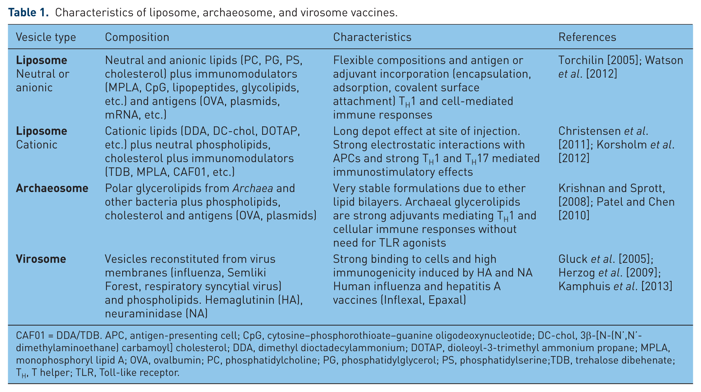
CAF01 = DDA/TDB. APC, antigen-presenting cell; CpG, cytosine–phosphorothioate–guanine oligodeoxynucleotide; DC-chol, 3β-[N-(N’,N’-dimethylaminoethane) carbamoyl] cholesterol; DDA, dimethyl dioctadecylammonium; DOTAP, dioleoyl-3-trimethyl ammonium propane; MPLA, monophosphoryl lipid A; OVA, ovalbumin; PC, phosphatidylcholine; PG, phosphatidylglycerol; PS, phosphatidylserine;TDB, trehalose dibehenate; TH, T helper; TLR, Toll-like receptor.
Liposome-based antigens
Liposome-mediated effects of antigen uptake, trafficking, processing and presentation
As the majority of vaccines are administered by intramuscular or subcutaneous injection, liposome properties play a major role in local tissue distribution, retention, trafficking, uptake and processing by APCs. Earlier studies showed clear size-dependent, but not unambiguous charge or lipid composition dependent effects at the injection site [Oussoren et al. 1997]. Newer studies with the cationic liposome formulation dimethyl dioctadecylammonium (DDA) plus trehalose dibehenate (TDB) (DDA/TDB, CAF01) showed no differences in liposome draining or antigen release from the injection site. However, differences in movement to regional lymph nodes (LNs) were noted [Henriksen-Lacey et al. 2010, 2011]. A cationic liposome pDNA vaccine of 500 nm and 140 nm size with encapsulated ovalbumin (OVA) encoding pDNA as antigen showed strongest retention at large vesicle size. Addition of poly(ethyleneglycol) (peg) coating resulted in enhanced lymphatic drainage, without improved immune response [Carstens et al. 2011]. Other pegylated DDA/TDB liposomes reduced the depot effect and altered the immune response, confirming these results [Kaur et al. 2012]. Badiee and colleagues evaluated liposomes of different sizes containing the surface glycoprotein of Leishmania (rgp63). Immunization with small liposomes induced a TH2 response, whereas large liposomes induced a TH1 response, higher interferon γ (IFNγ) levels and immunoglobulin IgG2a/IgG1 ratios [Badiee et al. 2012]. Adjuvant effects of neutral, positive or negative liposomes were evaluated when admixed with OVA, cationized OVA (cOVA) or Bacillus anthracis antigen by Yanasarn and colleagues. Immunization with OVA admixed with different liposomes generated different antibody responses. Interestingly, OVA admixed with negative 1,2-dioleyl-sn-glycero-3-phosphatidic acid liposomes was as immunogenic as OVA admixed with positive 1,2-dioleoyl-3-trimethyl ammonium propane liposomes. The cOVA antigen showed comparable adjuvant activities in all liposomes [Yanasarn et al. 2011]. Neutral phosphatidylcholine (PC)/cholesterol small unilamellar vesicles (SUV) also proved to be effective vaccine carriers. We evaluated a vaccine with peptides derived from the glycoprotein of the lymphocytic choriomeningitis virus (LCMV). Liposome-encapsulated peptides were highly immunogenic and elicited protective antiviral immunity by in vivo antigen loading of DCs. Encapsulated cytosine–phosphorothioate–guanine oligodeoxynucleotides (CpGs) further enhanced immune activation [Ludewig et al. 2000]. We also used the vaccine to prime a CD8+ T-cell response against 10 different hepatitis C virus (HCV) epitopes, resulting in strong CTL responses. Challenge experiments with Vaccinia virus expressing HCV epitopes emphasized the utility of neutral liposomes as HCV vaccine [Engler et al. 2004; Schwendener et al. 2010]. Moon and colleagues describe novel interbilayer-crosslinked multilamellar vesicles (MLVs) formed by crosslinking adjacent lipid bilayers within MLVs. These vesicles entrapped protein antigens in their core and lipid-based immunostimulatory molecules in the bilayers, forming a potent vaccine, eliciting strong T-cell and antibody responses [Moon et al. 2011].
Investigation of hemagglutinin (HA) adsorption versus encapsulation and coencapsulation of CpGs in 3β-[N-(N’,N’-dimethylaminoethane)-carbamoyl] cholesterol (DC-chol) liposomes showed that adsorbed HA was more immunogenic than encapsulated HA. Cholesterol enhanced the adjuvant effect and CpG-loaded liposomes were highly efficient at enhancing HA-specific humoral responses [Barnier Quer et al. 2012, 2013]. Covalent attachment of protein antigens to nanocarriers can disrupt protein structure and mask epitopes, altering the antibody response. Watson and colleagues used metal chelation via nitrilotriacetic acid (NTA) to attach antigens to liposomes. OVA and a HIV-1 gp41 (N-MPR) peptide were attached via NTA or covalent linkage. Attachment of N-MPR, but not OVA, elicited stronger antibody responses than antigen admixed with liposomes and covalent attachment was superior to NTA-anchored antigens [Watson et al. 2011].
Mannose receptors (MRs) expressed on macrophages and APCs mediate endocytosis and cooperate in antigen capture and presentation. MRs recognize carbohydrate moieties of many pathogens. Thus, targeting of glycosylated antigens or carrier systems to MRs is a method to develop vaccines [Irache et al. 2008]. To establish a human papilloma virus 16 (HPV16) cancer vaccine, Mizuuchi and colleagues generated oligomannose liposomes containing HPV16-E6 plasmid antigens (OML-HPV). HPV16-E6-specific CTLs were generated from HPV16-positive cervical carcinoma patients with OML-HPV, but not with standard liposomes [Mizuuchi et al. 2012]. OMLs in combination with entrapped dsRNA to induce antihuman parainfluenza virus 3 (HPIV3) immunity were studied by Senchi and colleagues [Senchi et al. 2013]. Hemagglutinin neuraminidase antigen was coencapsulated with adjuvant poly(I:C) into OMLs. Systemic and mucosal immune responses were generated and immune sera suppressed viral infection in vitro. Finally, Li and colleagues constructed a mannosylated liposome/protamine/DNA (Man-LPD) vaccine. Man-LPD exhibited higher intracellular uptake and transfection in vitro and induction of costimulatory molecules on bone marrow DCs [Li et al. 2013].
Peptides and proteins as antigens
The antigen location in liposomes influences immunogenicity. Both, entrapped or surface-attached antigens induce T-cell responses, the latter having advantages of availability for antibody or B-cell recognition, whereas encapsulated antigens require vesicle disruption to be accessible. The necessity of CD4+ T cells to induce memory CD8+ T cells was investigated in mice immunized with liposome surface-coupled OVA peptides. CTL responses were induced and confirmed in mice lacking CD4+ T cells, suggesting that CD4+ T cells were not required for memory CD8+ T-cell generation [Taneichi et al. 2010]. Phosphatidylserine (PS)-liposome conjugated antigens were efficiently captured by APCs, resulting in TH cell stimulation, validating PS as adjuvant for peptide vaccines [Ichihashi et al. 2013]. Takagi and colleagues coupled several HCV peptides to liposomes. One Db-restricted and three HLA-A(*)0201-restricted peptides conferred complete protection to immunized mice and long-term memory [Takagi et al. 2013].
Liposome-encapsulated protein antigens have been used frequently in earlier work. More recently, Nagill and colleagues compared encapsulated 78 kDa antigen of Leishmania donovani with antigen plus monophosphoryl lipid A (MPLA), resulting in decreased parasite burden after challenge [Nagill and Kaur, 2010]. In another study, Bal and colleagues coencapsulated OVA and the TLR ligand Pam3CysSK4 or CpGs in dioleoyl-3-trimethyl ammonium propane (DOTAP) liposomes. Encapsulation of both ligands did not obstruct activation of TLR-transfected cells and OVA/CpG liposomes shifted the IgG1/IgG2a balance to IgG2a, whereas Pam3CysSK4 was less efficient [Bal et al. 2011]. Hepatitis B surface antigen (HBsAg) encapsulated liposomes coupled with Ulex europaeus agglutinin 1 were developed by Gupta and Vyas. Lectinized liposomes were predominantly targeted to M cells on intestinal Peyer’s patches after oral immunization, yielding high antibody titers in mucosal secretions [Gupta and Vyas, 2011]. Another mucosal vaccine was described by Figueiredo and colleagues who encapsulated Streptococcus equi antigens in PC/cholesterol/stearylamine liposomes or chitosan nanoparticles. Intranasal immunization of mice elicited mucosal, humoral and cellular responses with higher serum IgA levels of the chitosan nanoparticles, due to enhanced mucoadhesive properties [Figueiredo et al. 2012]. Liposomes modified with pH-sensitive 3-methyl-glutarylated hyperbranched poly(glycidol) (MGlu-HPG) were used to encapsulate OVA. MGlu-HPG liposomes induced a strong immune response which was suppressed with anti-MHC-I/MHC-II antibodies [Hebishima et al. 2012]. Ding and colleagues developed so-called RAFTsomes by isolating membrane microdomains containing MHC-I and I-Ab restricted epitopes from OVA-primed DCs and reconstituted them on liposome surfaces. RAFTsome immunization gave high anti-OVA IgG1 levels and protection against OVA-expressing EG.7 tumor challenge [Ding et al. 2013].
Liposomal DNA vaccines
Nucleic acid vaccines are an alternative to attenuated bacterial antigens or protein or peptide vaccines. MLVs as inexpensive carriers were used by Rodriguez and colleagues to deliver DNA to mice with plasmids encoding bovine herpesvirus type 1. Vaccinated mice developed specific IgG responses [Rodriguez et al. 2013]. The M1 gene of influenza A virus was used by Liu and colleagues to construct a cationic liposome/DNA vaccine with a M1-encoding plasmid for oral vaccination, resulting in M1 gene expression in intestines of vaccinated mice and strong immune responses and protection against challenge infection [Liu et al. 2014]. Liposomes were also used to deliver plasmid DNA encoding heat shock protein 65 (hsp65) to treat the pulmonary fungal infection paracoccidiomycosis, resulting in protective immune response and reduced fungal burden [Ribeiro et al. 2013]. Amidi and colleagues proposed liposomes as artificial microbes that can be programmed to produce specific antigens for vaccination. A bacterial transcription and translation system together with a gene construct encoding β-galactosidase or a luciferase–nucleoprotein (NP) fusion epitope as antigens were entrapped in liposomes. Vaccination of mice showed that such antigen-producing liposomes elicited higher specific immune responses against the produced antigen than control vaccines [Amidi et al. 2011, 2012].
Liposomal messenger RNA vaccines
The immune system is naturally activated by foreign nucleic acids by inducing specific immune responses. Lack of persistence, genome integration and auto-antibody induction are advantages of mRNA and siRNA vaccines. Currently, mRNA vaccines are developed to treat various diseases, including cancers. Pichon and Midoux loaded mannosylated nanoparticles with mRNA encoding a melanoma antigen [Pichon and Midoux, 2013]. The mRNA was formulated with histidylated liposomes promoting endosome destabilization, allowing cytosolic nucleic acid delivery which enhanced anti-B16F10 melanoma vaccination in mice. A liposome encapsulated double-stranded RNA (LE-PolyICLC) was tested in the influenza (H5N1-HPIV) model by Li and colleagues. Intranasal LE-PolyICLC inhibited virus replication, reduced viral titers, increased survival of infected mice and attenuated pulmonary fibrosis [Li et al. 2011].
The MUC1 (BLP25) antigen
The MUC1 glycoprotein is often overexpressed and hypoglycosylated in tumor cells of cancers, making it an attractive target for immunotherapy (for other examples, see Table 2) [Acres and Limacher, 2005; Roulois et al. 2013]. MUC1 variable number tandem repeats conjugated to tumor-associated carbohydrate antigens (TACAs) break self tolerance in humanized MUC1 transgenic mice. Sarkar and colleagues formulated an anticancer vaccine composed of a MUC1 glycopeptide containing a GalNAc-O-Thr (Tn) TACA conjugated to a TLR ligand. Additional surface-displayed l-rhamnose (Rha) epitopes were included in 1,2-dipalmitoyl-sn-glycero-3-phosphatidyl-choline (DPPC) liposomes. Mice were immunized with a Rha-Ficoll conjugate followed by the vaccine, resulting in an increase in anti-MUC1-Tn more than eightfold, anti-Tn antibody titers and increased T-cell proliferation [Sarkar et al. 2013]. Another liposome vaccine containing the immunoadjuvant Pam3CysSK4, a TH peptide epitope and a glycosylated MUC1 peptide was reported by Lakshminarayanan and colleagues. Covalent surface linkage of all three components was essential for maximum efficacy [Lakshminarayanan et al. 2012]. The BLP25 liposome (L-BLP25) vaccine which targets MUC1 extended survival of patients with non-small cell lung cancer (NSCLC) and showed promise in prostate cancer [North and Butts, 2005; North et al. 2006]. Butts and colleagues conducted phase II/IIB studies to evaluate L-BLP25 in patients with stage IIIA/IIIB NSCLC. Patients received either L-BLP25 plus best supportive care (BSC) or BSC alone. Survival time and rates were longer in patients receiving the combination compared with BSC alone [Butts et al. 2010, 2011]. Wu and colleagues are conducting an ongoing L-BLP25 study (INSPIRE) in patients with NSCLC of East Asian ethnicity, which is the first large therapeutic cancer vaccine study in an East-Asian population [Wu et al. 2011]. Accordingly, a L-BLP25 study was conducted in Japanese patients with NSCLC showing consistency with studies of white patients [Ohyanagi et al. 2011].
Examples of liposomal veterinary vaccines.

DCPC, 1,2-dihexanoyl-sn-glycero-3-phosphocholine; DDA, dimethyl dioctadecylammonium; DOPE, 1,2-dioleyl-sn-glycero-3-phosphatidyl ethanolamine; DOTAP, dioleoyl-3-trimethyl ammonium propane; DPPC, 1,2-dipalmitoyl-sn-glycero-3-phosphatidyl choline; DPPE, 1,2-dipalmitoyl-sn-glycero-3-phosphatidyl ethanolamine; DPPS, 1,2-dipalmitoyl phosphatidylserine; DSPC, 1,2-distearoyl-sn-glycero-3-phosphocholine; IFN, interferon; Ig, immunoglobulin; LPS, lipopolysaccharide; MLV, multilamellar vesicle; n.a., not applicable; NDV, Newcastle disease virus; NFκB, nuclear factor κB; PC, phosphatidylcholine; PG, phosphatidyl glycerol; PS, phosphatidylserine; SA, starylamine; SUV, small unilamellar vesicle; TDP, trehalose dibehenate; TH, T helper.
Liposomes as carriers for adjuvants
Liposomal DNA as adjuvant
CpGs are adjuvants composed of unmethylated CpG dinucleotide sequences similar to those found in bacterial DNA. They trigger TLR9, activate DC maturation, increase antigen expression and induce TH1 immune responses [Shirota and Klinman, 2014]. Antigens and CpGs must be colocalized in one APC to generate optimal immune responses [Krishnamachari and Salem, 2009]. CpG encapsulation in liposomes of different properties altered antigen encapsulation efficiency, release and delivery rates, thus influencing the immune response. OVA/CpG coencapsulation augmented TH1 and cell-mediated immune response [Erikci et al. 2011]. Coencapsulation of OVA and Pam3CysSK4 or CpGs in cationic liposomes shifted the IgG1/IgG2a balance to IgG2a, showing that antigen/adjuvant coencapsulation shapes the type of immune response [Bal et al. 2011]. Nuclease-resistant phosphorothioate CpGs (PS-CpGs) or sensitive phosphodiester CpGs (PO-CpGs) were used by Shargh and colleagues in a leishmaniasis model. PO-CpGs or PS-CpGs were encapsulated in DOTAP liposomes for protection against nuclease degradation. Mice immunized with liposomal soluble Leishmania antigens (SLA) coincorporated with PO-CpGs or PS-CpGs showed no significant difference in immune response. Thus, nuclease-sensitive PO-CpGs can be used as adjuvants [Shargh et al. 2012]. Finally, CpGs incorporated in cationic DOTAP liposomes but not in neutral 1,2-dioleoyl-sn-glycero-3-phosphocholine (DOPC) liposomes provided complete protection against challenge with Burkholderia pseudomallei in a mouse model [Puangpetch et al. 2012].
Cationic liposome adjuvant vaccines
The introduction of positively charged compounds is a common method used to alter liposome properties. Cationic liposomes are frequently used as cell transfection reagents and vaccine adjuvants. Most cationic lipids form bilayer liposomes but often additional lipids are needed. The high surface density of positive charges increases liposome adsorption on negatively charged cell surfaces. Cationic liposomes penetrate into cells through specific mechanisms and activate different cellular pathways depending on cell type, cationic lipid nature, but also on formulation types and liposome size [Korsholm et al. 2012; Lonez et al. 2012].
The cationic adjuvant CAF01
CAF01 is a novel adjuvant composed of the synthetic immunostimulating mycobacterial cordfactor glycolipid TDB and the cationic membrane forming molecule DDA. TDB induces strong TH1 and TH17 immune responses and the C-type lectin Mincle is the receptor for APC activation. The adjuvant effect also requires MyD88 and Schweneker and colleagues identified the Nlrp3 inflammasome as mediator for TDB-triggered induction of immune response [Werninghaus et al. 2009; Desel et al. 2013; Schweneker et al. 2013]. Properties of cationic liposome-forming lipids were studied with rigid DDA or fluid dimethyldioleoylammonium (DODA) liposomes. When the antigen Ag85B-ESAT-6 was mixed with DDA/TDB or DODA/TDB liposomes, DDA liposomes formed a depot, resulting in continuous activation of APCs, whereas DODA liposomes were rapidly cleared [Christensen et al. 2012]. Milicic and colleagues explored modifications of DDA/TDB liposomes such as size, antigen association and addition of TLR agonists to assess their activity using OVA as antigen. SUV without TLR agonists showed higher antigen-specific antibody responses than MLVs. Addition of TLR3 and TLR9 agonists increased the adjuvant effects of MLVs but not of SUVs. The ability of DDA/TDB SUVs to induce CD8+ CTL responses without immunostimulators could avoid safety risks associated with TLR agonists [Milicic et al. 2012]. Yu and colleagues tested several adjuvants, including DDA-monophosphoryl lipid A (DDA-MPLA), DDA-TDB (CAF01) and DDA-monomycolyl glycerol (DDA-MMG, CAF04). Chlamydia antigens were used in a mouse genital tract infection model. DDA-MPLA and DDA-TDB elicited the best protective immune responses, characterized by CD4+ T cells coexpressing IFNγ and tumor necrosis factor α and by significantly reduced infection [Yu et al. 2012]. Ingvarsson and colleagues studied the parameters of CAF01 spray dried powder formulations using lactose, mannitol or trehalose as stabilizers. Immunization of mice with the tuberculosis antigen H56 demonstrated that spray drying with trehalose resulted in the best preservation of adjuvant activity [Ingvarsson et al. 2011, 2013]. Lindenstrom and colleagues showed that CAF01 vaccination in mice led to establishment of TH17 memory cells by retaining phenotypic and functional properties for 2 years. Challenge with Mycobacterium tuberculosis (MTB) 2 years later induced TH17 memory cells at levels comparable to TH1 memory cells [Lindenstrom et al. 2012]. A trivalent influenza vaccine (TIV) with CAF01 enhanced the immune response determined by HA inhibition and antibody titers, promoting strong TH1 responses. Maintenance of the TH1/TH17 cytokine profile over 20 weeks resulted in complete survival of H1N1 challenged mice [Rosenkrands et al. 2011]. A commercially available TIV was compared with the same vaccine mixed with CAF01 in ferrets. CAF01 induced increased influenza-specific IgA and IgG levels and promoted immunity and protection against challenge with H1N1 [Martel et al. 2011]. The combination of cationic liposomes and immunopotentiators such as MPL with DDA/TDB liposomes was tested in mice using OVA as antigen. DDA/TDB/MPL liposomes induced antigen-specific CD8+ T-cell and humoral responses [Nordly et al. 2011]. CAF01 was also used in a phase I trial with a therapeutic HIV-1 peptide vaccine. Safety and immunogenicity were assessed in individuals with untreated HIV-1 infection. Vaccine-specific T-cell responses were induced in 6 of 14 individuals, showing that therapeutic immunization with CAF01-adjuvanted HIV-1 peptide in humans is feasible [Roman et al. 2013]. In another clinical trial the potential of inducing T-cell immunity during chronic HIV-1 infection was investigated. Treatment-naive individuals with HIV-1 infection were immunized with peptides/CAF01. Specific CD4+ and CD8+ T-cell responses were induced in all individuals [Karlsson et al. 2013]. Kamath and colleagues reported that physical linkage between antigens and immunomodulators is required to elicit TH1/TH17 responses. Separate same-site administration of a mycobacterial fusion antigen and CAF01 failed to elicit TH1/TH17 responses. Tracking experiments showed that separate same-site administration elicited an early antigen-positive/adjuvant-negative DC population. Antigen targeting of LN DCs prior to their activation generated nonactivated antigen-pulsed DCs that recruited antigen-specific T cells and triggered proliferation, but interfered with TH1 induction in a dose-dependent manner. Thus, synchronization of DC targeting and activation is a critical determinant for TH1/TH17 adjuvanticity [Kamath et al. 2012].
In summary, CAF01-adjuvant liposomes prove to be a valuable vaccine formulation for different antigens.
CLDC adjuvant liposomes
Another widely studied cationic liposome complex contains the cationic lipid 1-[2-(oleoyloxy)-ethyl]-2-oleyl-3-(2-hydroxyethyl)imidazolinium-chloride and cholesterol. CLDCs are prepared by mixing liposomes with DNA. CLDC (JVRS-100, Juvaris BioTherapeutics, Burlingame, CA, USA) is a lyophilized powder composed of selected plasmid DNA complexed with liposomes. CLDCs facilitate APC uptake, activate TLRs and IFN production and stimulate the adaptive immune response. Several CLDC vaccines have been tested in various models. Gowen and colleagues analyzed liposomal delivery and CpG content of plasmid DNA with CLDCs. CpG-free or CpG-containing plasmids with and without liposomes, and poly(I:C) were evaluated to elicit protection against lethal Punta Toro virus challenge in hamsters. CLDC-containing CpG plasmid significantly improved survival, decreased viral loads and reduced liver damage [Gowen et al. 2009]. CLDC enhanced anti-simian immunodeficiency virus (SIV) immune responses induced by SIV vaccines. CLDC immunized rhesus macaques developed stronger SIV-specific T- and B-cell responses compared with controls, resulting in persistence and better memory responses [Fairman et al. 2009]. As no vaccines are available for common herpes simplex virus (HSV) infections CLDCs were evaluated for a HSV gD2 vaccine in a genital herpes guinea pig model. The CLDC/gD2 vaccine significantly decreased duration of acute and recurrent disease compared with gD2 alone. However, when evaluated as therapeutic vaccines they were ineffective, suggesting that such HSV-2 vaccines need improvement [Bernstein et al. 2010, 2011]. The protective effects of CLDCs against encephalitic arboviral infection were investigated in a Western equine encephalitis virus (WEEV) model. CLDC-vaccinated mice were challenged with virulent WEEV. CLDC pretreatment provided increased survival and higher cytokine levels, strong TH1 activation and protective immunity against lethal WEEF [Logue et al. 2010]. An influenza A virus vaccine adjuvanted with CLDC or alum was tested by Hong and colleagues. CLDC induced more robust adaptive immune responses with higher levels of virus-specific IgG2a/c and CD4+ and CD8+ T cells plus cross protection from lethal viral challenges [Hong et al. 2010]. In another influenza A vaccine study, Dong and colleagues showed that addition of CLDC (JVRS-100) to a H5N1 split vaccine induced higher virus-specific responses than adjuvant-free formulations. CLDC-vaccinated mice challenged with H5N1 had mild illness, very low viral titers, 100% survival and long-lasting protective immunity [Dong et al. 2012]. Strategies to improve influenza vaccine efficacy in older individuals are needed. Thus, Carroll and colleagues determined whether CLDC (JVRS-100) could improve the efficacy of the influenza vaccine Fluzone (Sanofi Pasteur, Lyon, France) in older rhesus macaques. Vaccination with Fluzone with or without CLDC and challenge with human H1N1 influenza virus showed that only the Fluzone/CLDC-vaccinated animals had lower virus replication. Thus, CLDC enhances immunogenicity and efficacy of a licensed vaccine in immunosenescent monkeys [Lay et al. 2009; Carroll et al. 2014]. CLDC (JVRS-100) was also evaluated as adjuvant for HBsAg in mice expressing hepatitis B virus (HBV). HBsAg+JVRS-100 elicited T- and B-cell responses, whereas HBsAg elicited only a B-cell response. However, the response by HBsAg+JVRS-100 was not sufficient to cause destruction of infected liver cells, but it suppressed HBV DNA noncytolytically [Morrey et al. 2011]. Similar results were obtained using the woodchuck model of HBV. HBV infection induced T-cell responses to Woodchuck hepatitis surface antigen (WHsAg) and selected WH peptides and expression of CD8+ CTL and TH1 cytokines. WHsAg plus CLDCs elicited antibodies earlier, in more woodchucks and with higher titers than WHsAg and alum [Cote et al. 2009]. There is a need for mucosal vaccines for pulmonary Yersinia pestis infections. The ability of an oral CLDC-adjuvanted vaccine against lethal pneumonic plague was investigated by Jones and colleagues. Oral immunization with Y. pestis F1 antigen combined with CLDC produced high titers of anti-F1 antibodies and long-lasting CD4+ T-cell-dependent protection from lethal pulmonary challenge with Y. pestis [Jones et al. 2010].
Other cationic lipid complexes
Several other cationic lipid adjuvant complexes were evaluated in various vaccine models. Phillips and colleagues tested an alphavirus vaccine composed of cationic lipid nucleic acid complexes (CLNCs) and the ectodomain (E1ecto) of WEEV. Interestingly, CLNC alone had therapeutic efficacy, as it increased survival of mice following lethal WEEV infection. Immunization with the CLNC/WEEV/E1ecto mixture provided full protection after challenge. Passive serum transfer from immunized to naïve mice conferred protection to challenge, indicating that antibody is sufficient for protection [Phillips et al. 2014]. Liposomes containing different cationic compounds and neutral DPPC were loaded with influenza HA by adsorption. DC-chol/DPPC liposomes with a high amount of DC-chol had stronger immunogenicity compared with less DC-chol and elicited higher antibody titers compared with the other compounds and nonadjuvanted HA. Liposome-adsorbed HA was more immunogenic than encapsulated HA and incorporation of cholesterol in DC-chol liposomes as well as CpGs enhanced adjuvancy [Barnier-Quer et al. 2013]. A similar study by Ma and colleagues showed that DOTAP/DOPC liposome regulated immune responses relied on surface charge density and might occur through reactive oxygen species (ROS) signaling. High charge density liposomes potently enhanced DC maturation, ROS generation, antigen uptake and production of IgG2a and IFNγ, whereas low-charge density liposomes failed to promote immune responses [Ma et al. 2011]. Lipid assemblies composed of a polycationic sphingolipid [ceramide carbamoyl spermine (CCS)] are effective adjuvants/carriers for several vaccines when complexed with cholesterol (CCS/C, VaxiSome, NasVax, Tel Aviv, Israel). Ferrets immunized intranasally with CCS/C-influenza vaccine produced higher HI antibody titers compared with controls. Following viral challenge, the vaccine reduced the severity of infection. Biodistribution studies showed that lipids and antigens are retained in nose and lung, increasing cytokine levels and expression of costimulatory molecules [Even-Or et al. 2011]. Chen and colleagues developed a cationic lipopolymer, the liposome–polyethyleneglycol–polyethyleneimine complex (LPPC) adjuvant for surface adsorption of antigens or immunomodulators. LPPC enhanced presentation on APCs, surface marker expression, cytokine release and activated TH1 immunity. With lipopolysaccharide (LPS) or CpGs, LPPC dramatically enhanced the IgA or IgG2A proportion of total Ig, demonstrating host immunity modulation [Chen et al. 2012]. Effects of pegylation of cationic DOTAP liposome vaccines on LN targeting and immunogenicity were studied by Zhuang and colleagues. Peg-DOTAP liposomes accelerated drainage into LNs, prolonged retention and APC uptake, increased anti-OVA antibody responses and modulated their biodistribution, which improved vaccine efficiency [Zhuang et al. 2012]. The activity of cationic vaccines can be hampered by immobilization in the extracellular matrix caused by electrostatic interactions. Thus, Van den Berg and colleagues found that surface shielding of DOTAP liposomes by pegylation improved antigen expression drastically. Mice vaccinated with pegylated pVAX/Luc-NP antigen containing liposomes elicited T-cell responses comparable to naked DNA, suggesting that charge shielding improves dermally applied vaccines [Van Den Berg et al. 2010].
Other adjuvants
Muramyl dipeptide
Muramyl dipeptide (MDP) originates from a bacterial peptidoglycan cell-wall fragment and is responsible for the activity of Freund’s complete adjuvant (FCA). After phagocytosis by APCs, MDP is detected by the NOD2 receptor that activates the immune response. Numerous MDP derivatives have been synthesized to evaluate their immunostimulatory effects and adjuvant activity [Traub et al. 2006; Ogawa et al. 2011]. It was recognized early on that liposomes were ideal carriers for MDP and its derivatives [Alving, 1991]. For example, the lipophilic MDP analogs muramyl dipeptide phosphatidyl ethanolamin (MDP-PE) and MDP glycerol dipalmitate were added to liposomal HBsAg formulations, both of which induced higher antibody titers, TH1 response and IFNγ levels [Jain et al. 2009]. Masek and colleagues used small Ni-chelating liposomes to attach His-tagged Candida albicans hsp (hsp90-CA) as antigen and to coincorporate the MDP derivative C18-O-6-norAbuMDP as adjuvant. The immune response was of TH1 and TH2 type, comparable to FCA, but without side effects [Masek et al. 2011]. Liposomes formulated as MDP with 1,2-dipalmitoyl-sn-glycero-3-phosphatidyl-ethanolamine are called mifamurtide (Mepact, Takeda, Osaka, Japan), which is an adjuvant to standard chemotherapy for osteosarcoma. A study by Anderson and colleagues in patients with metastatic osteosarcoma showed that mifamurtide had a manageable safety profile but that a randomized clinical trial is required to further determine its utility [Anderson et al. 2014].
Monophosphoryl lipid A
MPLA is the active moiety of the bacterial endotoxin LPS. TLR4 is the receptor for LPS forming a complex with MD2, representing the main LPS binding component. LPS supports the development of diverse TH cell types, depending on the tissue microenvironment. For instance, peripheral immunization with LPS drives TH1 priming in lymphoid tissue and TH17 priming in the intestinal mucosa [McAleer and Vella, 2010]. Several lipopeptides derived from microbial origin convey self-adjuvanting activity by TLR2 signaling, recognizing many PAMPs [Kawai and Akira, 2010]. MPLA liposomes have considerable potency and safety with a variety of candidate vaccines, including malaria, HIV-1 and several types of cancer [Alving et al. 2012; Zaman et al. 2013]. Combination of MPLA with the saponin QS-21 and immunostimulants in various adjuvant formulations forms the basis of Adjuvant Systems (AS; GlaxoSmithKline-Biologicals, Rixensart, Belgium) to promote protective immune responses. MPLA and aluminum salts are present in AS04, and both MPLA and QS-21 are present in AS01 and AS02, which are liposome- and emulsion-based formulations [Garcon and Van Mechelen, 2011]. Rizwan and colleagues analyzed the immunological activity of cubosomes containing MPLA and imiquimod. Cubosomes, a novel variation of MLVs, are composed of highly twisted lipid bilayers and nonintersecting water channels, providing increased encapsulation rates of lipophilic compounds. In MPLA cubosomes, sustained release of OVA was observed with induction of OVA-specific antibodies and CD8+ and CD4+ T-cell proliferation at higher efficiency than liposomes plus adjuvants and alum [Rizwan et al. 2013]. Immune response and protection induced by liposomal SLA formulated with lipid A trehalose dicorynomycolate (MPLA-TDM) was evaluated by Ravindran and colleagues. This vaccine induced strong and long-lasting protection against experimental visceral leishmaniasis [Ravindran et al. 2012]. An influenza vaccine with MPLA was developed by Adler-Moore and colleagues using the ectodomain of the M2e channel protein as a universal vaccine candidate. A liposomal M2e vaccine elicited anti-M2e antibodies that inhibited viral cell lysis and conferred complete protection to mice challenged with H1N1. Lymphocyte depletion markedly decreased protection, suggesting a primarily TH2-mediated immune response [Adler-Moore et al. 2011].
Listeriolysin
Cytolysins are virulence factors of various pathogenic bacteria. They form pores in target cell membranes, degrade membrane lipids or solubilize cell membranes. Bacteria use cytolysins to either inhibit functions of host immune cells or to gain access to intracellular niches. The bacterium Listeria monocytogenes can escape host immune defenses by lysis of the phagosomal membrane by use of listeriolysin O (LLO). LLO is used as vaccine adjuvant to provide cytosolic access for antigens in APCs [Dietrich et al. 2001]. LLO-based vaccines were reported by Mandal and Lee, who prepared OVA/LLO liposomes. OVA immunization resulted in higher CTL activity and high IFNγ production. The vaccine also conferred protection to mice from lethal challenges with antigen-expressing tumor cells [Mandal and Lee, 2002]. LLO liposomes were also used to deliver the LCMV NP to stimulate a NP-specific CTL response. Immunized mice generated high frequencies of NP-specific CD8+ T cells and full protection against a lethal intracerebral challenge with virulent LCMV [Mandal et al. 2004]. An anionic liposome–polycation–DNA complex combined with LLO was used as vaccine by Sun and colleagues to deliver OVA-cDNA. This formulation produced an enhanced CD8+ T-cell response, higher CTL frequency and IFNγ production after stimulation by an OVA-specific peptide [Sun et al. 2010]. Andrews and colleagues analyzed whether encapsulating CpGs in LLO liposomes would enhance cell-mediated immune response and skew TH1-type responses in a protein antigen-based vaccine utilizing LLO liposomes. Coencapsulation of CpGs in LLO liposomes activated the nuclear factor κB pathway, maintaining cytosolic delivery of antigen mediated by coencapsulated LLO. Immunization with OVA and CpG-LLO liposomes showed enhanced TH1 immune responses [Andrews et al. 2012].
Currently, 26 clinical trials are registered at ClinicalTrials.gov, a service of the US National Institutes of Health (see ClinicalTrials.gov with the search terms liposome AND vaccine).
Veterinary vaccines
Knowledge of molecular details of immune mechanisms is relatively scarce for veterinary and pet animals and special concerns regarding the use of vaccine adjuvants must be considered. Demands such as compatibility with human consumption, animal production, costs, challenges met by different species, vaccine administration for large numbers of animals and others must be evaluated [Heegaard et al. 2011; Underwood and Van Eps, 2012]. Table 2 summarizes some of the most recent experimental studies of liposome-based veterinary vaccines.
Therapeutic cancer vaccines
Although most cancers modify host proteins that can function as antigens, the development of effective vaccines against such antigens is hampered by the weak immune response and the immunosuppressive effects induced by cancers. Tumor-associated antigens include viral proteins (e.g. HPV), chromosomal translocation products (e.g. bcr/abl), overexpressed proteins likeHER2/neu, telomerase, MUC1 and others [Kozako et al. 2012]. In Table 3 some recent examples of experimental liposome-based cancer vaccines are listed.
Examples of liposomal therapeutic cancer vaccines.

bFGF, basic fibroblast growth factor; BCG, Bacille Calmette-Guerin; CpG, cytosine–phosphorothioate–guanine oligodeoxynucleotide; CTL, cytotoxic T lymphocyte; DC, dendritic cell; DDA, dimethyl dioctadecylammonium; DOG(Man)2, mannosylated dioleylglycerol; DOPE, 1,2-dioleyl-sn-glycero-3-phosphatidyl ethanolamine; DOTAP, 1,2-dioleoyl-3-trimethyl-ammonium-propane; HP16, human papilloma virus 16; IL, interleukin; MAP, multiple antigen peptide; MPLA, monophosphoryl lipid A; NK, natural killer; OVA, ovalbumin; PC, phosphatidylcholine; pegDSPE, pegylated 1,2-distearoyl-sn-glycero-3-phosphatidylethanolamine; PG, phosphatidyl glycerol; PIC, polyriboinosinic: polyribocytidylic acid; SA, stearylamine; SUV, small unilamellar vesicle; TC-1, type 1 T cell; TDB, trehalose dibehenate; TLR, Toll-like receptor.
Archaeosomes
Archaebacteria (Archaea) were discovered and classified by Woese and Fox as a new group of prokaryotes, besides the Eubacteria (Bacteria) [Woese and Fox, 1977]. Archaea contain DNA-dependent RNA polymerases and proteinaceous cell walls that lack peptidoglycan. Their cell membranes are composed of L-glycerol ether lipids with isoprenoid chains instead of D-glycerol ester lipids with fatty acid chains [Spang et al. 2013]. Archaeal lipids are uniquely constituted of ether-linked isoprenoid phytanyl archaeol (diether) or caldarchaeol (tetraether) cores conferring high membrane stability. Archaeosomes are liposomes prepared with archaeal glycerolipids. The head groups displayed on the glycerol lipid cores of archaeosomes interact with APCs and induce TH1, TH2 and CD8+ T-cell responses to the entrapped antigen. The immune responses are persistent and subject to strong memory responses [Krishnan and Sprott, 2008; Benvegnu et al. 2009]. The polar lipid from the archaeon, Methanobrevibacter smithii, has been well characterized for its adjuvant potential. It contains archaetidyl serine, promoting interaction with a PS receptor on APCs. These archaeosomes mediate MHC-I cross priming and promote costimulation by APCs without inflammatory cytokine production [Krishnan et al. 2000]. Patel and colleagues showed that archaesomes prepared from M. smithii lipids were suitable adjuvants for multivalent mucosal vaccines. Archaeosomes containing the encapsulated antigens OVA, bovine serum albumin and hen egg lysozyme conferred strong and sustained specific antibody responses to all three antigens [Patel et al. 2004]. Intranasal immunization of mice with the archaeal lipid mucosal vaccine adjuvant and delivery (AMVAD) system, obtained by interaction of archaeosomes/antigens with multivalent cations, induced robust mucosal antigen-specific IgA responses. AMVAD formulations are stable, safe and show protective efficacy in murine models of infection/challenge [Patel and Chen, 2010]. Archaeosomes prepared from lipids of the nonpathogenic bacteria Leptospira biflexa (leptosomes) and Mycobacterium smegmatis (smegmosomes) were used as adjuvants. Both vesicles caused strong APC activation, cytokine release and expression of costimulatory signals, which was significantly higher for smegmosomes compared with leptosomes. APC activation by both formulations induced immune responses in mice to entrapped OVA [Faisal et al. 2009, 2011]. Borrero and colleagues studied immune response and cross reactivity against MTB with archaeosomes containing a mixture of cell wall glycolipids such as phosphatidylinositol mannosides of M. smegmatis (Ms) and 1,2-distearoyl-sn-glycero-3-phosphocholine/cholesterol. Ms-containing liposomes induced a specific IgG response and recognition of MTB surface antigens, showing that immunogenic Ms glycolipids could enhance subunit vaccines against tuberculosis [Borrero et al. 2013]. The relation between archaeal lipid structures and their activity was explored by synthesizing novel head groups linked to archaeol. Archaeosomes consisting of various combinations of synthesized lipids with entrapped OVA antigen were used to immunize mice. Addition of the glycolipids gentio-triosyl archaeol, mannotriosyl archaeol or maltotriosyl archaeol to archaetidylglycero-phosphate-O-methyl (AOM) archaeosomes significantly enhanced CD8+ T-cell responses, but diminished antibody titers. All three triglycosyl archaeols combined with AOM resulted in additive CD8+ T-cell responses [Sprott et al. 2012]. Ansari and colleagues showed that archaeosome-entrapped secretory antigens (SAgs) of L. monocytogenes resulted in upregulation of TH1 cytokines and boosted protective effects by reducing listerial burden in infected mice. Archaeosome-entrapped SAgs enhanced CTL response and increased survival of immunized animals [Ansari et al. 2012]. Finally, Singha and colleagues used E. coli lipid liposome (escheriosome) based DNA delivery to induce superoxide dismutase (SOD) and interleukin (IL)-18-specific immune responses in murine Brucellosis. Escheriosome-mediated delivery of SOD- and IL-18-encoding DNA induced specific immune responses in immunized mice. Coexpression of SOD + IL-18 resulted in stronger IgG2a-type response compared with free SOD DNA [Singha et al. 2011].
Currently, no clinical trials with archaeosomal vaccines are registered at ClinicalTrials.gov (see ClinicalTrials.gov, search terms archaeosome AND vaccine).
In summary, vaccines prepared with archaeal lipids, the archaeosomes, represent a new interesting and promising alternative to classical liposomes and virosomes.
Virosomes
Virosomes are liposomes prepared by combining natural or synthetic phospholipids with virus envelope phospholipids, viral spike glycoproteins and other viral proteins. The first virosomes were prepared and characterized by Almeida and colleagues [Almeida et al. 1975], followed by Helenius and colleagues who incorporated Semliki Forest virus glycoproteins in liposomes [Helenius et al. 1977; Balcarova et al. 1981]. Significant progress was made with virosomes termed ‘immunopotentiating reconstituted influenza virosomes’ (IRIVs). IRIVs are SUVs with spike projections of the influenza surface glycoproteins HA and neuraminidase. The fusogenic properties of HA are their primary features. IRIVs allow antigen presentation in the context of MHC-I and MHC-II and induce B- and T-cell responses [Gluck, 1992, Gluck et al. 2005]. The first virosome-based influenza vaccine used in humans was Inflexal V (Crucell NV, Leiden, The Netherlands), which has an excellent tolerability profile and immunogenicity in healthy and immunocompromised people [Herzog et al. 2009]. Another virosome vaccine containing inactivated hepatitis A virus (HAV), Epaxal (Crucell NV, Leiden, The Netherlands), was developed as hepatitis A vaccine. It is excellently tolerable and highly immunogenic, conferring protection of at least 9–11 years in vaccinated individuals [Ambrosch et al. 1997; Gluck and Walti, 2000; Bovier et al. 2010]. Immunogenicity and safety of Epaxal was evaluated in Thai children with HIV infection. Prevalence of HAV protective antibodies was 100% after vaccination, showing that Epaxal is an effective HAV vaccine for HIV-infected children [Saksawad et al. 2011]. Another vaccine contains an aspartyl proteinase 2 (Sap2) of Candida albicans incorporated into IRIVs. Following intravaginal administration, anti-Sap2 antibodies were detected in vaginal fluids of rats, inducing long-lasting protection [De Bernardis et al. 2012]. Walczak and colleagues demonstrated that a heterologous prime boost with Semliki Forest virus encoding a fusion protein of E6 and E7 of HPV16 and virosomes containing the HPV16-E7 protein resulted in higher numbers of antigen-specific CTL in mice than homologous protocols [Walczak et al. 2011]. Today, a second generation of influenza virosomes has evolved for various preclinical and clinical stage vaccine candidates. Additional components are included to optimize particle assembly and stability and to enhance immunostimulatory effects [Moser et al. 2013]. GPI-0100, a saponin derivative, enhanced immunogenicity and protective efficacy of a virosomal influenza vaccine, providing full protection of infected mice at extremely low antigen doses [Liu et al. 2013]. A combination of reconstituted respiratory syncytial virus (RSV) envelopes with incorporated MPLA (RSV-MPLA) virosomes was studied by Kamphuis and colleagues in enhanced respiratory disease prone rats. Vaccination with RSV-MPLA induced higher antibody levels and protection against infection [Kamphuis et al. 2013]. Jamali and colleagues developed a DNA vaccine using cationic influenza virosomes (CIV). CIV-delivered epitope-encoding DNA induced equal numbers of IFNγ and granzyme B-producing T cells than a 10-fold higher dose of naked pDNA [Jamali et al. 2012]. Another DNA/virosome vaccine was reported by Kheiri and colleagues, who prepared a vaccine complex containing an influenza NP-encoding plasmid that induced much higher T-cell responses and protection than plasmid alone [Kheiri et al. 2012]. In clinical trials, IRIVs have shown vast potential for delivery of peptides derived from Plasmodium falciparum antigens [Peduzzi et al. 2008]. An IRIV-formulated fusion protein composed of two malaria antigens was described by Tamborrini and colleagues. Compared with other vaccines, the adjuvant-free formulation elicited specific IgG1 antibody profiles in mice and cross reactivity with blood-stage parasites [Tamborrini et al. 2011]. Virosomes containing surface HIV-1 gp41-derived P1 lipid conjugated peptides (MYM-V101) as prophylactic HIV-1 vaccine were prepared. MYM-V101 was safe and well tolerated when administered by intramuscular and intranasal routes in healthy women. P1-specific serum IgGs and IgAs were detected in all recipients but P1-specific TH1 responses were not found [Leroux-Roels et al. 2013].
Currently, several clinical trials with virosome vaccines are registered at ClinicalTrials.gov (see ClinicalTrials.gov, search terms virosome AND vaccine).
Conclusion
The enormous versatility of liposomes and the related archaeosomes and virosomes endows them as highly valuable carrier systems for vaccines. Besides improving antigen stability and presentation to immunocompetent cells, depending on their specific properties including composition, size and surface properties, these nanocarriers also possess the ability to overcome biological barriers, such as skin and mucosa, and provide controlled and slow release of antigens. Together with the ability to induce strong immune responses provided by coformulated adjuvants, liposome-based vaccines provide properties that are fundamental for the development of modern vaccine formulations. It is predictable that these delivery systems will be increasingly applied in the near future with success, leading to major improvements in vaccine development.
Footnotes
Funding
This research received no specific grant from any funding agency in the public, commercial, or not-for-profit sectors.
Conflict of interest statement
The author declares that there is no conflict of interest.