
Abstract
Foreign body ingestion is a common occurrence in the pediatric population, often involving the gastrointestinal tract. However, the presence of foreign bodies in the oral cavity, particularly within the buccal mucosa, is relatively rare. This case report describes an unusual presentation of a foreign body embedded in the buccal mucosa and discusses the diagnostic and management challenges associated with such cases. A 10-month-old female child with no significant previous medical history presented with recurrent buccal abscess. Following the removal of the foreign body (grass fragments), the child experienced a complete recovery.
Introduction
Foreign bodies in the ENT (Ear, Nose, and Throat) region, oral cavity, and airway present unique challenges in clinical practice. Common scenarios involve aspiration or ingestion of foreign bodies, leading to potential complications such as airway obstruction or injury to surrounding structures. In the ENT region, foreign bodies may be found in the ear canal or nasal passages, often causing discomfort and hearing impairment. In the oral cavity, ingested objects can pose risks of choking or injury to the gastrointestinal tract. Foreign body ingestion is a well-documented phenomenon in children, especially in the age group under consideration. 1 Children are naturally curious, and their exploratory behaviors may lead to ingestion of various objects which can go unnoticed by caregivers. Foreign body ingestion is a frequent occurrence among children, with a wide range of objects ingested, from coins to toys. 2 Prompt recognition and intervention are crucial in managing these cases. Timely and appropriate removal of foreign bodies is essential to prevent complications and ensure optimal patient outcomes in this specialized area of medical care. Most foreign bodies pass through the gastrointestinal tract without causing significant harm. However, foreign bodies lodged in the oral cavity, especially within the buccal mucosa, are uncommon. This case report aims to highlight the unique challenges in diagnosing and managing a foreign body embedded in the buccal mucosa of a pediatric patient.
Case presentation
A 10-month-old female child, with no significant previous medical history, presented to the ENT clinic with complaints of right parotid swelling and fever for 5 days. The parents noted that the swelling had gradually increased in size and was associated with difficulty in opening the child’s mouth. On physical examination, the child appeared clinically stable, and weighed 9 kg. There was noticeable swelling on the right side of the face. Intraoral examination revealed a localized, erythematous swelling on the buccal mucosa, approximately 1 cm in diameter. The surrounding mucosa appeared normal. The child was reluctant to open her mouth, making a thorough examination challenging. Considering the clinical presentation, a decision was made to perform needle aspiration but there was no aspirate. The child was admitted and started on intra venous (IV) Ceftriaxone 50 mg per kg body weight administered twice a day. After 5 days the swelling and other clinical symptoms resolved and hence, discharged. After 1 week, they presented with similar right sided cheek swelling which was fluctuant in consistency. After taking written informed consent from the guardian/Legally Authorised Representative (LAR), a decision was made to perform incision and drainage of the buccal abscess. Under Total Intra Venous Sedation (TIVA), an incision was made on the buccal mucosa, and around 5 ml of frank pus was drained. The pus culture showed Escherichia coli sensitive to Ceftriaxone, resistant to Penicillin. The child was again admitted for IV ceftriaxone. The child was discharged after 5 days upon resolution of the symptoms. After a month since incision and drainage, the parents brought the child to the ENT clinic again with right-sided buccal swelling with discharging sinus over the skin. Due to the recurrent nature of presentation, facial magnetic resonance imaging (MRI) scan was done to rule out collaural fistula which revealed “blind tubular non enhancing structure within the superficial lobe of right parotid gland with a single external opening into the dimpled skin of the right side of the face, postero-lateral to the right masseter muscle and inferior to the right ear canal.” Intra oral incision and drainage was done again which had similar intra operative findings. The pus culture report was not different either. The child was again admitted for IV ceftriaxone. The child was discharged after 5 days upon resolution of the symptoms. After a month since incision and drainage, the parents brought the child to the ENT clinic again with right sided buccal abscess. After taking written informed consent from the LAR, a decision was made to perform incision and drainage of the buccal abscess. Under TIVA, an incision was made on the buccal mucosa, and around 5 ml of frank pus was drained. Unlike in the past, this time, few pieces of grass were also found along with the pus (Figure 1). The child was again admitted for IV ceftriaxone. The child was discharged after 5 days upon resolution of the symptoms. Due to the recurrence of the illness, the child was kept on close follow up monthly for 6 months. In subsequent monthly visits, the child has had full resolution with no recurrence.
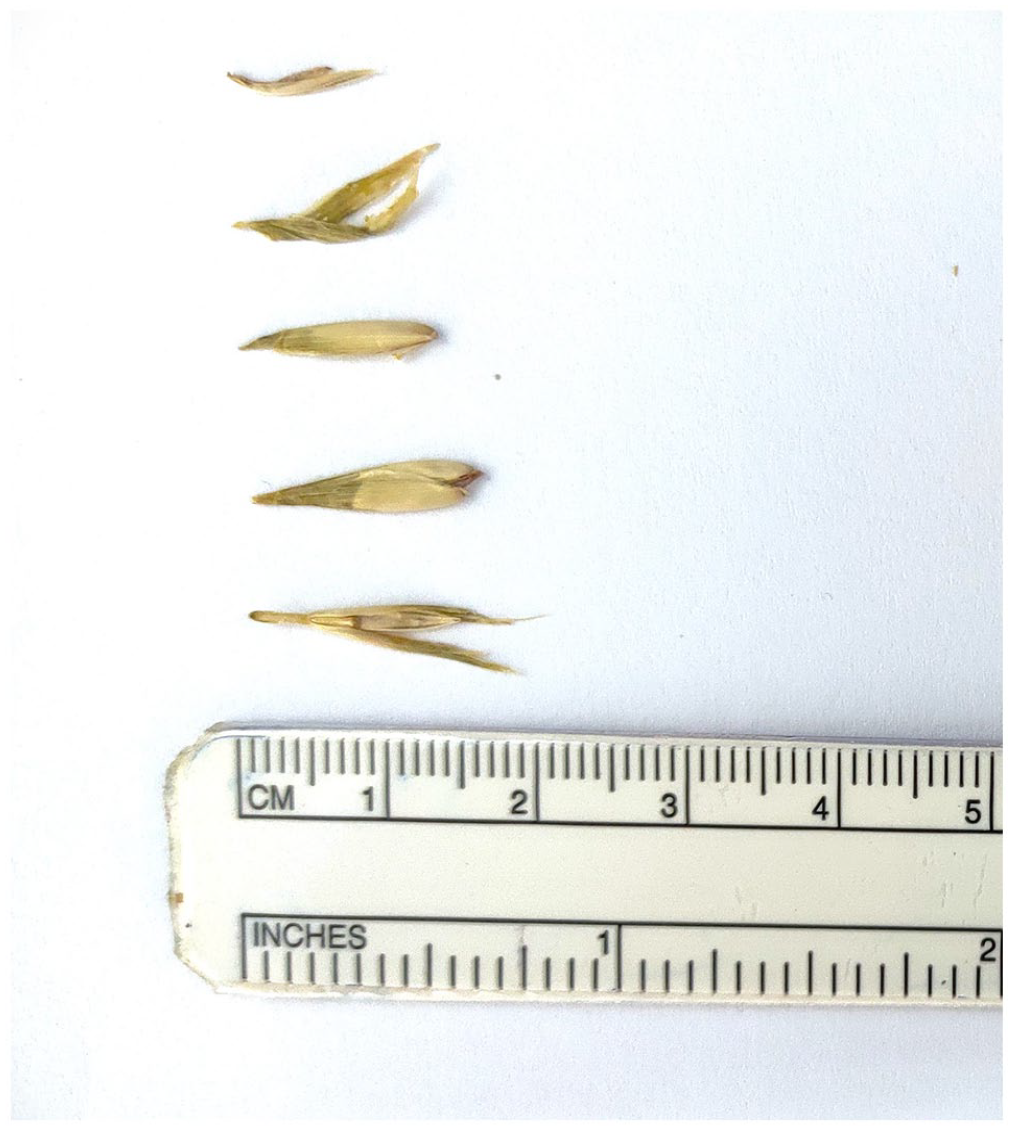
Foreign body (grass fragments) retrieved from the buccal mucosa of the patient.
Discussion
Recurrent buccal abscesses in pediatric patients often pose diagnostic challenges, with varied etiologies ranging from infectious to anatomical factors. 3 This discussion delves into a rare presentation of recurrent buccal abscesses in a 10-month-old female child, where foreign body grass fragments within the buccal mucosa were identified as the unusual cause.
While foreign bodies as a cause of recurrent abscesses are not unheard of, the presence of grass fragments in the buccal mucosa is an uncommon occurrence, highlighting the importance of a meticulous diagnostic approach. While there have been occasional mentions of organic foreign bodies, such as grass, in the eyes in the literature, such occurrences are not commonly reported in the ENT region. Foreign bodies in the buccal mucosa are rare, and their diagnosis can be challenging due to the limited visibility and cooperation of pediatric patients. 4 The lack of a clear history of foreign body insertion further complicates the diagnostic process.
Imaging studies play a crucial role in identifying the presence, type, and location of the foreign body. While plain radiographs are commonly used to detect radiopaque foreign bodies, small fragments of organic foreign bodies may not be easily apparent. 5 Ultrasonography can provide valuable information in cases where radiographs are inconclusive or when a detailed assessment of soft tissues is required. 6 The combined use of these imaging modalities allows for a comprehensive preoperative assessment.
The utilization of MRI for buccal abscesses is not commonly heard of (Robinson et al.) 7 but in our patient, it played a crucial role in unraveling the mystery behind the recurrent abscesses. The MRI revealed a blind tubular structure within the superficial lobe of the right parotid gland, with an external opening into the skin. This finding provided valuable insights into the foreign body-related etiology, emphasizing the significance of imaging studies in cases of persistent or recurrent symptoms.
The recurrent nature of buccal abscesses in the presented case initially led to a focus on infectious etiologies. The decision to perform needle aspiration, although inconclusive, was a logical step in ruling out purulent infections. However, the absence of aspirate prompted further investigation, revealing the unexpected presence of grass fragments during subsequent incision and drainage procedures. This underscores the importance of considering unconventional foreign bodies in the differential diagnosis of recurrent abscesses. 8
Conclusion
This case report highlights the importance of considering foreign bodies in the buccal mucosa as a potential cause of facial swelling and pain and in the differential diagnosis of recurrent buccal abscesses in pediatric patients. The presence of grass fragments, an unusual foreign body, emphasizes the need for a comprehensive diagnostic evaluation, including imaging studies, to uncover such atypical causes. Recognition of foreign body-related recurrent abscesses is essential for effective management and prevention of further episodes. A thorough clinical examination, supplemented by appropriate imaging studies, is crucial for accurate diagnosis and planning of the management strategy. In the absence of ENT or oral surgeons, healthcare providers, particularly those in emergency and pediatric settings, should be aware of the potential for foreign bodies in atypical locations, such as the buccal mucosa, and be prepared to employ a variety of diagnostic and therapeutic modalities to ensure optimal patient outcomes.
Footnotes
Acknowledgements
The authors are grateful to the patient for consenting to be a part of the study. We would also like to extend our gratitude to all the consultants and residents of the department for their guidance and inputs.
Author’s contribution
UP: contributed to concept and design of case report, literature review, and final manuscript writing. SM: supervised the overall progress of the case report and contributed to the final approval of the version to be published.
Declaration of conflicting interests
The author(s) declared no potential conflicts of interest with respect to the research, authorship, and/or publication of this article.
Funding
The author(s) received no financial support for the research, authorship, and/or publication of this article.
Ethical approval
Our institution does not require ethical approval for reporting individual cases or case series.
Informed consent
Written informed consent was obtained from a legally authorized representative(s) of the minor subject for publication of this case report.