
Abstract
In the treatment and management of severe wounds, microsurgical repair remains the gold standard. However, it is difficult to transfer free tissue from a Tanzanian perspective due to donor site morbidity, longer operational times, bulky forms, recipient vessel stress, sophisticated surgical expertise, and high costs of the equipment. Meanwhile, the reverse sural flap has been considered as the ultimate tissue restoration technique. This study reviews a case of a 35-year-old man who was admitted at Mbeya Zonal Referral Hospital in the Department of Orthopaedics, Trauma, and Neurosurgery in Mbeya, Tanzania. The patient had a 9-day injury to the rear of his right ankle due to a cut he suffered after tripping over a toilet seat in the washroom. After the diagnosis, the plan involved surgical debridement, tendon repair, and reverse sural flap rotation. Despite the difficult working environment requiring advanced learning experience, our patient fully recovered after 3 weeks. Proving that reverse sural flap is one of the few available possibilities in the protection of vital structures such as bone or tendons, in the distal areas of the leg, ankle, and foot especially when the medical treatment facility lacks a strong microsurgery team and equipment.
Introduction
Flap surgery is a plastic and reconstructive surgical treatment that entails taking any type of tissue from a donor’s site and transferring it to a recipient’s site with a continuous blood supply. 1 When the residual tissue cannot sustain a graft, reverse sural flap can be done to fill in the defect such as a wound after an injury or surgery. 2 Ponten described the sural fasciocutaneous flaps for the first time in 1981. 3 Later, Donski and Fogdestam first introduced the distally based sural flap in 1983. 4 They explained the anatomy and gave a detailed account of the surgical technique. To reconstruct the distal region of the legs and feet, the reverse flow sural flap has been highlighted as a viable approach. 5 According to research, 6 this flap has had a positive effect and has been the only available soft-tissue reconstruction technique for the distal leg around the ankle and proximal foot. For a patient with distal leg or ankle soft-tissue defect as the case with our patient, reconstruction of the ankle results to a psychological and functional relief. According to Dr Jamey D’O’Neill, acknowledging these mistakes may improve the first results and promote their continuous use. 7 The tibia and tendons are easily exposed since the leg has a thin layer of subcutaneous fat and a small number of muscles. 8 The skin in this area is hard to detect and has little elasticity. Soft-tissue loss and exposed fractures are common side effects of injuries. The sural flap functions as an axial flap receiving resources from the vascular plexus, the superficial sural artery and arteries that follow the minor saphenous vein. 9 The artery that runs parallel to the medial superficial sural nerve can be exploited to ensure that blood flow is restored. This vein must be kept at least as high as the fibular artery’s calibre perforator. 10 In the distal regions of the legs and feet, the reverse flow sural flap is one of the few therapy alternatives. 11
Surgery for soft-tissue defect reconstruction in the foot and ankle poses a significant challenge. According to Dr David Denny, while surgeons are learning how to employ this flap, they make several common mistakes. 12 The surgeon provides some recommendations on how to use the flap to overcome these challenges.13,14 The main disadvantage of the sural flap is the reverse venous flow with frequent venous congestion. Surgeons have made several attempts to overcome this problem including super draining the vein through supercharging it or using intermittent drainage by venous cannulation. Plastic surgeons face significant challenges during reconstructing foot and ankle deformities. In this area, free flaps are the gold standard; however, to execute them successfully, it requires microsurgical expertise and a long-duration process. Any complications are likely to limit skin transplants, local flaps, and distant flaps. In this case report, we present the results of an attempt of overcoming these obstacles. These attempts include delayed procedure that led to successful flap especially with a large-sized flap and ischemic conditioning of the flap before finishing. This report also highlights the significance of this study in Tanzanian perspective and Africa as a whole. Soft-tissue reconstruction in the Tanzania setting and sub-Saharan Africa excluding South Africa is not a common procedure as it needs dedicated efforts in convincing practitioners that lower limbs soft-tissue defect should not be ignored, leaving the patient to suffer. Rather for the distal leg, ankle, and foot, reversed sural flap has proven to be the best option. This report provides highly valuable information to practitioners in Tanzania and other low-income countries.
Case report
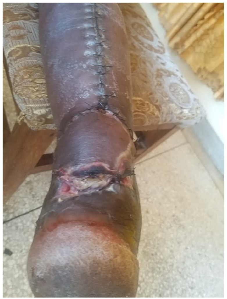
A 35-year-old man was admitted to Mbeya Zonal Referral Hospital’s (MZRH) in the Department of Orthopaedics, Trauma, and Neurosurgery in Mbeya, Tanzania. The patient had a 9-day injury to the rear of his right ankle. The patient had a 9-day injury to the rear of his right ankle due to a cut he suffered after tripping over a toilet seat in the wash room. He had attended Chunya District Hospital where debridement was done and the wound stitched. After a few days of swelling around the ankle and intense pain, Chunya District Hospital referred the patient to our facility. His right ankle had a cut on the backside with an exposed transected Achille’s tendon and some sloughs (Figure 1(a)). The Achille’s tendon had been traumatically ruptured, resulting in the loss of large amounts of soft tissue in the distal area of the leg and bone exposure without fracture. Upon examination, the patient seemed to have been in good health with stable vitals and good medical history. The plan involved surgical debridement, tendon repair, and reverse sural flap rotation.

(a) His right ankle has a cut on the backside with an exposed transected Achille’s tendon and some sloughs and (b) dissection of the fascio-subcutaneous margin around the flap’s skin enhances circulation and prevents the need for a skin graft by leaving a lane of skin over the pedicle.
The patient was put in a prone position. Doppler ultrasonography was used to identify perforating arteries from the peroneal artery along the lower leg’s posterolateral intermuscular septum. Wound debridement was the first step in the procedure. The wound size was then estimated, as was the distance between the rotation point (pivot, 5 cm above the lateral malleolus) and the wound, to determine the pedicle size. The distance between the medial and lateral medial head of gastrocnemius muscle was palpated to aid in the transfer of these measures to the central axis of the posterior part of the leg (Figures 1(b) and 2(a)). An incision was made in the upper margin of the flap, 5 cm distal to the popliteal crease and the subcutaneous area was dissected with shears in a bevelled and ascending manner to construct a fascio-cutaneous margin larger than the piece of skin. The medial sural nerve and the minor saphenous vein were ligated (Figures 1(b) and 2(a)). These structures were in the superficial plane in the dissections in the medial third of the leg, but in the upper third of the leg, they were in the subfascial plane. The flap was rotated to cover the defect on the posterior aspect of the ankle. The dissection of the flap was only resumed after these structures were maintained in these circumstances. The dissection, which went from proximal to distal, encompassed deep fascia, medial sural nerve, and minor saphenous vein (Figures 1(b) and 2(a)). These procedures were completed in one session.

(a) The ideal method for generating a fasciosubcutaneous flap in wounds in the heel or over the lower Achilles tendon is to fold it over its rotation point like a booking sheet. The skin graft should be applied to the fascial side, which should be pointing upwards and (b) final wound closure, coverage of skin graft and the pedicle flap.
As seen in Figures 3–5, the patient began to improve significantly after the surgery on the ninth day. His wounds were virtually completely gone 3 weeks after the surgery. However, some necrosis were observed on the margins of the flap a few days after the surgery whereupon debridement was performed, and the patient was kept on antibiotics to full recovery.
Wounds begin healing after 9 days.

Wound continues to heal after 14 days.

The patient has fully recovered and is beginning to walk.
Discussion
Reverse sural flap is a useful reconstructive method in patients with soft-tissue defects of the distal leg, ankle, and foot. It takes less time, easier to conduct, and is more reliable than more complicated procedures that require lengthy general anaesthesia. Dissection is simple, the profile and volume are low, and the major lower extremities arteries are preserved. In practice, the geometric contour of the defect determines flap size and pivot point position. For a successful flap, it should not be under pressure or any mechanical forces.14,15 The reverse sural flap has proven to be far more reliable than the lateral supra malleolar flap. Since its initial description nearly 30 years ago, several revisions to the operative technique have been proposed, and the reverse sural flap is now considered an accepted and popular method for coverage of soft-tissue loss in the distal third of the leg, ankle, heel, and foot.
The patient who underwent this procedure was able to recuperate completely after 3 weeks, according to the visits. As a result, this study reveals that once surgeons have passed the learning phase and the technique has been perfected, the outcome tends to improve and the outcomes are quite positive. However, for these findings to be statistically significant, a larger number of instances must be reviewed. Because the learning phase influences the results, we cannot say that the proposed changes, such as maintaining a fasciocutaneous margin that exceeds the skin flap and preserving a lane of skin over the pedicle, are accountable for the improved results. Our findings support prior research findings, emphasizing the necessity of paying close attention to technical details when constructing the flap and avoiding the most common mistakes made during the learning period. The most crucial details that should be addressed, as well as the most common mistakes made by surgeons learning these techniques.
There are numerous limitations to this study. Because it is a follow-up case on a single patient. Some complications observed in a case follow-up involved peripheral flap- recipient interphase dehiscence, necrotic flap margins due to a big-sized flap and wound infection, these were all resolved by debridement and secondary suture, which, along with the data presented by Baumeister et al., 15 represents a more realistic view of the complication rate that should be expected with use of this flap. Finally, any operation has a learning curve, and it is possible that the surgeons in this study improved their reverse sural flap process with time, which may have contributed to the lower incidence of venous congestion in the late group of patients. Although this is a possibility, the complication rate was not significantly different across the groups, supporting the theory that increased pedicle width or delayed technique enhances flap survival via improving venous drainage.
Conclusion
The risk of problems can be reduced by dissecting fasciocutaneous margins, maintaining a lane across the pedicle and using a skilled surgeon. The reverse flow of the sural artery is one of the few ways of handling similar complications on essential structures such as bones or tendons in the distal area of the leg, especially where strong microsurgery team is unavailable. Because the procedure is easy and quick, this flap is also a good choice for institutions that undertake microsurgery.
Footnotes
Author contributions
J.R.M. conducted the operations. J.R.M. interpreted the data. C.N.M. prepared the original manuscript. All co-authors contributed to subsequent revisions of the manuscript. All authors read and approved the final manuscript.
Declaration of conflicting interests
The author(s) declared no potential conflicts of interest with respect to the research, authorship, and/or publication of this article.
Ethics approval
Our institution does not require ethical approval for reporting individual cases or case series.
Funding
The author(s) received no financial support for the research, authorship, and/or publication of this article.
Informed consent
The patient provided consent for publication of this case report including images.