
Abstract
Gastrointestinal stromal tumors are the most common subepithelial tumors of the gastrointestinal tract. Gastrointestinal stromal tumors are commonly detected in the stomach followed by the small intestine. Surgery for gastrointestinal stromal tumors affecting the duodenojejunal junction is technically challenging because of the anatomical complexity of this area. A 56-year-old woman visited the outpatient clinic with the chief complaint of melena associated with dizziness from 5 days ago. Abdominal computed tomography revealed an enhancing small bowel mass measuring 4 cm × 5 cm in the left upper abdomen abutting the duodenojejunal junction. She underwent successful laparoscopic small bowel segmental resection of the duodenojejunal junction area. Laparoscopic segmental resection with side-to-side duodenojejunostomy for a gastrointestinal stromal tumor at the duodenojejunal junction is an advanced and challenging procedure requiring experience and a good surgical technique.
Introduction
Gastrointestinal stromal tumors (GISTs) are the most common mesenchymal tumors of the gastrointestinal (GI) tract. GISTs are commonly detected in the stomach and the small intestine. Approximately 5% of GISTs are located in the duodenum and are associated with an increased risk of GI bleeding. 1 Surgical resection with a negative margin is the treatment of choice for primary GISTs. To date, the optimal surgical treatment of duodenal GISTs remains doubtful owing to the technical complexity in obtaining wide surgical margins at this site. Recently, laparoscopic surgery is widely used to resect gastric and small bowel GISTs and has shown oncological outcomes that are similar to those associated with open surgery. 2 In addition, this approach provides all the benefits of minimally invasive surgery, including lesser postoperative abdominal pain, a shorter hospital stay, and better cosmesis. Laparoscopic surgery is gaining popularity for the treatment of GISTs; however, performing an anastomosis after segmental resection of the duodenojejunal junction (DJJ) for GISTs may be technically demanding, although it is not so frequent. We present the case of a woman who presented with a bleeding duodenal GIST and underwent successful laparoscopic resection.
Case presentation
A 56-year-old woman without a significant medical history was admitted to the hospital with upper GI bleeding manifesting as generalized weakness and melena. Her general physical and systemic examinations were normal. Upon admission, her blood pressure was 110/70 mm Hg, pulse was 88 beats/min, and the hemoglobin level was 6.6 g/dL. GI endoscopy showed no mucosal lesion (Figure 1(a) and (b)). Abdominal computed tomography (CT) showed as well-defined exophytic space-occupying mass measuring 40 mm × 45 mm at the DJJ. No lymphadenopathy or metastatic disease was identified. The radiologist suspected the lesion was a GIST or some other small bowel tumor (Figure 2(a)–(c)). Biopsy such as endoscopic ultrasound-guided fine-needle aspiration is essential for definite diagnosis, but we decided the use of minimally invasive operation in the diagnosis is preferred as it minimizes the biopsy-related risk and danger if malignant cell dissemination. The patient was scheduled for laparoscopic tumor resection.
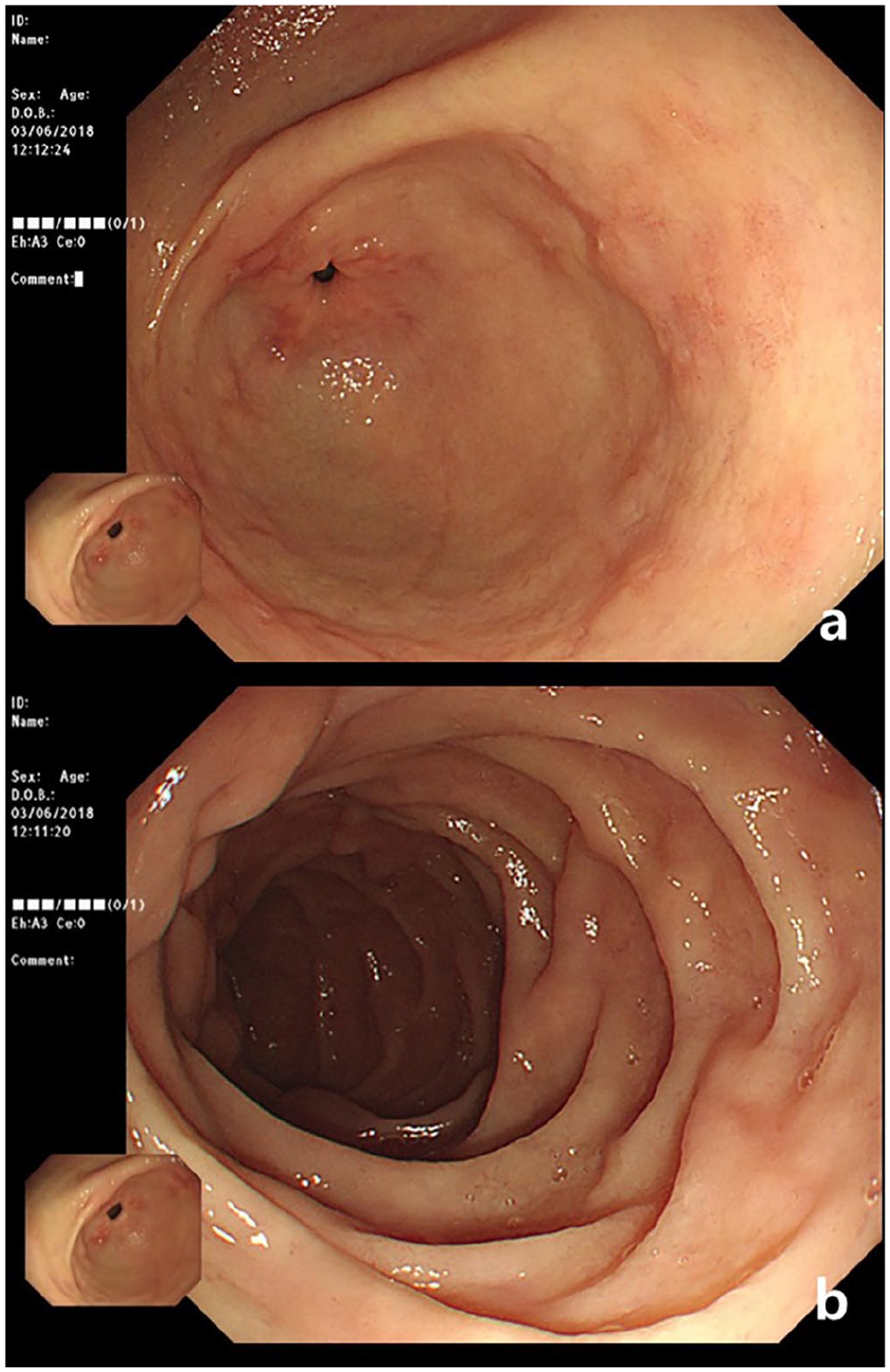
(a, b) Gastrointestinal endoscopic images show no mucosal lesion.

(a—Axial view, b—Coronal view, c—Sagittal view) CT images show a large well-defined mass measuring 4 cm × 4 cm originating from the DJJ without evidence of lymphadenopathy or metastatic disease (white arrowheads). The mass is abutting the first jejunal branch of the superior mesenteric artery and vein (white arrow). CT: computed tomography, DJJ: duodenojejunal junction.
Under general anesthesia, with the patient in a trendelenburg position, three ports were inserted: the infraumbilical area for the camera (12 mm), three fingers above the umbilical level lateral to the right midclavicular line (5 mm), three fingers above the umbilical level lateral to the left midclavicular line (12 mm). After laparoscopic exploration and traction of the omentum and the transverse colon toward the diaphragm, the DJJ was mobilized after dissection of the ligament of Treitz. The branches from the first jejunal vessels were carefully divided (Figure 3(a)–(c)). The DJJ proximal to the tumor was excised with a linear stapler (Echelon Flex™ Endopath® Stapler 60 mm Blue Cartridge (Ethicon Endo-Surgery, J&J)), and segmental resection was performed to include the area affected by the tumor by transecting the jejunum distal to the tumor with a linear stapler after obtaining a tumor-free margin (Figure 3(d)). To restore bowel continuity, the second and third parts of the duodenum were exposed by dissecting the transverse mesocolon up to the midcolic vessels, and after adequate mobilization, the distal loop of the jejunum was anastomosed with the third part of the duodenum in a side-to-side manner using a linear stapler (Figure 3(e) and (f)). The point of entry of the stapler was laparoscopically sutured. The surgical specimen was placed in a retrieval bag and removed from the abdominal cavity (Figure 3(g)).

(a) Intraoperative images show a mass measuring 4 cm × 4 cm originating from the serosal surface of the DJJ at the anti-mesenteric surface. (b) The first jejunal branch of the superior mesenteric vein is observed in this image (white arrow). (c) The ligament of Treitz and the feeding vessels from the first jejunal branches are dissected (white arrow). (d) The third part of the duodenum is transected and stapled after obtaining a tumor-free margin. (e) The second and third parts of the duodenum are exposed by dissecting the posterior leaflet of the transverse mesocolon up to the midcolic vessels (white arrow = second part of the duodenum, black arrow = pancreas). (f) Laparoscopic side-to-side duodenojejunostomy is performed after resection of a GIST (Echelon Flex™ Endopath® Stapler 60 mm Blue Cartridge (Ethicon Endo-Surgery, J&J)). (g) Images obtained after segmental resection of the small bowel (white arrow = distal end of jejunum, black arrow = proximal end of jejunum). DJJ: duodenojejunal junction; GIST: gastrointestinal stromal tumor.
Histopathological examination of the specimen revealed a high-grade GIST (mitotic count 20 per 50 high-power field) showing an uncommitted cell subtype that stained positive for c-Kit. The tumor measured 45 mm in its largest diameter. Surgical resection margins were clear, and the mass was diagnosed as a malignant GIST. No complication occurred during the procedure. The patient’s postoperative course was uneventful. Oral intake was initiated on the third postoperative day, and the patient was discharged on the fourth postoperative day.
Discussion
Duodenal GISTs are relatively rare tumors. GISTs most commonly originate in the stomach (40%–60%) and the small intestine (30%–40%), with only 3%–5% affecting the duodenum. 3 Duodenal GISTs clinically present as variably sized lesions and show mucosal ulceration. Clinically, patients with duodenal GISTs commonly present with GI bleeding or abdominal pain. GISTs of the distal duodenum may remain undetected at GI endoscopy. Alternative diagnostic modalities include CT, magnetic resonance imaging, barium studies, or ultrasonography.
Surgery is the mainstay of treatment for resectable GISTs. The tumor should be removed en-bloc with its pseudocapsule to obtain an adequate resection margin. Duodenal GISTs may be treated using options ranging from minimally invasive to major procedures. Based on the size and the location of the tumor, various surgical procedures including pancreaticoduodenectomy, partial duodenectomy, or pancreas-sparing duodenectomy are performed for duodenal GISTs. Limited resection should be considered for duodenal GISTs when technically feasible. Treatment of a GIST at the DJJ is technically challenging even via open surgery because resection and reconstruction around the DJJ is difficult secondary to the anatomical complexity of this area. The fourth part of the duodenum lies in close proximity to the point where the inferior mesenteric vein drains into the splenic vein. Moreover, the upper end of the root of the mesentery attaches around the DJJ. The DJJ is suspended by the ligament of Treitz which extends from the duodenojejunal flexure to the right crus of the diaphragm. This anatomically complex area prevents optimal exposure of the operation field and optimal circumferential visualization of the intestine. Thus, anastomosis is technically difficult. Although several techniques have been described to restore bowel continuity following surgical manipulation of the DJJ, hand-sewn end-to-end duodenojejunostomy or end-to-side anastomosis with staplers is the technique commonly used in such cases. 4 Laparoscopy is not yet well established for GISTs involving the DJJ. Following the recent advances in laparoscopic surgery, reconstruction after resection of a DJJ GIST can be performed even in this anatomically complex area using a minimally invasive approach. Laparoscopic reconstruction was performed in our patient using a side-to-side duodenojejunostomy that offered all the advantages of minimally invasive surgery. An important concern regarding the resection of GISTs is oncological safety. The consensus is that the surgical treatment of choice for GISTs is tumor resection with negative surgical margins and prevention of intraoperative tumor spillage. 5
Conclusion
Laparoscopic limited resection in this anatomically complex area is feasible and safe and reduces postoperative morbidity without compromising oncological results. Thus, this technique is a feasible alternative to the traditional open approach. However, patients deemed suitable to undergo this procedure should be chosen carefully. Laparoscopy is contraindicated in patients with large tumors secondary to the risk of tumor rupture and the consequent high risk of relapse. A larger cohort of patients is needed to draw definitive conclusions; however, a laparoscopic segmental resection with side-to-side duodenojejunostomy might potentially be a useful treatment option in patients with GISTs located at anatomically complex areas such as the DJJ. Of note, this technique offers all the benefits of minimally invasive surgery.
Footnotes
Author contributions
S.L. and J.K. participated in the surgical treatment, prepared the manuscript, and performed the literature search; J.L. participated in manuscript revision and the final review. All authors read and approved the final manuscript.
Declaration of conflicting interests
The author(s) declared no potential conflicts of interest with respect to the research, authorship, and/or publication of this article.
Ethical approval
Our institution does not require ethical approval for reporting individual cases or case series.
Funding
The author(s) disclosed receipt of the following financial support for the research, authorship, and/or publication of this article: This paper was supported by the Wonkwang University in 2020 (S.Y.L).
Informed consent
Written informed consent was obtained from the patient who participated in this study.