
Abstract
Objectives:
Candida glabrata is becoming one of the most prevalent pathogenic yeasts in cases of oral diseases. Mucositis is an recurrent oral infection in immunocompromised patients, and the actual guidelines recommend the use of fluconazole (Flu) for many cases. However, the azole resistance by C. glabrata is renowned, causing a reduced therapeutic response, especially when it occurs in biofilms. In this study, we performed an in vitro evaluation of an alternative pharmacotherapy for C. glabrata biofilm infections, combining ascorbic acid (AA) with Flu. AA is recognized for degrading β-glucans, an important compound of the biofilm matrices, which prevent drug diffusion.
Materials and Methods:
Routine clinical 30 or 40 mg/l doses of Flu were applied to C. glabrata biofilms simultaneously with 200 or 300 mg/l of AA.
Results:
The results showed that this combination effectively promoted the degradation of the biofilm network, but unfortunately, also stimulated the growth of the yeasts population due to release of several glucose monomers during β-glucans hydrolysis.
Discussion:
AA lead to the hydrolysis of the β-glucans of the matrix, liberating glucose molecules which are used as carbon souce by the yeasts, thus suppressing the desired antifungal effect of the drug combination with Flu.
Conclusions:
Unlike to what happens in treatment of bacterial infection, AA should not be used together with Flu in the treating oral mucositis caused by Candida.
Introduction
Fungal infections are a major clinical health problem that affects more than 300 million people annually. 1 Candida glabrata is a commensal yeast living in human mucosal surfaces (e.g. mouth, esophagus, intestine) but can easily turn into a pathogen, especially in immunocompromised individuals, instigating a high rate of morbidity and mortality.2–5 In clinical practice C. glabrata is the second most prevalent pathogen in humans in the United States, and the third in Europe, after Candida albicans and Candida parapsilosis.3,6 This species is highly resistant to antifungal agents7–9 and has high capacity to produce biofilms,10–12 which are extremely refractory to antimicrobial therapy, thus being very difficult to treat with the traditional antifungal therapies.10,13–20 Formation of bioflims by Candida species in mouth and/or esophageal mucosae is very common, with C. glabrata21–23 being a very important contributor to human oral disease due to a high level of antifungal resistance.24–26 Biofilms are biological communities with an extraordinary degree of organization, in which microorganisms form structured, coordinated, and functional communities, embedded in an extracellular matrix.24 –26 These structures are known to induce high rates of morbidity and mortality, especially in immunocompromised patients,27–29 creating a dangerous prospect of ineffective therapies against infectious diseases caused by Candida. The National Institutes of Health signposts that biofilms are directly or indirectly responsible for over 80% of all microbial infections.30–32 Therefore, there is an urgent need to overcome the numbers related to biofilm-associated C. glabrata infections and to understand the mechanisms behind the response of C. glabrata biofilms infections to antifungal treatment.
Mucositis (or oropharyngeal candidiasis) is a frequent infection among immunocompromised patients,33,34 which is characterized by the presence of creamy, white plaques on the tongue and buccal mucosa that generally leave a raw, painful, and ulcerated surface when scraped. Although not being a severe infection normally, it can be uncomfortable and detrimental to the patient’s nutritional status thanks to the diminished food and liquid intake. The latest 2016’s guidelines recommend the treatment general mucositis infections with oral fluconazole. 35 However, the number of effective antifungal drugs is limited, and resistance to frequently used agents is emerging. 24 Specifically, the resistance of C. glabrata to the azole class of drugs has been well described. 25
Candida biofilm matrix is mainly composed of exopolysaccharides, the most predominant being β-glucans. These
Ascorbic acid, also designated as vitamin C, has being used concomitantly in antibiotherapy for its capacity to boost the immune system and role as a helper in the treatment of several infections, for several years. 42 AA’s concentration in phagocytes and lymphocytes is very high compared with the level in plasma, indicating that AA may have functional roles in these immune system cells, for example, increasing the functioning of phagocytes, the proliferation of T-lymphocytes and the production of interferon, and decreasing the replication of viruses. 43 Furthermore, its metabolism is affected by various infections (e.g. common cold, pneumonia resultant from many microorganisms, scurvy or Helicobacter pylori infections), proposing that there might be treatment benefits in the association of this compound to the regular pharmacotherapy.44–47 Furthermore, AA’s metabolism also has the capacity to induce formation of hydroxyl radicals (OH•), which are reactive oxygen species that may oxidize biomolecules such as proteins, DNA, and other biopolymers (e.g. β-glucans).
So, the goal of this study was to check the in vitro efficacy, already demonstrated on the use of AA with antibiotics, will also be the same with an antifungal agent. Therefore, AA will be used concomitantly with Flu to treat C. glabrata biofilm oral infections.
Materials and methods
Organisms and growth conditions
C. glabrata ATCC2001 (reference strain from the American Type Culture Collection) was used in the course of this study. For each experiment, C. glabrata ATCC2001 was subcultured on Sabouraud dextrose agar (SDA; Merck, Darmstadt, Germany) for 24 h at 37°C. Cells were then inoculated in Sabouraud dextrose broth (Merck, Darmstadt, Germany) and incubated for 18 h at 37°C under agitation at 120 rpm. After incubation, cells were harvested by centrifugation at 3000 g for 10 min at 4°C and washed twice with phosphate buffered saline (PBS, pH = 7.5). Pellets were then suspended in RPMI 1640 (Sigma-Aldrich, Roswell Park) and the cellular density was adjusted to 1 × 10⁵ cells/ml using a Neubauer counting chamber. 48
Flu and AA
Flu was kindly provided by Pfizer®. AA was purchased from Sigma-Aldrich (Roswell Park). Aliquots of 5000 mg/l were prepared using dimethyl-sulfoxide (DMSO), and the final concentrations used (30 and 40 mg/l for Flu and 200 and 300 mg/l for AA) were prepared with RPMI 1640. Controls were performed with DMSO in order to assure that the concentration used was not toxic (concentrations below 1% (v/v) DMSO).
Flu and/or AA effect on C. glabrata biofilm
Biofilm formation
Cell suspension was prepared and washed as described above and resuspended in RPMI 1640. Then, standardized cell suspensions (200 µl) were placed into selected wells of 96-well polystyrene microtiter plates (Orange Scientific, Braine-l’Alleud, Belgium). As a negative control, RPMI 1640 was used without cells and antifungal agent. As positive control, only cell suspensions were tested without antifungal agent. After 24 h, 100 µl of RPMI 1640 was removed and an equal volume of fresh RPMI 1640 plus the antifungal agent (2× concentrated) or the combination of flu and AA was used. The plates were incubated at 37°C for an additional 24 h period, a total of 48 h at 120 rpm. In addition, in order to assess the effect of glucose in the medium, the assay was performed in the same conditions explained above using RPMI supplemented with 2% of glucose.
Biofilm cultivable cells and biomass determination
The number of cultivable cells in the bioflim was determined by the enumeration of colony forming units (CFUs). For that, after the period of biofilm formation, all medium was aspired and the biofilms were washed once with 200 µl of PBS to remove non-adherent cells. After biofilms were scraped from the wells and the suspensions were vigorously vortexed for 2 min to disaggregate cells from the matrix. Serial decimal dilutions in PBS were plated on SDA and incubated for 24 h at 37°C. The results were presented as total of CFUs per unit area (Log10 CFUs/cm2). Total biofilm biomass was quantified by crystal violet (CV) staining. After the biofilm formation, the medium was aspirated and non-adherent cells removed by washing the biofilms with sterile ultra-pure water. Biofilms were then fixed with 200 µl methanol, which was removed after 15 min of contact. The microtiter plates were allowed to dry at room temperature, and 200 µl of CV (1% v/v) added to each well and incubated for 5 min. The wells were then gently washed twice with sterile, ultra-pure water and 200 µl of acetic acid (33% v/v) was added to release and dissolve the stain. The absorbance of the obtained solution was read in triplicate in a microtiter plate reader (Bio-Tek Synergy HT, Izasa, Lisbon, Portugal) at 570 nm. The results were presented as percentage of biomass. 48 All the assays were performed in triplicate and on three separate occasions.
Statistical analysis
Results were compared using one-way analysis of variance (ANOVA), Dunnett’s and Bonferroni’s multiple comparisons test, using GraphPad Prism 5 software. All tests were performed with a confidence level of 95%.
Results
The present study aimed to evaluate an alternative treatment for oral mucositis related to C. glabrata biofilms using an combination of Flu and AA, similar to what has been used for treatment of bacterial infections for years. To determine the influence of AA in the fungistatic activity of Flu, C. glabrata ATCC2001 biofilms were grown for 24 h and then different concentrations of these agents were added and allowed to incubate for an additional period of 24 h. The working concentrations, for both Flu and AA, were chosen according to their use in clinical practice, since the goal was to overtake the biofilm resistance of C. glabrata, using Flu in the same doses, but with another compound as an adjuvant.
Figure 1 shows the percentage of biomass of C. glabrata ATCC2001 when exposed to different concentrations of AA combined with Flu, in a pre-formed 24-h-biofilm. Controls were performed with the two agents alone. After 24 h of exposure, AA alone had no effect on the reduction of the biofilm biomass, compared with the lower dose of Flu. Furthermore, AA showed to be an enhancer of C. glabrata’s biomass (Figure 1). On the other hand, combining 30 mg/l of Flu with both concentrations of AA, the biomass presented a statistically significant reduction (p < 0.001), compared with the use of Flu alone, however, in a very low percentage: 10–20%. Moreover, when the Flu concentration was higher, this effect was not noticed. An increase in biofilm biomass was also observed in the presence of the higher concentration of AA when used with the 40 mg/l of Flu. In order to explain the results obtained regarding the lack in the drug activity of Flu, viability of cells after treatment was also assessed (Table 1) for the drugs alone. It was verified that using AA, the viable cells increased (around 10% and 15% for 200 and 300 mg/l of AA, respectively), when compared with the controls. Flu was unable to eradicate 50% of C. glabrata viable cells even at higher concentrations (Table 1). In fact, when the highest concentration of Flu (40 mg/l) was used, it was only possible to reach a reduction of 7.0% in terms of biofilm cell viability (Table 1). With 30 mg/l, the values were rather superior (9.1%), but with no biomass reduction (Figure 1), thus not therapeutically interesting.
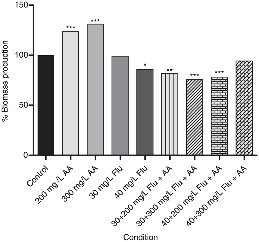
Percentage of biomass detected using CV staining with Flu and Flu + AA in biofilms of Candida glabrata ATCC2001 (*p < 0.05; **p < 0.01; ***p < 0.001). The control is considered to have 100% of biomass production.
CFUs count (Log10 CFUs/cm2) when using AA and Flu alone in biofilms of Candida glabrata ATCC2001 after 24 h and percentage of CFU reduction.
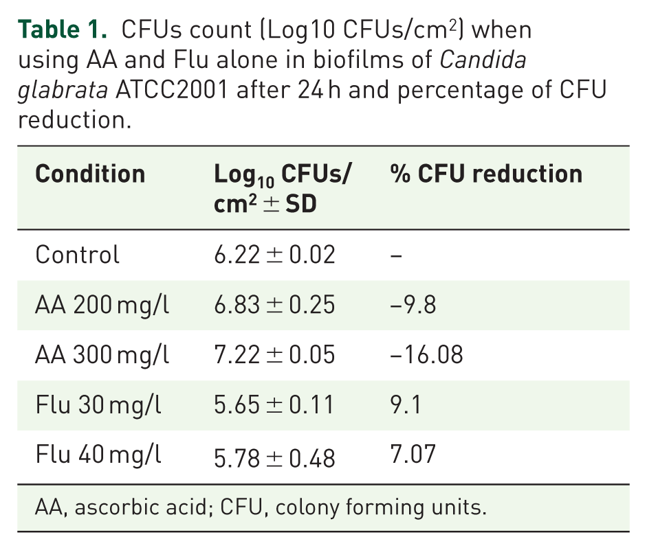
AA, ascorbic acid; CFU, colony forming units.
To verify if the possibility of whether the increased glucose concentration resulting from the β-glucans hydrolysis was contributing to the increase in the C. glabrata population, another assay was performed. The RPMI was supplemented with 2% of glucose for three selected conditions: 200 mg/l of AA, 30 mg/l of Flu, and 30 mg/l of Flu + 200 mg/l of AA (Figure 2). With extra glucose in the medium, the growth in control was 10% more than the control without glucose. Also, significant results were obtained when comparing the use of 200 mg/l of AA with 30 and 40 mg/l of Flu alone (p < 0.01 and p < 0.001, respectively). No significant differences were obtained when comparisons with the drug combinations were made (Figure 2).
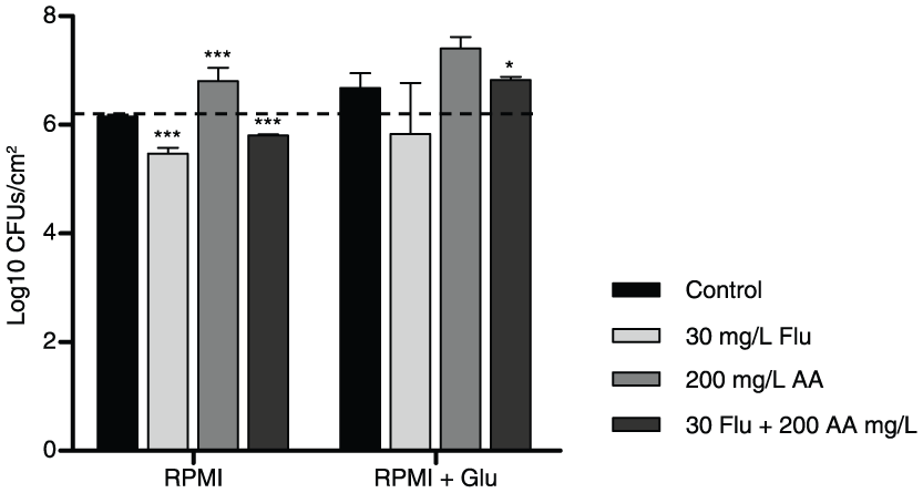
Cell production with Flu and Flu + AA in biofilms of Candida glabrata ATCC2001 (*p < 0.05; ***p < 0.001), by CFUs count (Log10 CFUs/cm2).
Lastly, concerning the drug combination (30 mg/l Flu + 200 mg/l AA), results show that the presence of glucose (Figure 2) in the medium was clearly harmful, since there was no variation in CFU count (thus, no cell death) comparing with the respective control. Additionally, when comparing this result with the control without glucose, there was an increase in cell population (Figure 2).
Discussion
Mucositis associated to erythematous ulcerations in the oral cavity causes pain, xerostomia, dysphagia, and lastly septicaemia. 49 It disturbs functions such as drinking, eating, speaking, dental and other mouth care practices, and it affects not only nutrition and quality but also can be life threatening.23,49
Despite the recognized resistance profile of Candida biofilm cells to antifungal agents,15,17,18 mucositis still has good outcomes with the use of azoles, particularly Flu.23,33,35,50 The knowledge that the therapeutic response of C. glabrata oral biofilms to Flu is lower than the other Candida species (e.g. C. parapsilosis or Candida guillermondii)24–26 is the main reason for the development of the present study, in which clinical doses of Flu are associated with AA.
It was verified (Figure 1) that AA alone had no effect on the reduction of the biofilm biomass, compared with 30 mg/l of Flu, and it showed an increase in the growth of C. glabrata’s biomass. Generally, no significant differences were obtained when comparisons with the drug combinations were made. With the aim of explaining these results, cell viability after treatment with the drugs alone was also assessed (Table 1). It was then possible to observe, contrarily to what was expected, an increase in the cell viability, showing a possible degradation and consumption of β-glucans. In fact, other researchers described that during the degradation of AA, the hydrolysis into glucose monomers occurred.51,52 Xu et al., 53 have also showed a similar situation. The authors tested a considered antifungal drug compound, β-1,3 glucanase, in a C. albicans strain, but instead of having a reduced yeast count, there was a proliferation in C. albicans, which can be explained by the same reasons mentioned above. Similar to the goal of this work, Al-Fattani and Douglas 54 tested various enzymes to degrade biofilms and noted that they were also easily detached from biofilms, but as specific hydrolase enzymes, they released many glucose monomers. Despite this fact and since the authors used amphotericin B as the antifungal agent, the results for the CFU count were favorable.
The chemical events underlying this process were explained through the β-glucan degradation pathways.55–58 Thereby, there are, mainly, two paths: (1) the oxidative cleavage of β-glucan is initiated by the removal of a hydrogen atom from the anomeric carbon (C1) of the polysaccharide inducing the formation of an alkyl radical, and there are two possible courses for the cleavage of the glycosidic bond with the generation of a lactone in C1. The glycosidic bond may fragment due to delocalization of the unpaired electron, leading to the release of a β-glucan fragment with a lactone, and of a β-glucan fragment with an alkyl radical, which may react with O2 to form the corresponding peroxyl radical, which can further undergo transformations. Also, the glucan with an alkyl radical on the C1 can also suffer hydrolysis, which would point to the release of a non-radical β-glucan fragment (glucose monomers) and a β-glucan fragment containing a radical at C1; (2) the formation of peroxyl radical following the alkyl radical in C1. The carbon-centered radical in C1 would react rapidly with O2 to give a peroxyl radical, which can further combine with another peroxyl radical and fragment via an alkoxyl radical (R–O•). 46 The alkoxyl radical formed would undergo a β-fragmentation, liberating a β-glucan fragment with a lactone in C1 and a β-glucan fragment with an alkoxyl radical at C3. Other pathways can contribute to the scission of β-glucan, since the non-selective attack of •OH radicals most likely also generate radicals at other locations besides C1.44,59
In fact, a loss of viscosity in the biofilm was observed during the biofilm manipulation whenever AA was used. The biofilm was more flexible and easily breakable, which is related to the formation of hydroxyl radicals during β-glucan degradation.44,57,60,61 Actually, this glucose hydrolysis reaction is used as an alternative method to the DuBois et al. 62 for carbohydrate quantification.63,64
In the concomitant use of Flu and AA (30 mg/l Flu + 200 mg/l AA), the addition of glucose to the RPMI was very unfavorable. No cell death was achieved (there was no variation in the CFU count), compared with the respective control. In fact, there was even a small increase in cell population (Figure 2) because of the consumption of the free glucose derived from the hydrolysis of the β-glucans and/or the supplementation of the medium, which disrupted the Flu fungistatic activity.
To conclude, Flu is a drug that is significant in the treatment of mucositis. Although with certain side effects, it is generally well tolerated, it has low toxicity, and the usual therapeutic regimen is very appealing to the patient. Unfortunately, once more, the final data presented indicate that this drug might not work for infections derived from C. glabrata biofilms, as previous work from our group have demonstrated. The matrix of Candida biofilms is a solid barrier to the diffusion of drugs into the cells. Thus, the use of a drug combination in which one drug would degrade this matrix and the other drug would be fungicidal or fungistatic could be a good strategy and improve the therapeutic response in fungal infections. Flu has been shown to be recalcitrant in vitro due to the lack of drug penetration. 41
AA leads to hydrolysis of β-glucan which forms glucose that is later used as carbon source by C. glabrata, enabling the cells to colonize and infect. Thus, their virulence was not annulled by Flu in this drug combination. Though this outcome might be possible in other Candida strains and/or species, this work has a clear limitation because of the use of a single strain and thus the results cannot be directly generalized to all C. glabrata strains.
Footnotes
Acknowledgements
The authors thank the Project ‘BioHealth – Biotechnology and Bioengineering approaches to improve Programa Operacional Regional do Norte’ (ON.2 – O Novo Norte), QREN, FEDER. The authors would also like to thank Pfizer(R) for the kind donation of Fluconazole.
Funding
The author(s) disclosed receipt of the following financial support for the research, authorship, and/or publication of this article: This work was supported by the Programa Operacional, Fatores de competitividade – COMPETE and by national funds through FCT – Fundação para a Ciência e a Tecnologia on the scope of the projects FCT PTDC/SAU-MIC/119069/2010, RECI/EBB-EBI/0179/2012 and PEst-OE/EQB/LA0023/2013 and Célia F. Rodrigues’ SFRH/BD/93078/2013 PhD grant.
Conflict of interest statement
The author(s) declared no potential conflicts of interest with respect to the research, authorship, and/or publication of this article.