
Abstract
We describe an improved technique for the reconstruction of a neglected patellar tendon rupture, using semitendinosus-gracilis grafts and FiberWire augmentation.
Case report
A fit and well, non-smoker, 23-year-old male motorbike courier was referred to the senior author (DC) for the management of a neglected patellar tendon rupture, 10 weeks following a low-speed road traffic collision.
Surgical technique
Pre-operatively, lateral radiographs of both knees were performed in order to estimate the Insall–Salvati ratio and use the measurement from the uninjured side as a guide during the reconstruction. 1 The Insall–Salvati ratio measured 1.0. The patient was consented for a patellar tendon reconstruction using hamstring graft and possible Z lengthening of the quadriceps tendon.
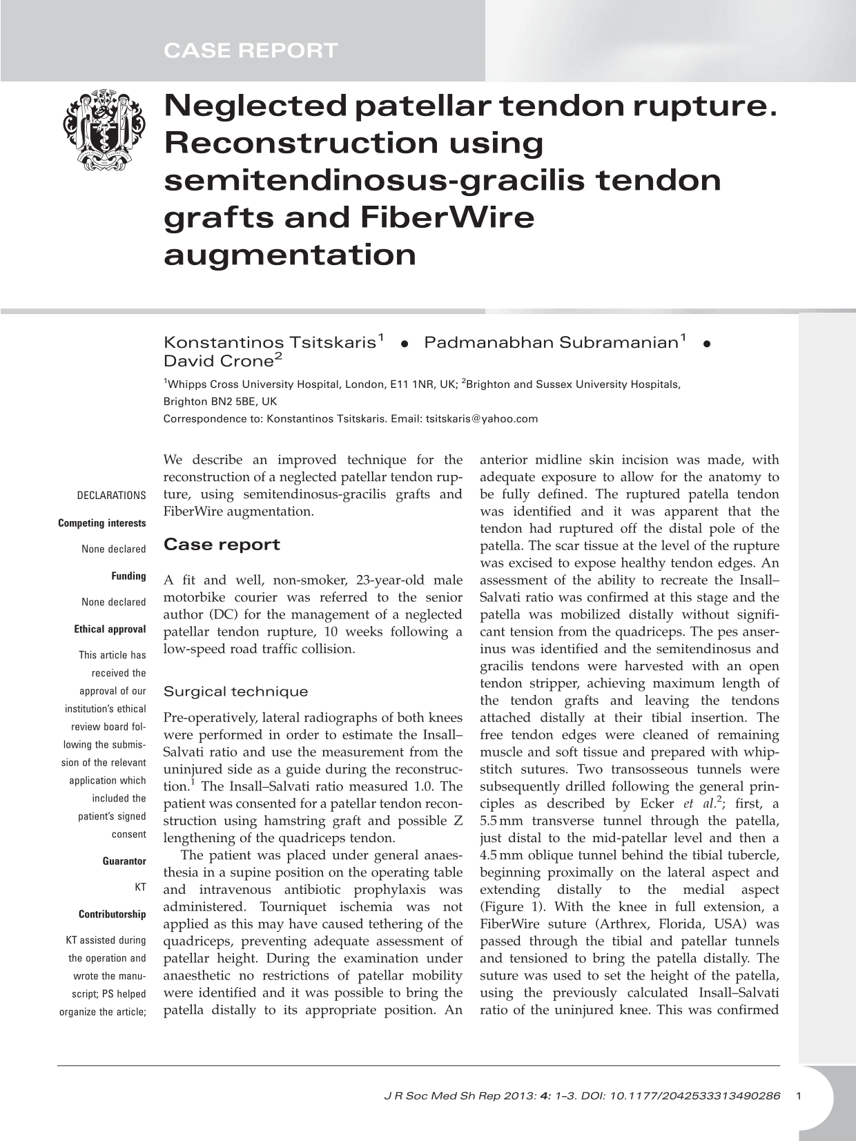
The patient was placed under general anaesthesia in a supine position on the operating table and intravenous antibiotic prophylaxis was administered. Tourniquet ischemia was not applied as this may have caused tethering of the quadriceps, preventing adequate assessment of patellar height. During the examination under anaesthetic no restrictions of patellar mobility were identified and it was possible to bring the patella distally to its appropriate position. An anterior midline skin incision was made, with adequate exposure to allow for the anatomy to be fully defined. The ruptured patella tendon was identified and it was apparent that the tendon had ruptured off the distal pole of the patella. The scar tissue at the level of the rupture was excised to expose healthy tendon edges. An assessment of the ability to recreate the Insall–Salvati ratio was confirmed at this stage and the patella was mobilized distally without significant tension from the quadriceps. The pes anserinus was identified and the semitendinosus and gracilis tendons were harvested with an open tendon stripper, achieving maximum length of the tendon grafts and leaving the tendons attached distally at their tibial insertion. The free tendon edges were cleaned of remaining muscle and soft tissue and prepared with whipstitch sutures. Two transosseous tunnels were subsequently drilled following the general principles as described by Ecker et al.
2
; first, a 5.5 mm transverse tunnel through the patella, just distal to the mid-patellar level and then a 4.5 mm oblique tunnel behind the tibial tubercle, beginning proximally on the lateral aspect and extending distally to the medial aspect (Figure 1). With the knee in full extension, a FiberWire suture (Arthrex, Florida, USA) was passed through the tibial and patellar tunnels and tensioned to bring the patella distally. The suture was used to set the height of the patella, using the previously calculated Insall–Salvati ratio of the uninjured knee. This was confirmed using C-arm image intensifier with the knee at 30° of flexion (Figure 2). The free end of the semitendinosus tendon was passed medio-laterally through the oblique tunnel behind the tibial tubercle using the whipstitch and a suture passer. It was then passed latero-medially through the transverse patellar tunnel, again using a suture passer. The free end of the gracilis tendon was then passed medio-laterally through the transverse tunnel in the patella. The remaining tendons were then overlapped and sutured to each other. The tendon edges at the site of the original tendon rupture were also sutured (Ethibond suture, Ethicon, Gargrave, UK). After the repair was completed, the knee could be flexed to 90° passively without any gapping at the rupture site and the final patellar height was assessed at 60° of knee flexion and found to be symmetrical to the uninjured side. The wound was closed in layers (Vicryl and Monocryl sutures, Ethicon, Livingston, UK), after repair and reefing of the injured extensor retinaculum.
The FiberWire has been passed through the patellar tunnel and is subsequently brought through the tibial tunnel with a suture passer. The distal pole of the patella is identified with the arrow. The patella height is set according to the Insall–Salvati ratio, using the FiberWire.

Postoperatively, the knee was immobilized in a plaster cast at 20° of flexion. The patient was discharged the second postoperative day, not weight bearing. At the two-week follow-up, the cast and sutures were removed and the knee was placed in a hinged brace allowing flexion from 0 to 20°. Quadriceps isometric exercises were initiated at this stage. Further follow-up appointments were arranged at two weekly intervals with a 20° increase of the knee flexion on every occasion, ultimately achieving 120° of flexion in 12 weeks. At the three-month follow-up, initiation of weight bearing was permitted along with closed chain knee exercises under the supervision of a physiotherapist. At the six-month follow-up, the patient was able to walk unaided and without a limp. He had full active knee extension and 130° of flexion, achieving pre-injury functional levels, including return to sport. His Lysholm knee score was 87 points.
Discussion
A neglected rupture of the patellar tendon is a rare occurrence and the exact incidence remains unknown. Several methods of treatment for this challenging problem have been previously described. Simple reapproximation of the torn ends and direct repair augmented by cerclage wire was successfully employed in four cases by Casey and Tietjens. 3 Other authors 4 have used autogenous semitendinosus-gracilis tendon grafts adopting the principles of the Ecker et al. 2 technique, but without the use of a Steinman pin for patellar traction. Instead, they used circular wire through the patellar and tibial tunnels, in order to obtain satisfactory patellar height. Milankov et al. 5 used a contralateral bone-patellar tendon-bone graft followed by double-wire loop reinforcement. Furthermore, bone-patellar tendon-bone allograft, 6 Achilles tendon allograft 7 and synthetic materials 8 have also been used yielding satisfactory results.
Our improved method follows the principles of the technique as initially described by Ecker et al. 2 and subsequently adapted by other authors 4 . Chen et al. 4 have recently described the value of the use of a semitendinosus-gracilis tendon graft and the preservation of its distal insertion. They also described the principle behind the use of a tension-reducing wire as an aid to natural recovery and proper patellar tracking. The advantage of our approach to the same clinical problem lies with the use of a FiberWire suture, instead of metal wires or Steinman pins. The FiberWire suture serves a dual purpose; initially it establishes a satisfactory patellar height for the semitendinosus-gracilis tendon graft reconstruction, then it serves as augmentation for the reconstruction; crucially, further operative intervention for its removal is not required.
One of the challenges during the treatment of neglected patellar tendon ruptures is the difficulty in achieving the correct patellar height. This is usually due to adhesions and quadriceps contracture or atrophy that may ultimately cause proximal migration of the patella. Several methods have been previously described in order to adequately mobilize the patella and relocate it to its anatomical position. Mandelbaum et al. 9 recommended Z lengthening of the quadriceps tendon and Z shortening of the patellar tendon with augmentation using the semitendinosus and gracilis tendons. In cases with severe contracture of the quadriceps tendon, external fixation with pins and wires 10 and the Ilizarov method have been employed. 11 In our case, the patella proved mobile enough so as not to require additional procedures for its mobilization distally. In case of inability to achieve the patellar height, we suggest that our proposed surgical technique may be adjusted with the addition of Z lengthening of the quadriceps tendon. We recommend that this step is included in the consent form and that the same rehabilitation protocol is followed.
In conclusion, the use of a semitendinosus-gracilis tendon graft with FiberWire augmentation provides a practical solution for the treatment of a neglected patellar tendon rupture. We believe that this technique is easily reproducible and warrants further investigation.