
Abstract
We present the case of an amateur boxer who presented to the otolaryngology department with a large sino-nasal haemorrhagic polyp.
DECLARATIONS
None declared
None declared
Written informed consent has been obtained from the patient for publication of this article.
AA
I declare that the above four authors were the only contributors to the report. AA was the primary contributor with RP and MC being the secondary contributors. AP was the paper supervisor and made final changes to the draft prior to submission.
Case presentation
A 32-year-old Polish Caucasian man was referred to the otolaryngology clinic by his general practitioner with a two-month history of right-sided nasal obstruction and epistaxis. The patient was an amateur boxer and had sustained many blows to the nose and face, but could not recall any major recent injuries. He had no symptoms of rhinorrhoea, postnasal drip, hyposmia or facial pain. The patient denied suffering from dysphagia, dysphonia or weight loss.
The patient was hypertensive (blood pressure of 167/95 when checked in the preassessment clinic) but was not on medication for this and was noted to have a high body mass index (35.9 kg/m2). He had no previous surgical history, was on no regular medications and had no drug allergies. There was no relevant family history. The patient was an ex-smoker.
Flexible nasendoscopic examination revealed a large right-sided nasal mass arising from the right middle meatus with some contact bleeding. Examination of the left nasal cavity, postnasal space and larynx was normal. There was no significant septal deviation. Systemic examination was unremarkable. Blood tests revealed a normal haemoglobin, platelet count, clotting and renal function.
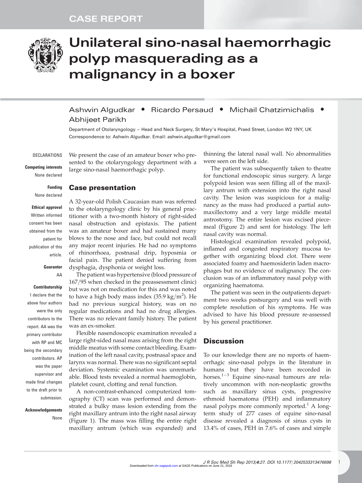
A non-contrast-enhanced computerized tomography (CT) scan was performed and demonstrated a bulky mass lesion extending from the right maxillary antrum into the right nasal airway (Figure 1). The mass was filling the entire right maxillary antrum (which was expanded) and thinning the lateral nasal wall. No abnormalities were seen on the left side.

Coronal section computerized tomography of the nasal mass arising from the right maxillary antrum
The patient was subsequently taken to theatre for functional endoscopic sinus surgery. A large polypoid lesion was seen filling all of the maxillary antrum with extension into the right nasal cavity. The lesion was suspicious for a malignancy as the mass had produced a partial automaxillectomy and a very large middle meatal antrostomy The entire lesion was excised piecemeal (Figure 2) and sent for histology. The left nasal cavity was normal.

Photograph of the lesion after surgical excision
Histological examination revealed polypoid, inflamed and congested respiratory mucosa together with organizing blood clot. There were associated foamy and haemosiderin laden macrophages but no evidence of malignancy. The conclusion was of an inflammatory nasal polyp with organizing haematoma.
The patient was seen in the outpatients department two weeks postsurgery and was well with complete resolution of his symptoms. He was advised to have his blood pressure re-assessed by his general practitioner.
Discussion
To our knowledge there are no reports of haemorrhagic sino-nasal polyps in the literature in humans but they have been recorded in horses.1–3 Equine sino-nasal tumours are relatively uncommon with non-neoplastic growths such as maxillary sinus cysts, progressive ethmoid haematoma (PEH) and inflammatory nasal polyps more commonly reported. 1 A long-term study of 277 cases of equine sino-nasal disease revealed a diagnosis of sinus cysts in 13.4% of cases, PEH in 7.6% of cases and simple sino-nasal polyps in 2.5% of cases. 2 Histological examination in horses has revealed that PEH showed recent and older haemorrhage, as did sinus cysts, but support for a common aetiology between sinus cysts and PEH was absent. 3
Histological assessment of nasal polyp haematomas in horses has revealed highly vascular lesions with a distinctive histopathology resulting from repeated local haemorrhage, breakdown of haemoglobin and organization. 4 In addition to massive haemosiderin deposition, marked ferruginous and calcareous encrustation of connective tissue fibres (with such fibres appearing to be the principal stimulus to giant cell formation) has also been noted in equine specimens with no evidence of exogenous material or that the polyp haematomas resulted from infection. 4 Similar histological features were noted in the specimen from the patient in this case.
The patient in this case was an amateur boxer and so had suffered chronic trauma to the nose and face. He was also an uncontrolled hypertensive. The most likely aetiology in this case was therefore a pre-existing unilateral inflammatory polyp which through repeated trauma in the presence of a high blood pressure led to haematoma formation possibly in association with further polyp growth. Subdural haematomas in boxers have been widely reported in the literature 5 but sino-nasal haematomas in boxers have not.
The patient described in this report had not suffered from any episodes of epistaxis but had he presented to the emergency department with unilateral epistaxis, blunt trauma would clearly be the most likely aetiology. However, whatever the cause of the epistaxis is believed to be nasendoscopy should be performed in all patients with significant bleeding (not necessarily at the time of acute presentation) to ensure sino-nasal lesions are not missed. Any unilateral nasal mass should be treated with suspicion and biopsied (as performed in this case) to ensure that a malignant or premalignant lesion is excluded.
Other local causes of epistaxis include foreign bodies, inflammatory reactions (rhino-sinusitis with or without polyp formation or acute upper respiratory tract infections), granulomatous diseases (Wegener's granulomatosis, sarcoidosis), nasal insufflations of drugs (both prescribed and recreational) and postsurgical. Blood tests should always be performed in epistaxis patients to ensure that the bleeding has not occurred secondary to an underlying coagulopathy or blood dyscrasia. Other systemic factors that may contribute to epistaxis include hypertension, drugs (aspirin, Clopidogrel) and pregnancy.
In this case preoperative imaging through non-contrast-enhanced CT clearly identified the anatomical boundaries of the right maxillary antrum mass. However more information on the nature of the lesion could not be obtained through non-contrast CT. There have been reports of increased uptake of 18F-FDG (fluorodeoxyglucose) in positron emission tomography/CT in benign lesions such as inflammatory masses and haematomas 6 and so there may have been a role for this in the preoperative diagnosis of this case. Ultimately however surgical excision of the lesion was needed for symptomatic relief with histological examination required for definitive diagnosis.
In conclusion haemorrhagic sino-nasal polyps have been described in equine populations but to our knowledge have not been previously reported in humans. Unilateral sino-nasal masses must always be treated with suspicion and should generally always be biopsied. This case describes how a large unilateral sino-nasal polyp haematoma in a hypertensive boxer was successfully surgically excised with complete resolution of the patient's symptoms.
Footnotes
Acknowledgements
None