
Abstract
Peripheral neuropathies are disorders affecting the peripheral nervous system. Among them, Charcot-Marie-Tooth disease is an inherited sensorimotor neuropathy for which no effective treatment exists yet. Research on Charcot-Marie-Tooth disease has been hampered by difficulties in accessing relevant cells, such as sensory and motor neurons, Schwann cells, and myocytes, which interact at the neuromuscular junction, the specialized synapses formed between nerves and skeletal muscles. This review first outlines the various in vivo models and methods used to study neuromuscular junction deficiencies in Charcot-Marie-Tooth disease. We then explore novel in vitro techniques and models, including complex hiPSC-derived cultures, which offer promising isogenic and reproducible neuromuscular junction models. The adaptability of in vitro culture methods, including cell origin, cell-type combinations, and choice of culture format, adds complexity and excitement to this rapidly evolving field. This review aims to recapitulate available tools for studying Charcot-Marie-Tooth disease to understand its pathophysiological mechanisms and test potential therapies.

Keywords
Introduction to Charcot-Marie-Tooth disease
Peripheral neuropathies are among the most common inherited neurological diseases. They constitute a vast and heterogeneous group of rare disorders affecting peripheral nerves. Inherited peripheral neuropathies (IPNs) have a genetic cause and range from purely motor (Hereditary Motor Neuropathy) to purely sensory (Hereditary Sensory Neuropathy), but many patients are affected by an intermediate sensory-motor form known as Charcot-Marie-Tooth disease (CMT). CMT overall prevalence was estimated in the 1970s at 1:2500. 1 However, broader population studies have lowered the prevalence between 1:5,000 and 1:10,000 in European populations and 1:3,300 worldwide. 2
First described in 1886, CMT is characterized by distal motor and/or sensory deficits in all four limbs, following a distal-to-proximal pattern. CMT is a heterogeneous neurological disease characterized by a wide range of clinical manifestations that can vary significantly in onset, severity, and progression. Symptoms usually begin during childhood or early adulthood, although it can occur anytime from infancy to later in life. Over time, the disease leads to a gradual deterioration in muscle function, typically spanning over several decades. 1 The variability in severity and type of symptoms underscores the heterogeneous nature of CMT. Most common motor symptoms include muscle weakness and wasting in the feet and lower legs, seen in up to 99% of patients. 3 Patients frequently develop foot deformities, such as high arches (pes cavus) and curled toes (hammertoes). Therefore, patients experience frequent tripping, reduced walking speed, foot drop, and shorter stride lengths. In addition to motor symptoms, sensory issues commonly arise, typically in the lower legs and feet, with numbness, tingling, and reduced sensitivity to touch. These symptoms may progress to the hands. Pain is another key feature of CMT, often manifesting as a mix of neuropathic and nociceptive pain, which mainly affects the lower back and joints. 4 Other complications may include scoliosis, hand tremors, fatigue, hearing loss, and in rare cases, vocal cord paralysis. Chronic pain, especially in the extremities, is reported by many CMT patients, typically moderate in severity and of neuromuscular origin.4–6 Although CMT does not typically reduce life expectancy, it has a profound impact on quality of life, particularly by affecting balance, mobility, and manual dexterity.
Given that CMT is a complex neurological disease, its classification is essential for a better understanding of its pathophysiology and advancing research. It can be classified into subgroups based on inheritance pattern (either autosomal dominant (AD), autosomal recessive (AR), X-linked dominant (XD) or X-linked recessive (XR)) and nerve conduction velocity (NCV) testing, an electrodiagnostic that helps distinguish between demyelinating and axonal forms of the disease. Indeed, one of the major breakthroughs in CMT classification was made in 1968, by Dyck and Lambert and followed a decade later by Harding and Thomas through their electrophysiological studies.6,7 They observed differences in NCVs among CMT patients, distinguishing two major subtypes among them, either exhibiting demyelinating or axonal peripheral neuropathies based on these measurements. The first group is characterized by an NCV < 38 m/s and is referred to as CMT1 or “demyelinating CMT,” while the second group has an NCV ⩾ 38 m/s, similar to that of unaffected individuals, is known as CMT2 or “axonal CMT.” These differences arise from the specific types of cells that are damaged. CMT1, the most common form affecting approximately 70% of patients, is characterized by Schwann cell (SCs) alterations with progressive demyelination of axons. Myelin plays a crucial role in protecting and speeding up the transmission of electrical signals which explains the reduced NCV.8–10 In contrast, CMT2 primarily affects the neurons themselves, which explains why NCV values remain unchanged, 11 but with a reduced action potential (AP) amplitude. Additionally, several studies have identified a third group, called “intermediate CMT” (CMTI), which includes clinical cases that fall between the axonal and demyelinating forms, sharing characteristics of both. The NCV for CMTI ranges from 25 to 45 m/s.
With advancements in genetic testing, different subtypes of CMT have been identified, each associated with specific inheritance patterns and affected genes. For instance, CMT can result from dominant or recessive mutations. CMT4 corresponds to an autosomal recessive form of demyelinating CMT, with a prevalence of 1% to 9% depending on geographical regions in genetically identified CMT.12,13 When the CMT-causing gene is located on the X chromosome, the disease is referred to as CMTX. CMT has extremely heterogeneous genetic causes. The most common genes responsible for CMT are PMP22 (CMT1A), GJB1 (CMT1X), MPZ (CMT1B), and MFN2 (CMT2A) representing 90% of mutations found in patients diagnosed subjected to genetic testing. 12 Moreover, different alterations in the same gene can lead to distinct forms of CMT (demyelinating, axonal, or intermediate) and distinct modes of transmission, as is the case for GDAP1. Altogether, over 100 genes have been identified as CMT-causing genes. 14 This wide spectrum of genes intervene in ubiquitous and fundamental functions of the cell (i.e. myelination, transcription factors, adhesion, mitochondrial fission/fusion, vesicular trafficking, cell signaling and others).15,16
A striking observation is that despite the ubiquitous and crucial nature of mutated genes, deleterious conditions occur only in the peripheral nervous system (PNS), particularly affecting motor neurons (MNs) and their supporting SCs. These cells, along with skeletal muscle (SkM) fibers, are implicated in neuromuscular junctions (NMJs), which are one of the main impacted components of all forms of CMT.17–19
NMJs are specialized synapses formed between lower MNs nerve terminals and motor end plates of SkMs. The MN cell body is located in the dorsal root ganglion (DRG) and innervates striated muscles. The NMJ corresponds to the physical junction, where APs are translated into muscle contractions. When APs reach the presynaptic terminal, a transient and local increase in calcium concentration triggers the exocytosis of acetylcholine (ACh)-loaded vesicles into the synaptic cleft. Upon binding to acetylcholine receptors, sodium influx triggers the release of calcium from the sarcoplasmic reticulum which translates macroscopically into contraction of SkM fibers. ACh is then degraded in the synaptic cleft by ACh-esterase.
The NMJ also includes non-myelinating SCs known as terminal SCs (tSCs, also referred to as perisynaptic SCs or teloglia). Myelinating SCs, on the other hand, are essential supporting cells wrapping around the axon and allowing the propagation of APs via saltatory conduction. Another cell type, known as kranocytes, has been identified as fibroblast-like cells that cap the NMJ and play a role in synaptic regeneration. 20
Although the NMJ is not the primary site of pathology, the axonopathy or demyelinating damage caused by CMT directly impacts this structure and leads to secondary myopathic consequences. Indeed, both CMT1 and CMT2 patients exhibit myopathic changes, 21 which are more pronounced in CMT2. Interestingly, NMJ dysfunction has been reported in patients with CMT2D and CMT2M using repetitive nerve stimulation and single fiber EMG, 22 even if NMJ defects have been scarcely directly observed. 23 Moreover, extensive studies on animal models have established this phenomenon, showing structural and morphological changes, synaptic dysfunction, and destabilization of NMJs.17–19,24,25 An observational study on neuromuscular function in CMT1 and CMT2 patients as well as in healthy subjects, named ESTABLISH, has been conducted (NCT04980807). Although the results have not yet been published, this study confirms the growing interest of the scientific community in this crucial aspect of the pathology. Indeed, because the NMJ is the functional unit necessary to trigger the movement via the recruitment of muscles, muscle weakness and atrophy are an indirect proof of its contribution in some serious and invalidating effects in CMT. Studying it in vivo and in vitro definitely helps to understand the pathophysiological characteristics and evaluate the effects of therapeutic agents that could act directly upon it.
Despite significant advances in the field over the past few years, therapeutic options for patients with CMT remain limited. 26 The genetic heterogeneity of the disease and the diversity of pathophysiological mechanisms involved make it challenging to address a single therapeutic strategy. To better understand CMT disease and to test potential therapeutic approaches, it is necessary to generate a wide variety of models as close as possible to human pathology.
This review endeavors to comprehensively explore the various methodologies employed in both in vivo and in vitro models to deepen our understanding of CMT mechanisms. Moreover, we highlight significant advancements, such as the utilization of human induced pluripotent stem cells (hiPSCs) and cutting-edge microfluidic technologies, which have revolutionized the creation of more physiologically relevant in vitro models. By exploring these advancements, we endeavor to unveil a spectrum of possibilities that could pave the way for the development of a highly representative CMT model.
1. Conventional in vivo models
In the past, animal models have proven crucial in investigating neuropathy onset, molecular determinants, pathogenesis, and identifying potential druggable targets. We will address these in vivo models through the scope of the different techniques and methods used to study the NMJ pathophysiology in CMT.
Non-mammal models
Saccharomyces cerevisiae (S. cerevisiae; yeasts) is a simple organism used to study CMT at the genetic and molecular levels. S. cerevisiae are ideal for gene complementation assays27–29 and are often used to study CMT-mutation consequences,30–32 and to perform large-scale drug screening. 33 However, yeasts are single-cell organisms for which the study of NMJ is unattainable. Caenorhabditis elegans (C. elegans), Danio rerio (D. rerio; zebrafish), and Drosophila melanogaster (D. melanogaster) are commonly used models of NMJ diseases. They are well-characterized, genetically and molecularly traceable animals that show short generation times and are inexpensive to breed on a large scale, thereby increasing experimental throughput.34–36
Behavioral assays
One of the simplest yet indirect ways to assess NMJs’ integrity is to perform behavioral assays. These can vary considerably among species. The locomotor capacities of C. elegans worm models of different CMT forms were measured using crawling or swimming/thrashing assays at the larval and adult stages, respectively.37–39 Similarly, D.melanogaster can be subjected to either crawling or climbing assays depending on their developmental stage. Ali et al. tested both crawling and climbing (negative geotaxis assay) locomotor capacities of their pan-neuron-specific dSLC25A46a knock-down flies modeling CMT2, exhibiting locomotive defects. These results, supplemented by NMJ and mitochondrial visualization, lifespan analysis, learning assays, and ATP/Reactive Oxygen Species (ROS) production analysis, highlighted the role of dSLC25A46ain mitochondrial dynamics, revealing the contribution of its loss-of-function in the neurodegeneration characteristic of the disease. 40 Muscle contraction assays in larvae can also be performed by counting forward body wall contraction exhibited for a given period of time. 41 Using transgenic D. rerio lines overexpressing the human HARS1 (CMT2W) mutant in which sensory and motor divisions are tagged with GFP and RFP, respectively, Mullen et al. 42 linked abnormal PNS morphology with functional consequences through impaired touch-evoked escape response and swimming behavior of the fishes. These tests are widely used in D. rerio studies. 43 Other methods, such as the vibrational startle response state, visual motor response test, swimming endurance, and swimming posture are available to assess the locomotive behavior of D. rerio larvae.44,45 These locomotive assays are indirect but enable to draw conclusions regarding the presence of nervous system defects. In top-bottom approaches, such findings often prompt researchers to investigate the cause at cellular and molecular levels. In addition, in the context of large drug screening, phenotypic changes in motility can help discriminate between potentially efficient compounds. 39
NMJ visualization methods
At the cellular level, a convenient way of appreciating NMJ morphology and physiology is to immunolabel of pre- and/or post-synaptic proteins. Qualitative and quantitative immunolabeling techniques inform on the number and size of synaptic boutons, as well as the density of the active zone and length of synaptic branches. MNs reaching their target, and therefore establishing NMJs, can be quantified using transgenic nematodes expressing GFP in MNs subpopulations.37–39 As the organization of the PNS is well established and conserved in C. elegans, abnormalities are easy to detect (Figure 1). However, this method, although simple due to the transparency of worms, does not hint at morphological and functional defects other than misguidance and/or incapability to form synapses. To specifically visualize the NMJ, the gold standard involves using α-bungarotoxin (BTX), which selectively binds to AChR on muscle cells. For D. rerio, this approach can be enhanced by staining muscle cells with fluorescent phalloidin conjugates and using D. rerio lines expressing GFP under the control of an Hb9 promoter, as demonstrated by Vettori et al. In this CMT2A model, caudal primary MNs in transparent D. rerio embryos appeared disorganized, with less arborization; phalloidin staining permitted to enlighten a reduced width of MFN2 mutant’s muscle fiber compared to control D. rerio. 46 An alternative strategy, when combined with BTX, involves marking the presynaptic SV2 protein.45,47 In addition, standardized, high-resolution quantitative imaging techniques have been developed, providing useful parameters such as axonal length, AChR clustering, and AChR-axon overlap, rendering high-throughput drug screening amenable in D. rerio models.43,48 Regarding D. melanogaster, their nerves can be specifically stained by anti-horseradish peroxidase antibodies. 49 The presynaptic active zone and the postsynaptic muscle folds can be stained with anti-Bruchpilot (Brp) and anti-Disk Large Protein (DLG1) antibodies. This combination of antibodies is the basis of many studies on NMJ in D. melanogaster.40,41,50–52 Although not described in studies of NMJs in the context of CMT, it is worth noting that the use of ultrastructural microscopy can provide insight into the morphological changes of the NMJ (i.e. size, density of active zone) and organelles (i.e. mitochondria, microtubules). Nonetheless, electron microscopy is a valuable tool for studying myelin abnormalities in demyelinating neuropathy models. 53

Non-exhaustive listing of techniques and assays performed to study NMJ integrity in traditional models of Charcot-Marie-Tooth disease. Studies on CMT have primarily focused on C. elegans, D. rerio, D. melanogaster, and rodents. Non-mammalian models are advantageous for large-scale breeding, but rodents remain a preferred model for peripheral neuropathies. NMJ integrity is crucial for CMT research. Behavioral tests, though indirect, allow quick screening of locomotor defects. NMJ visualization can be achieved by immunolabelling, electron microscopy, or transgenic lines with fluorescent proteins, with D. rerio and C. elegans being particularly suited due to their transparent skin. Less commonly used techniques include electrophysiology, with rodents’ muscle activity assessed via electromyography. Pharmacological assays and omics approaches have also yielded interesting findings.
Electrophysiological NMJ assessment
Although not reported in the context of CMT, electrophysiological recordings are technically possible and allow appreciation of the functionality of NMJs in D. rerio. 54 Notably, NMJ electrophysiological activity was recorded in nine nematode lines harboring different mutations in CMT-causing genes. The frequency of spontaneous post-synaptic currents (PSCs) in mutant nematodes was lower than that in wild type (WT) animals. 39 Spontaneous PSC frequency depends on post-synaptic membrane integrity and cholinergic receptor activity, leading to the conclusion that these structures might be affected, in accordance with morphological and behavioral data. 39 In D. melanogaster, the Giant Fiber system is a well-characterized neuronal circuit mediating the escape response in flies. Briefly, giant fibers originate from the brain and form a synapse with both an interneuron and a MN, which in turn projects to the tergotrochanteral muscle. Electrophysiological recordings permit to gather features such as response latency and ability to follow stimuli one-to-one at high frequency, representing synaptic strength and reliability, respectively. 55 D. melanogaster NMJ physiology is also assessable via excitatory junction potential electrophysiological recordings. 51
Pharmacological assays
Pharmacological assays of neuromuscular function in nematodes were performed using aldicarb, (an AChE inhibitor) and levamisole (a selective agonist of AChRs). Inhibition of AChE by aldicarb leads to spastic paralysis, the onset speed of which depends on the rate of ACh release from the presynapse. Thus, an increased sensitivity to aldicarb reflects presynaptic defects. In contrast, levamisole-induced paralysis translates to post-synaptic (muscle) defects. For example, hPDK3R158H C. elegans (CMT6X) were found to be hypersensitive to aldicarb and insensitive to levamisole, indicating that locomotor abnormalities observed are due to neuronal defects, confirming the axonal nature of neuropathy. 56 Levamisole was also used to determine whether the functionality of the body wall muscles of the nine nematode lines generated by Soh et al. 39 was compromised, which was indeed the case.
It is important to note that studies often reach their conclusions by combining the different approaches described above.
Mammal models
Rodents, including Mus musculus (mouse) and Ratus norvegicus (rat), remain the gold standard for disease modeling, 57 and CMT is not ruled out. 58 Fundamentally, the techniques used to study NMJ are the same as those used for non-mammalian models. However, the study of rodent NMJ is extremely widespread and enables detailed studies of NMJ integrity in an extensively characterized organism.
NMJ integrity, behavioral and electrophysiological tests: The example of GDAP1 deficiency
To investigate the functional consequences of loss of GDAP1 function in vivo, Gdap1 KO mice (Gdap1-/-) were subjected to the rotarod test, gait, and footprint analyses, all of which pointed out the defects in locomotor capacities of this model. 24 Subsequently, electrophysiological recordings along the sciatic nerve confirmed the occurrence of axonal neuropathy concomitant with an absence of demyelination, as assessed by ultrastructural analysis. Analysis of MN numbers and morphology confirmed MN degeneration while immunolabelling of β-tubulin and AChR (α-BTX) revealed a reduced occupancy of the NMJs in 12-month-old Gdap1-/- mice and abnormal tangle-like structures in terminal nerves. Additional investigations of GDAP1 loss were carried out on cultured DRG sensory neurons or MNs from Gdap1-/- mice. Electron microscopic ultrastructural analysis of cultured MNs extracted from KO mice revealed the presence of abnormal mitochondria in the proximal and distal regions of the mice sciatic nerves. Finally, mitochondrial protein expression levels were measured in various tissues of 5-month-old Gdap1-/- mice. Limited energy provision linked to dysfunctional mitochondria was found in the cerebellum and peripheral nerves of Gdap1-/- mice. This study reports a model that has been used in numerous other studies since.59–61 This model was also used for the final in vivo assessment of drug efficiency in a wide screening of already-approved FDA drugs. In this study, motor performance and muscle strength were used as indicators of drug ability to reverse the disease course. 62 It is worth noting that a wide variety of motor behavior assessments exists in rodents (Figure 1). Interestingly, GDAP1 has also been associated with a gain-of-function phenotype. Evidence regarding whether alterations result in gain or loss of function are sometimes intricate. Therefore, these findings have been listed for each gene discussed in this review according to the corresponding subtype (Supplemental Table 1).
NMJ visualization methods and myelination insights
The pathophysiological involvement of NMJ can be a consequence of both axonal and demyelinating forms and its understanding has significantly evolved over time. Specifically, understanding how NMJ abnormalities and demyelination interact can clarify the underlying disease mechanisms, highlight the temporal sequence of pathological events, and help identify early therapeutic targets.
Since D. melanogaster and C. elegans differ in terms of myelination mechanisms and organization, those models are not proper to study the context of CMT disease. However, in other animal models, myelin abnormalities can, as in humans, manifest as reduced myelin thickness 63 and characteristic onion bulb formation resulting from continuous demyelination and remyelination processes (as seen in CMT1B models). 64 Myelination alterations can be effectively visualized using transmission electron microscopy (TEM), while instability in its protein and lipid structure are easily observable via immunocytochemistry or immunohistochemistry (as observed in PMP22-/- mice). 65
Regarding NMJ visualization methods, NMJ imaging and morphological analysis in CMT2D mice can unveil important parameters such as synapse elimination, endplate morphological maturation, NMJ growth, and denervation. 17
Although demyelinating forms ultimately lead to NMJ problems, 21 studies directly examining both myelination and NMJ integrity are scarce. Indirect links have been identified, particularly in CMT4C models, where distinct studies have reported NMJ abnormalities on one hand, and demyelination along with characteristic onion bulb formations on the other.
In axonal forms of CMT, where the primary defect lies within the axon itself, the effects on the NMJ are more direct. Studies on CMT2 have highlighted connections between NMJ alterations and denervation, changes in axon diameter, and myelin abnormalities, as seen in models such as CMT2D (GARS1) and DGAT2 zebrafish.47,66
In conclusion, although the direct approach to NMJ analysis in CMT research was complex, it has yielded valuable insights. The combined analysis of NMJ function and myelination now provides a comprehensive perspective on the interplay between axonal integrity, myelination, and NMJ function across CMT models, enhancing both our understanding and the therapeutic potential for CMT.
Pharmacological assays
Regarding pharmacological studies, the neurotoxic agent acrylamide can be used to explore subclinical phenotypes, as has been performed in mice genetically modified with loss-of-function mutations in Lrsam1, which showed no evident impairments in neuromuscular performance or NMJ morphological, functional, or anatomical PNS changes. 67 However, challenging LRSAM1-mice with acrylamide revealed increased neurological dysfunction, tremors, and dyscoordination, indicating that their axons are more prone to degeneration. 67 Moreover, homozygous mice presented with significantly reduced NCV arising directly from acrylamide treatment. Heterozygous mice showed significantly reduced motor axon numbers, whereas homozygous mice had a significantly decreased motor axon area compared with treated WT controls. 67 Although part of a study on the demyelinating form of CMT4C caused by mutations in SH3TC2, the applied proteomics analysis carried out by Cipriani et al. is of major importance in the field of study of NMJ pathologies, which requires mention in this section. By selecting key proteins for maintenance, function, and axonal preservation, this study revealed changes of 50% in the abundance of the selected proteins, probably explained by compensatory mechanisms and prevention of NMJ breakdown. 68
Omics approaches
Omics approaches, more particularly transcriptomics and proteomics, offer a comprehensive view of the molecular changes associated with diseases. Transcriptomics examines the full set of mRNAs, revealing genes with altered expression, while proteomics analyzes the complete set of proteins, providing insights into affected signaling pathways and cellular processes. Cipriani et al. 68 utilized transcriptomics and proteomics to study Sh3tc2∆cEx1/∆Ex1 mice, a model of CMT4C. Their transcriptomic analysis revealed an increased expression of AChRγ, a marker of denervation, and NGF, which may be involved in the enhanced nerve branching observed in these mice. The proteomic analysis of the sciatic nerves showed an upregulation of extracellular matrix proteins, suggesting an attempt to stabilize the NMJ in response to pathology. 68 Proteomics has also underlined the impact of florfenicol and MitoQ pharmacological derivatives on the sciatic nerves of GDAP1-null mice. 62 Both treatments prevented the decline in proteins involved in energy metabolism and antioxidant responses observed in untreated mice. Additionally, they reduced oxidative stress in nerve proteins These omics approaches are virtually transposable to any disease model.
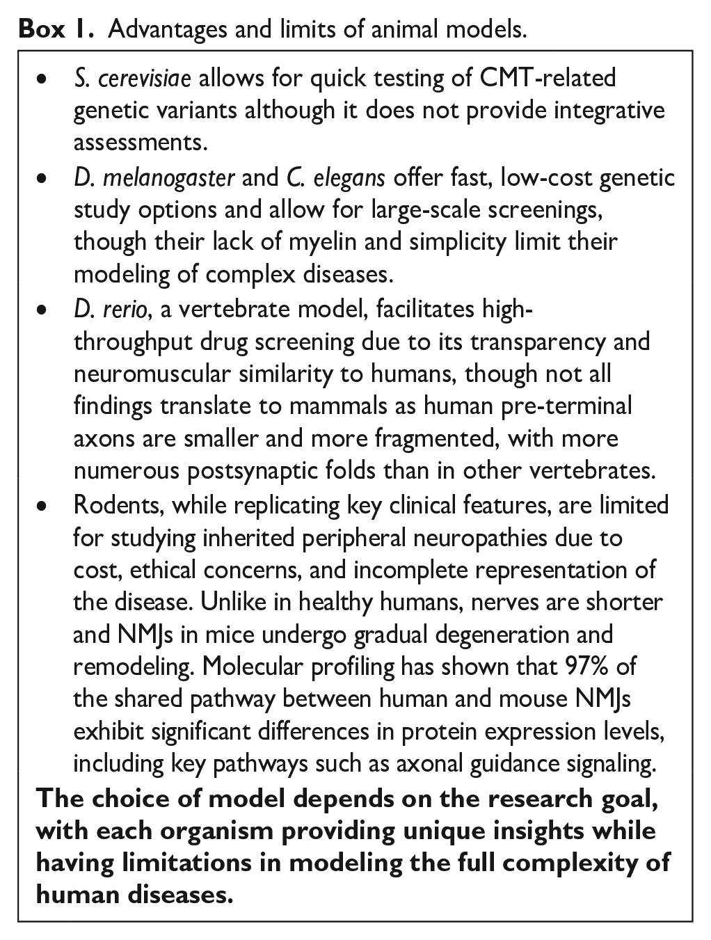
Taken together, C. elegans, D. rerio, D. melanogaster, and rodents provide a wide collection of living organisms in which NMJs studies are feasible, each with its own advantages and limitations (Box 1). These models are particularly valuable for investigating the mutations encountered in CMT (Figure 1). Large animal models have also been studied69,70; however, despite positive arguments, their use is questionable from an ethical point of view. In any case, ethical considerations must be underlined, and compliance with 3R rules (Replace, Reduce, Refine) needs to be applied rigorously when animals are used as models.
Advantages and limits of animal models.
2. In vitro culture models: from monocultures to tissue bioengineering approaches
CMT has an impact on the cells of the PNs all the way to the NMJ, extending from MNs and their supporting cells, SCs and to the SkM fibers. Creating a functional in vitro NMJ is currently a focal topic in tissue bioengineering. Several emerging models hold the promise of enabling the simultaneous study of all cell-specific defects resulting from CMT gene mutations. In this context, we summarize the recent efforts made to develop CMT models that closely mimic in vivo settings.
Level 1: Progression of motor neurons, skeletal muscles, and Schwann cells monocultures
NMJs in vivo display a tripartite architecture, composed of MNs, SkM fibers, and SCs. The culture of these components individually is well-established in laboratories, whether they are obtained from cell lines, primary cultures, or induced pluripotent stem cells.
Motor neurons, Schwann cells and skeletal muscle fibers: Cell lines and primary culture
Extracting MNs from patients or healthy controls poses technical and ethical challenges, restricting resources to postmortem tissues, which only reflect severe end-stage disease. This limitation hinders the study of disease progression and therapeutic testing for CMT. To address this issue, in vitro CMT studies initially utilized primary animal cells or human cell lines. MNs were obtained by dissecting DRGs from rodents, a procedure applicable to specific disease models, 71 or from the ventral spinal cord of embryonic rats to obtain enriched primary MN cultures, further down transfected to study CMT mutations. 72 Civera-Tregón et al. 61 utilized the Gdap1-/- model described earlier to obtain easy to handle embryonic MNs primary cultures for mitochondrial defect studies related to GDAP1 deficiency. Human cell lines (such as SH-SY5Y or COS7) have also long been used to investigate gene mutation consequences in PNS disorders, including CMT.73–76
Human primary SCs cultures were first described in 1980, obtained from peripheral nerve biopsies on 40 patients with various peripheral neuropathies.
77
However, large-scale human primary SCs cultivation is challenging, and these cells have a finite capacity for proliferation and passaging.
78
Thus, SC isolation and culture protocols are mostly available for rodents,
79
with primary SC cultures from adult and newborn CMT1A rats’ sciatic nerves.
80
For example, Trembler-J mice (
Axonal loss and demyelination likely contribute to denervation and NMJ fragmentation, also known as the dying-back process, and linked to muscle atrophy in a CMT2A mouse model. 83 Although muscles are considered as secondary actors in CMT pathophysiology and therefore no CMT-specific muscle tissue monocultures have been developed, the potential role of satellite cells in some CMT forms cannot be dismissed. 84 Muscle tissues can easily be obtained either by culturing cell lines such as mouse myoblasts (C2C12) or by using human muscle precursor cells and primary myoblasts.85,86 However, muscle stem cells obtained from biopsies are rarely pure and rapidly lose their stem cell properties. 87
Induced pluripotent stem cells-derived models
About two decades ago, pluripotent stem cell research began to significantly advance cell type generation. CMT studies utilized human embryonic stem cells to produce MNs 88 and SCs. 89 Human induced pluripotent stem cells (hiPSCs) that share biological properties with embryonic stem cells without raising ethical issue have recently been contemplated with growing interest. Initially derived from mice, hiPSCs methods quickly adapted to human cells.90–92 The first study that successfully generated MN lines from CMT2E (NEFL) and CMT2A patients’ hiPSCs in 35 days correlated with the mouse model’s pathophysiology, showing intermediate neurofilament accumulation in CMT2E MNs, reduced AP threshold, and abnormal channel current properties. 93 Other researchers developed scalable methods to obtain MNs from hiPSCs via embryonic body formation, highlighting how slight changes in concentrations and exposure times impact differentiation. 94 Guo et al. 95 later refined the protocol, achieving a highly pure culture with 70%–95% of cells expressing MN markers. Indeed, A major challenge of hiPSCs-derived MNs is achieving homogeneous and pure cultures. Sorting methods like sedimentation field flow fractionation (SdFFF) 96 or magnetic activated cell sorting (MACS) 97 can enhance differentiation outcomes. Time required to obtain mature CMT MNs was also reduced, reaching as little as 20 days.98,99 Additionally, adding retinoic acid and SAG (smoothened agonist) at specific times can refine the rostro-caudal identity of MN populations during differentiation. 100
Differentiating hiPSCs from CMT patients into SCs is attainable, though efficiency, reproducibility, and maturity remain issues.101–104 Observing myelination of iPSC-derived MN axons by iPSC-derived SCs in culture would be ideal in the context of studying CMT disease, but this remains challenging in 2D cultures. Several studies have reported hiPSC-derived cell lines for various CMT-causing alterations, 105 as well as successful differentiation into MNs or SCs (Table 1).
Updated list of iPSC models of Charcot-Marie-Tooth disease derived from patient cells.

dHMN: distal hereditary motor neuropathies; NCSCs: neural crest stem cells; NPC: neural progenitor cells; N/I: no information or not assessed; PBMC: peripheral blood mononuclear cells; Px: patient x; yo: year old.
Article were selected based on a screening of PubMed database using the following key words: « iPSCs », « induced pluripotent stem cells », « Charcot-Marie-Tooth disease », « CMT ».
Compound heterozygous variants are signalled with a “+” in between the variations. Genetic alterations are stated as provided in the referenced paper, with the date of publication corresponding to the first article that introduced the cellular model.
As stated before, although muscle cells are not primarily affected in CMT, defects in MNs and SCs lead to muscle weakness and atrophy. To our knowledge, no SkM cell model from CMT patient hiPSCs exists, though healthy hiPSC lines have been differentiated into SkMs.133–136
Level 2: Co-cultures
Motor neurons and skeletal muscle fibers to establish a neuromuscular junction stricto sensus
In vivo settings involve numerous cell types that interact to form functional and complex NMJs. Although monocultures of MNs and SCs alone provide useful insights into axonal and demyelinating forms, efforts should focus on establishing a NMJs closely resembling those of patients. Moreover, MNs input is essential for proper muscle maturation and functional NMJ generation.137–139 Strickland et al. 140 reviewed NMJ co-culture models according to cell origin and structural complexity. Additionally, Barbeau et al. 141 have outlined an historical overview of in vitro NMJ models.
Homologous healthy rodents NMJ have been created using cell lines 142 and primary cells.143,144 Rodent ESC or iPSC-derived MNs and/or SkMs models have also been developed, including chimeric cultures with at least one cell component derived from human stem cells. 145 Co-culturing immortalized human SkMs with rat embryonic spinal cord explants formed functional NMJs with SCs for up to 14 days.146,147 Guo et al. 148 were the first to report that synapses between human spinal cord stem cell lines-derived MNs and rat SkMs could be formed under serum-free conditions in vitro. Shortly after, they presented an all-human NMJ composed of MNs and SkMs both derived from human stem cells, in a defined serum-free system. 149
A model combining MNs and myotubes from the same hiPSC line emerged later. 150 hiPSC-derived MNs were able to induce myotubes to elicite APs and contraction upon ACh or electrical stimulations. Nerve-muscle contacts, marked by AChR-α clusters, developed fully after 21 days. Co-localization of myotubes, MNs and synaptic markers was also observed. Thus, Demestre et al. 151 confirmed that hiPSC-derived MNs and SkMs could form functional NMJs, resembling those observed in heterologous NMJs. Other studies reported functional NMJs derived from single hiPSCs lines in MN-SkM mixed co-culture of independently differentiated MNs and SkMs, where MN electrical stimulations evidenced NMJ formation. 151 These human NMJ models are crucial for studying normal development, disease mechanisms, and new drug targets involving NMJ dysfunction and degeneration. However, to our knowledge, such MNs-SkMs co-culture have not been reported in the field of CMT disease. Establishing such models could be particularly relevant in a CMT context.
In pathological models, MNs derived from myotonic dystrophy type 1-hiPSC cell line were co-cultured with myotubes generated from a non-diseased human muscle cell line, Mu2b3. 152 While NMJs modeling myasthenia gravis (MG) are frequently cited, Amyotrophic Lateral Sclerosis (ALS) models are also documented.153,154 In 2021, an isogenic, MG patient specific NMJ was created allowing disease diagnosis and therapeutic evaluation using minimal blood serum, thanks to previously established functional quantification methods.155,156 In this study, optogenetically modified hiPSC-derived MNs were cultured with non-optogenetically hiPSC-derived SkMs attached to pillars in a compartmentalized system. Using an optical stimulation platform, NMJs responses were recorded and NMJs function could be evaluated in a robust and reproducible manner. 156 While the previous model aimed at diagnosing MG, other models have been designed and tested with the aim of becoming high-throughput therapeutic testing platforms. For instance, a MG co-culture model of primary SkM and WT hiPSC-derived MNs was used to investigate the effects of anti-nAChR antibodies. 157
These studies demonstrated that MNs and myotubes could be differentiated from the same individual and co-cultured to form functional NMJs. However, despite the advances and technologies developed for other pathologies, no co-culture modeling of CMT NMJs has been attempted. Additionally, determining the perfect media that permit the survival, growth and functionality of both cell types remains challenging, as maintaining the contractile capability of SkMs and the electrical activity of MNs is crucial.
Co-culturing motor neurons and Schwann cells
Incorporating SCs into NMJ models is not trivial as they are essential for accurate neuron physiology (e.g. saltatory nerve conduction, axon caliber, etc.), and their absence results in inaccurate NMJ models. 158 Different strategies exist to study MN-SC interactions. The first in vitro MN-SC model, proposed in 2009, demonstrated myelin sheath formation and nodes of Ranvier development in embryonic rat MN and neonatal SC co-cultures. 159 Using IFRS1 and NSC-34 mouse cell lines, Takaku et al. 160 observed axon-SC interactions, reminiscent of myelinated axons. Comparisons between 2D co-cultures of dissociated neurons from mouse embryo DRG and mouse sciatic nerve SC, and 3D adult mouse-derived DRG explants, highlighted the 3D DRG ex vivo model’s advantages in terms of reduced complexity, culture duration, and animal usage. 161 However, this strategy cannot be used for studies on human SC/MN interactions.
The first hiPSC-derived sensory neurons and rat SCs co-culture proved that rat SCs were able to myelinate human sensory neurons, and more importantly to enhance neuronal health and alignment of SCs to axons. 162 However, establishing such models with human SCs is more delicate, as myelination capacity of human SCs is decreased compared to rodent SCs. 163 A serum-free co-culture of hiPSC-derived MN and primary human SC displayed a 65% myelination efficiency, evaluated through the ratio of myelin segments to aligned SCs (assessed by MBP/neurofilament and S100b/neurofilament immunostainings, respectively), along with nodes of Ranvier formation. 164 Nonetheless, achieving the same efficiency with hiPSC-derived SC is more complex. 78 Enhancing myelination capabilities of hiPSC-derived SCs could be achieved by adding supportive molecules. Addition of fetal bovine serum, 165 c-AMP (via Krox20 activation)166,167, ascorbate, 164 or CoenzymeQ10 168 have been shown to positively influence myelination. Another approach, as discussed below, is to offer a 3D matrix to MN-SCs co-culture.
Level 3: Complex neuromuscular junctions
Although NMJ integrates MNs and SkMs, other cells intervene in the synapse physiology. 169 SCs are crucial for NMJ development and synaptogenesis,170–172 maturation, 173 functioning, 174 maintenance and repair.175,176 Their presence is associated with the stability and longevity of NMJs in vitro. 177 Barik et al. 178 highlighted their role in AChR cluster maturation, NMJ formation and maturation by selectively removing SCs at key time points in mice. Moreover, the role of myelinating and non-myelinating SCs has been repeatedly demonstrated in vitro,147,179–182 as they cap motor nerve terminals and myelinate axons in vivo. Including SCs in NMJ models would allow deeper study of their interactions with other cells and decipher their role in pathophysiology of the demyelinating forms of CMT, aiming to develop therapies targeting SCs dysfunctions. 183 Adding muscle tissues to the equation to study CMT will provide a means of assessing the functionality of the CMT MNs and SCs. Adding muscle tissues to CMT studies would assess the functionality of CMT MNs and SCs. Creating a tri-culture model of MNs, SkMs, and SCs to represent a human NMJ is technically challenging, with only a few successful functional tripartite NMJ studies.
A tri-culture mimicking NMJ in a microfluidic system was established using a rat-derived B35 neuronal cells, rat S42 SCs line, and mouse C2C12 myoblasts, where SC inclusion improved maturation and underscored the synergistic relationship among cells. 184 Numerous studies have documented myelinating co-cultures of neurons from various sources and rat SCs under 2D conditions,162,168,185 yet establishing such co-cultures with hiPSC-derived SCs remains challenging. Limitations include few protocols for SC differentiation from hiPSCs and the necessity to develop a medium accommodating all cell types. Few models have integrated healthy iPSCs-derived MNs, SCs and SkMs to create a tripartite NMJ (Tables 2 and 3). Hörner et al. created a functional NMJ by culturing hiPSCs-derived MNs and SCs with C2C12 mouse myoblasts within 9 days. SCs and MNs, which were previously characterized, were seeded on 2-day-old C2C12 myotubes. NMJ formation was assessed by immunostaining of AChRs. Cluster colocalizations of AChR-MNs were reported, and SCs aligned with neurites. Additionally, the presence of SCs and MNs synergistically increased myogenesis. The main challenge consisted in developing a tri-culture medium to meet the diverse needs of the different cell types. SCs displayed a bipolar, elongated spindle shaped morphology, decreased proliferation, and increased P0 and S100-positive cells, indicating better differentiation with the tailored tri-culture medium. 179 Hörner et al. went also established an in vitro NMJ derived entirely from hiPSCs. Interestingly, Hörner’s protocol for NMJ triculture permitted to control the purity and identity of cell populations prior to co-culture settings. Lin et al. employed an alternative approach to induce human NMJ formation within a single well using an hiPSC-line transiently expressing Dox-inducible MYOD1 coupled to mCherry. Doxycycline (dox), when added to hiPSC cultures, initiated differentiation into myotubes after 10 days. The culture medium was switched to MNs and SCs differentiation medium. NMJ presence was confirmed by AChR clustering on myotube surfaces, juxtaposed on the axon terminal, clustering of synaptic nuclei and formation of end plate structures, in addition to immunostaining and gene expression assays. TEM captured synaptic boutons capped by SCs and myelinated axons integrated with myotubes.180,186 Further tests showed NMJ functionality through muscle contraction in response to Ca²⁺ concentration and optogenetic activation of MNs. This healthy human NMJ model was adapted to a pathogenic condition by knocking down the survival motor neuron protein, hence recapitulating spinal muscular atrophy (SMA).180,186 Descriptive abnormalities and functional deficits observed recapitulated in vivo phenotypes of SMA and confirmed the interest of hiSPCs derived tri-cultures of MNs, SCs, and SkMs to evaluate the function of NMJ. While 2D NMJs described by Hörner et al. 179 detached due to massive contractions after a few days, 3D NMJs could be maintained for up to 100 days. 186
Tri-cultures containing motor neurons, skeletal muscle cells and Schwann cells to study the neuromuscular junction.

AChR: Acetylcholine Receptor; αBTX: α-Botulinum Toxin; ChAT: Choline O-Acetyltransferase; CPG: central pattern generator; DM1: Myotonig dystrophy; DMD: Duchenne muscular dystrophy; Fast MyHC: fast myosin skeletal heavy chain; FOXP1: Forkhead Box P1; FSHD2: facio scapulo humeral dystrophy; hiPSCs: human induced pluripotent stem cells; hPSCs: human pluripotent stem cells; LGMD2A: limb girdle muscular dystrophy 2A; MEA: multi electrode array; MG: myasthenia gravis; MN: motor neuron; N/I: no information or not assessed; SC: Schwann cells; SEM: scanning electron microscopy; shRNA: short hairpin RNA; SkM: skeletal muscle cells; SMA: spinal muscular atrophy; SMI32: non-phosphorylated neurofilament; TEM: transmission electron microscopy; TTX: tetrodotoxin; vAChT: vesicular ACetylcholine transporter.
If differentiated cells had been prepared previously and thawed when tri-culture is needed.
hiPSCs-derived SkMs were not from the same line as other cell types included in the triculture. They were obtained by following Chal et al.’s 133 differentiation protocol.
Main microfluidic systems commercially available that could allow to recreate neuromuscular junctions to study CMT.

N/I: no information or not assessed.
In studies focusing on the simultaneous generation of hiPSCs-derived MNs and SkMs, the presence of SCs was detected through immunostaining (S100β and MBP positive cells) and TEM in both 2D 187 and 3D formats.181,186 These “same dish NMJ” formed innervated and functional SkM tissue, confirmed via multi-electrode array (MEA), calcium imaging and biochemical stimulations181,187 (Table 2). These techniques, along with optogenetics, electrical stimulations, and patch clamp methods, are essential for assessing NMJ functionality. 194 Additionally, contraction strength can be quantified by calculating the muscle contraction-induced displacement of pillars to which the muscle bundle is attached. 153 Only one study examined the interaction of self-organizing neurons, muscle cells and other cell types, including myelinating SCs, in a CMT1A context. 115 A 3D organoid culture approach, favoring axon myelination by SCs, was chosen. Myelination was confirmed by electron microscopy, appearing impaired in the CMT1A model but not in the isogenic-corrected control or the CMT2A model. The defects could be corrected by short-hairpin RNA or a combinatorial drug. No other complex in vitro CMT models derived from hiPSCs have been developed. As highlighted by Van Lent’s study, such models could yield more predictive and physiologically relevant results for the various disease variants and facilitate therapeutic testing.
3. Structural complexity of Charcot-Marie-Tooth disease neuromuscular junction models: Insights into peripheral nervous system modelization with microphysiological systems
Challenges of classical 2D culture systems for NMJ studies
Classical 2D culture is a fundamental and straightforward method for in vitro analysis. NMJs were initially modeled by myotubes and MNs plated above. 148 Numerous studies involving MNs and SkMs (from mice or hiPSCs) employed these monolayer cultures to explore synaptogenesis, NMJ functionality and formation.150,195 Refinement of 2D culture includes using patterned surfaces or stimulating NMJ electrically or optogenetically. 196 However, maintaining MN/SkM co-cultures in a 2D conformation over time is challenging179,197 and the flat, rigid substrate restricts cellular migration, potentially hindering cell interactions and altering cell morphology and gene expression. 196 Traditional high-density 2D models do not allow clear visualization and orientation of axons. Functional analysis of individual MN-myotube is challenging, although plating at a lower density can mitigate this issue. 198
Emergence of microphysiological systems (MPS) in neuromuscular research
Microphysiological systems (MPS) encompass 3D culture, organoid/spheroid, and microfluidics composed of human or animal cells, tissue explants, or stem-cell derived “organoid” formations. Defined by international experts, MPS are systems “capable of emulating human (or any other animal species’) biology in vitro at the smallest biologically acceptable scale.” 199 In neuromuscular models for CMT research, 3D models have been described in two studies. Beyond the NMJ organoid from Van Lent et al. , Maciel et al. developed CMT2E spheroids derived from patients carrying mutations in either NEFL or MFN2.116,123 These spheroids/organoids facilitate cell-cell interaction and heterogeneous populations, providing a more accurate representation of human physiology. Regarding MPS, a compartmentalized microfluidic system elucidated pathomechanisms associated with CMT2B and its causing mutation gene Rab7. DRG neurons from adult rats and rat embryos, modified to express EGFP-Rab7, were plated into a compartmentalized microfluidic system, segregating axon terminals from cell bodies. Axonal transport of endosomes containing EGFP-Rab7 was observed with a high temporal resolution imaging technique (pseudo-TIRF). 200
To enrich the argumentation, the subsequent section explores strategies and techniques in the MPS field to recreate healthy human NMJs or model other peripheral neuropathies.
Designing effective NMJ-Chips
To create functional, operable NMJ models, the cell type layout on the chip must be defined. Spheroids and organoids are able to recapitulate NMJs, with Osaki’s work providing solid evidence. For instance, Faustino Martins et al. reported hiPSCs-derived self-organizing trunk neuromuscular organoids, reaching maturity 50 days post-differentiation. These organoids featured myelinating and terminal SCs and were maintained for up to 150 days. 181 If cell types are generated independently, MNs (in 2D or 3D) are typically placed in a compartment connected by channels to the muscle bundle compartment. SCs can be added in various ways.165,179
Stimulation techniques and functional readouts in NMJ-Chip designs
Designing a NMJ-chip involves careful consideration of MN stimulation methods, as it affects the overall design. Optogenetic activation of MNs via light induces muscle contraction, confirming cell interaction without requiring electric isolation between MN and SkM compartments.153,156,201 Alternatively, if genetic manipulation of the cell line is not desired, microchannels between compartments are required, as demonstrated by Santhanam et al., 190 where MNs were stimulated using an electrode in the MN compartment. Readout of NMJ functionality is essential. Osaki et al., 153 measured muscle bundle contraction force in response to MN opto-stimulation through the displacement of PDMS pillars. The study, comparing healthy and ALS-NMJ, showed reduced muscle contractions and other NMJ defects in the ALS model, validating the “ALS-on-a-chip” model for ALS pathogenesis studies and therapeutic testing. 153 Indeed, cantilevers are commonly used for real-time myotubes contraction measurements, assessing NMJ functionality. This “ALS-on-a-chip” is a derivative of organs-on-chips (OOCs), that have been primarily developed to furnish accessible models of ALS. 202 Another study reported a microfluidic device comprising a custom MEA. This upgrade permitted, for the first time, the recording of muscle AP triggered by evoked neuronal electrical stimulation of the human NMJs in culture. 203 Several adaptations of chips to MEA systems are already commercialized by several biotech companies. 204
Incorporation of Schwann cells and myelination in NMJ models
Adding SCs enhances NMJs functionality and is all the more important for the study of demyelinating pathologies such as CMT. It seems necessary for cells to be included in a matrix, enabling them to interact and rearrange. For example, co-cultures of iPSC-derived SC and MN in a 3D collagen sponge model has enabled to obtain neurites with a 0.2 μm thick myelin sheath, 205 though nodes of Ranvier were absent, likely due to an incomplete maturation. 205 Compared to 2D systems, SCs cultured in matrices such as gelatin methacryloyl, fibrin hydrogels and bioprinted alginate-gelatin hydrogel exhibited improved cell viability, proliferation, migration, differentiation, and neurotrophic factor production.206–209
hiPSC-derived NMJ organoids also exhibited myelinated neurites, offering functional assessment in CMT1A organoids compared to controls. 116 In a sophisticated 3D biomimetic in vitro peripheral nerve model, 4.7% of axons were myelinated. 165 Altogether, these studies indicate that providing a scaffold/matrix is essential to obtain myelination with cells derived from hiPSCs. Regarding myelin observation in spheroids, TEM can easily be employed with cryosections of spheroids/organoids. However, if neurons are placed in a microfluidic chip, experimenters will be subjected to a dilemma between visualization of axons with TEM techniques (which prohibits the use of glass coverslips) and functional recording. Different strategies have been employed to circumvent this problem. Santhanam et al. 190 developed two distinct chips to analyze NMJs in a cytological and functional manner. AxoSim’ nerve-on-chip is particularly innovative, allowing MN/SC spheroids to grow in matrigel within hydrogel, enabling both TEM and optic microscopy observations 165 (Table 3). Finally, an alternative to TEM for myelin observation is the use of 3D confocal Raman microscopy, a viable alternative for transmission electron microscopy analysis, which gives access to the biochemical nature of the sample.
Enhancing myotube maturation through 3D culture and stimulation
Alignment of myotubes is important for coordinated, force generating muscle contraction but is difficult to achieve under standard 2D conditions.148,198,210 SkMs grown in 3D exhibit advanced structural and functional characteristics compared to those cultivated in 2D conditions. 201 Indeed, 3D engineered muscle tissues provide an optimal culture environment for SkM tissues, the latter showing enhanced contractile output over time. 97 Interestingly, parameters influencing hiPSC-derived SkM organoids maturity and functionality include cell population heterogeneity (including neuronal and mesenchymal populations), three-dimensionality (tissue cultivated on metal holders) and maturation medium supplementation with triiodo-L-thyronine. 211 A recent study on self-organizing NMJ in 2D also supported the benefits of cellular heterogeneity, maintaining cultures for over 100 days. 188 Nanoscale topographical stimulations can further increase myogenic cell alignment and improve functional maturation compared to unpatterned surfaces.212–215 Another way of optimizing co-cultures is to electrically stimulate MNs and myotubes cultures, thus promoting maturation of both cell types and NMJ development, 216 stability, 217 and functionality. 218 Overall, it has been demonstrated that myotubes formation and maturation improve with chemical, mechanical, and electrical stimulations.
Alternative approaches to spheroids and microfluidic chips for NMJ models
Other solutions apart from spheroids/organoids and compartmentalized microfluidic chips are available to combine MN spheroids and muscle bundles. For example, Rimington et al. 219 cultivated primary human derived muscle cells in molds for 14 days before adding hiPSC-derived MN spheroids in a second layer of extracellular matrix, similar to perimysium, forming functional NMJ within 14 days in co-culture. In conclusion, the crucial point in the design of an MPS device is the outcome and type of data generated. Research on MPS has been one of the most prolific areas within the field of tissue engineering over the past decade, 199 and there are now turnkey commercial proposals for the cultivation of muscles, nerves and NMJ (Table 3).
As a final word, the advantages and limits of each culture setting (2D, 3D, and microfluidic chip) must be acknowledged and considered (Box 2).
Benefits and challenges of 2D, 3D, and microfluidic chip.

4. CMT pathological processes at the NMJ: a contribution of all models
Animal models have been pivotal in expanding our understanding of NMJ pathology in CMT. These models have provided valuable insights into NMJ indirect anatomical and developmental changes following denervation.Compared to in vitro models, denervation is more easily observed in animal models, such as D. melanogaster and mice.52,220 For instance, in Pmp22-Tg mice, denervation was accompanied by MNs axonal sprouting, which may indicate a compensatory mechanism for muscle reinnervation. 220 More direct NMJ-related phenomena were also observed in vivo in Sh3tc2∆Ex1/∆Ex1 mice, which model CMT4C. Specifically, both pre- and post-synaptic discontinuities were detected. These anatomical defects were associated with the overexpression of the AChR-γ subunit, considered as a denervation marker. However, the observed increase in branching suggests the appearance of adaptive changes. 68 Furthermore, multiple NMJ-related pathological processes were observed specifically in vivo such as delayed maturation 25 and synaptic transmission issues.25,56 Additionally, D. melanogaster carrying alterations in dSLC25A46 and GARS exhibited reduced NMJ size and number.40,50,52
On the other hand, other pathological phenomena observed in various CMT subtypes in vivo were found to be correlated with in vitro models. For example, protein accumulation at the pre-synaptic terminal can disrupt NMJ functionality. This was observed in both the D. melanogaster CMT2D model 41 and in CMT2E mice. 221 Similar defects were, later on, detected in hiPSC-derived MNs cultures carrying the same NEFL mutations,93,116 as well as in NEFL spheroids. 123 Mitochondrial dysfunction is another critical pathological process at the NMJ associated with CMT. Mitochondrial defects have been documented in various animal models, including dSLC25A46a and dSLC25A46b knockdown flies and Gdap1 KO mice.24,40,50,61 These defects are characterized by altered mitochondrial morphology and impaired transport, which can destabilize the NMJ. For example, the CMT2A D. rerio model showed reduced mitochondrial retrograde transport velocity, a finding that was later confirmed in in vitro models using hiPSC-derived motor neurons with disrupted mitochondrial morphology and distribution.45,93,119,121 In Gdap1-/- rodents, defects included reduced NMJ occupancy, axonal tangles, motor neuron loss, and mitochondrial dysfunction with oxidative stress, findings also mirrored in hiPSC-derived MNs.24,99 Finally, a more indirect but significant pathological process observed in CMT is myelin alteration. Dysmyelination leads to reduced stimulus transmission to MNs and, ultimately, NMJ dysfunction. This phenomenon has been observed in multiple in vivo rodent models and patient nerve biopsies as well as in in vitro complex models. For instance, in CMT1A in vitro models, organoids recapitulated myelin abnormalities, such as an increased G-ratio and irregular myelin periodic line spacing, findings that align with rodent observations and patient biopsies.115,222
In vivo animal models are crucial for studying the NMJ pathophysiology in CMT, as they enable direct observation of complex phenomena such as denervation, axonal sprouting, pre- and post-synaptic disruptions, AchR-γ subunit overexpression, delayed maturation, synaptic transmission defects, myelination defects and reduced NMJ size and density. These pathological processes affecting the NMJ are illustrated in Figure 2. While in vitro models are more ethical and offer distinct advantages, they must be rigorously validated to ensure they accurately replicate disease-related phenomena and reflect human pathophysiology. Therefore, integrating both in vivo and in vitro models is essential to comprehensively understand the full spectrum of CMT-associated pathologies.115,116

Integrative view of the observations and processes identified in the pathophysiology of NMJ dysfunction induced by CMT disease. Blue boxes represent findings obtained from in vivo models, whereas purple boxes indicate those observed in both in vivo and in vitro models. The mechanisms illustrated in this figure ultimately lead to muscle atrophy and the characteristic muscle weakness associated with the motor forms of CMT.
5. Insights into the therapeutic perspectives for CMT: Focus on the NMJ
Currently, there is no curative treatment for CMT, and treatments mainly focus on the management of CMT symptoms. However, a large number of in vivo preclinical studies have been conducted and documented, particularly in the fields of gene therapy (aimed at upregulating or downregulating the production of a protein of interest) and small molecule trials (such as the promising PXT3003 combinatorial therapy 223 ), as highlighted in recent reviews. 224 Animal models, particularly rodents, are an essential preclinical tool for evaluating the efficacy and safety of clinical approaches. The impact on the NMJ can be assessed through both direct and indirect methods, discussed in Section 1. As shown in Table 4, these models have been used to test various therapeutic approaches for treating CMT. Regarding treatments targetting the NMJ and its transmission failures directly, new approaches are being investigated. Salbutamol, a beta-2 adrenergic receptor agonist known to improve the structural and functional integrity of the NMJ in congenital myasthenic syndromes, did not lead to significant muscle strength improvements in a cohort of 15 patients presenting dHMN including 3 CMT2D and 2 CMT2M patients, despite a subjective improvement of fatigue. 22
Preclinical approaches tested on in vivo models and the methods used to investigate, either directly or indirectly, the NMJ.

AChR: acetylcholine receptor; BB: beam balance test; BT: bar test; E: electrophysiological assessments; GA: gait analysis; GT: grip test; HP: hot plate; IF: immunofluorescence; IP: inclined plane; MSQ: myofiber size quantification; N/I: no information or not assessed; R: Rotarod; RS: Randall Selitto test; SWR: spontaneous wheel running; TN: thermal nociception test; VF: Von Frey’s test; WB: weight bearing.
This table presents a non-exhaustive list of preclinical trials conducted on rodent CMT models. Models are named according to the princeps paper.
Other diseases, such as myasthenia gravis, benefit from molecules acting at the NMJ. For example, NMD670, a muscle specific chloride ion channel (CIC-1) inhibitor, has been shown to enhance neuromuscular transmission and, ultimately, skeletal muscle function as demonstrated in a phase I clinical trial in myasthenia gravis. 244 This compound will soon be tested in a phase II trial to assess its efficacy, safety, and tolerability in ambulatory adults with CMT (SYNPASE-CMT 245 ;). Other molecules, such as Pyridostigmine and Nifedipine, both used to enhance neuromuscular excitation in SMA, could benefit from repositioning to help alleviate symptoms related to the neuromuscular abnormalities observed in CMT.
Discussion and perspectives
Animal models, particularly genetically engineered ones, have traditionally been used to study IPNs due to their ability to overcome limitations in research materials. These models provide valuable data on physiological and behavioral mechanisms and are still models of choice to study PNs. Rodents, notably, are extremely widespread models.
The in vivo part of this review focused on studies assessing NMJ integrity mainly in axonal CMT mice models. However, demyelinating CMT forms also warrant comprehensive investigation of NMJ characteristics. Indeed, Court et al. 20 hypothesized that myelin impairments in the pre-terminal region of axons could lead to NMJ damage with functional consequences. To address this, they examined muscles of periaxin KO mice, a common CMT4A model, and found that preterminal demyelination led to abnormal innervation patterns with presence of supernumerary branches, thinning of axon branches and focal swellings. Moreover, KO mice NMJs showed compromised electrophysiological features, with impaired neuromuscular transmission at >30 Hz frequencies. Similar conclusions regarding NMJ affections in demyelinating forms of CMT have been reported in SH3TC2 deficient mice or Trembler-J/PMP22-Tg mice, modeling CMT4C and CMT1A, respectively.68,220,246
While animal models offer valuable insights, they come with ethical and biological limitations. First, from an ethical point of view, the use of animals in science is increasingly regulated, hence the 3Rs rule: reduce, refine, replace. Mice do not fully capture the length dependent symptoms of axonal CMT due to their shorter stature compared to humans. Moreover, animal models may not accurately replicate the pathogenesis seen in humans due to genetic, metabolic, 247 and developmental difference, which can impact drug testing outcomes, thus limiting the transferability of preclinical research results to clinical settings. 248 For example, human NMJs are more stable than mice’s, 249 with smaller, more fragmented pre-terminal axons and more post-synaptic folds.250,251 Molecular profiling of human versus mice NMJs revealed that despite 36 molecular pathways being similar, 97% of these pathways show statistically significant differences in protein expression levels known to be important for synapse formation and maintenance. 250 As another example of the difficulty to compare human and animal physiology, Drosophila’s major NMJ neurotransmitter is glutamate rather than acetylcholine. 252 Intriguingly, NMJ neurotransmitter release displayed sexual dysmorphism in adult Caenorhabditis elegans, 253 which is for now contrasting with human physiology. Altogether, physiological differences between in vivo models and humans represent a significant limitation and must be considered.153,254
On the scale of a disease such as CMT, establishing a model for each gene and mutation involved requires a considerably large number of models, and inevitably as many animals, 255 which raises ethical concerns as well. Moreover, axonal CMT forms are more difficult to reproduce in rodent models, as they do not fully recapitulate the clinical and pathological findings of human patients. 58 Currently, the enormous heterogeneity of CMT disease renders drug screening incredibly fastidious, theoretically requiring the creation of new models for each genetic alteration. Developing complex in vitro hiPSC-derived NMJ would not only accelerate the process, but allow the creation of clean, high-throughput platforms for testing new and promising therapeutics. This advancement would result in significant reduction in time, cost and the number of animals used.218,256
In vitro systems, particularly those utilizing hiPSCs, offer promising alternatives to in vivo models. These systems allow the generation of NMJs, containing MNs, SkMs and SCs all derived from the same individual. Studying all cell types found to interact in vivo is crucial for a rigorous representation of the cellular mechanisms and pathological aspects observed in CMT. hiPSCs, derived from easily obtainable sources, provide ethical, isogenic, and scalable options for generating NMJ components. Nevertheless, avoiding animal models does not absolve researchers from ethical consideration, which must remain an integral part of research on patient-derived cells. 257
The most widely used method to obtain cellular models is to use somatic cells of patients and reprogram them (Table 1). Yet, this method tends to be time and resource consuming and iPSCs differentiation protocols can influence the transcriptional profile of hiPSC-derived cells, such as myogenic progenitors. 258 Epigenetic memory can affect iPSC ability to differentiate into specific cell type. 259 Another concern with the use of hiPSCs is their lack of maturity. To address this, methods of artificial aging such as progerin expression 260 or trans-differentiation would further strengthen the model. Trans-differentiation refers to the direct differentiation of patient-derived somatic cells into the desired cell type without passing through a state of pluripotency, thereby retaining aging-associated transcriptomic signatures. 261 For example, fibroblasts can be trans-differentiated into MNs, 262 SCs263,264 or SkMs. In fact, MNs resulting from a trans-differentiation were shown to retain characteristics of aged cells, such as heterochromatin and nuclear organization, extensive DNA damage and increased senescence-associated SA-ß-Gal activity. 265
CRISPR technology offers a promising and innovative avenue for generating CMT models by introducing specific CMT mutations into a “healthy” hiPSCs line or by correcting mutations in hiPSCs derived from CMT patients.124,131 Developing disease-specific models, along with their isogenic controls offers genetic consistency and allows for rigorous comparison and analysis, helping distinguish the direct effects of the specific CMT mutations from other genetic variations. The choice between using isogenic models and traditional controls is critical: isogenic controls, which are genetically identical to the CMT models except for the specific mutation, provide a unique advantage by eliminating background genetic variability that might otherwise obscure or amplify disease phenotypes. In contrast, traditional controls may introduce confounding genetic differences that can influence results. Although introducing mutations into a control lineage provides crucial insights into the pathophysiology of the specific mutation, it may overlook interactions that could play an important role in the disease. For a long time, the CRISPR approach proved costly in terms of time and money, resulting in low editing rates and a high risk of indels, linked in particular to DNA double helix breakage. The refinement of genome editing techniques, in particular the base editing strategy, 266 now enables efficient and precise editing that is less deleterious to cells. This approach holds tremendous potential for advancing our understanding of CMT pathogenesis, especially linked to point alterations 115 and developing targeted therapies. However, caution is required to address off-target effects and epigenetic states.267–270
Maintaining in vitro NMJ over time is necessary, as it allows the study of a mature and functional system, since pathogenesis in CMT disease often manifests progressively over time due to genetic and environmental factors, leading to an increasing pathogenicity. 2D systems often do not permit to maintain complex structures such as NMJs for extended periods and do not fully recapitulate physiological settings. So far, only one CMT1A organoid NMJ and one CMT2E spheroid NMJ have been reported,115,123 but 3D culture, as any other culture format, has its drawbacks. The lack of control over the architectural organization of cells significantly impacts reproducibility. 271 Drawbacks for 3D models are also the difficulty in visualizing and imaging NMJ, performing precise electrophysiological recordings, 272 and the unequal oxygen and nutrient distribution, potentially leading to cell death. This challenge could be addressed by implementing continuous nutrient provision and waste removal strategies. 273 Sometimes, the 3D culture format is not compatible with approaching large-scale therapeutic tests, requiring the use of 96-well plates. For example, Faustino Martins et al.’ 181 protocol permits to generate organoids measuring about 5 mm in diameter after 50 days in culture.Urzi et al. 188 adapted this protocol to produce 2D self-organizing neuromuscular organoids. The transition from 3D to 2D, which may seem obvious from a theoretical point of view, nevertheless required the addition of 2 SMAD inhibitors. 188 The optimized protocol enabled the carry out of therapeutic tests and, most importantly, was not subjected to the detachment regularly observed in 2D muscle or NMJ cultures.179,197 Nonetheless, spheroids and organoids are valuable resources for studying cell-to-cell interaction in physiological or pathological conditions.
The methods used to obtain NMJs may also differ. While some protocols, such as Hörners’, assemble the cell types in a second step, after obtaining them separately, others opt for the self-organizing method. Whether in 2D184,187,188 or 3D,115,181 it would appear that the cells can influence themselves to differentiate via autocrine and paracrine signals into the different cell types without the need to add exogenous factors. 188
In addition to MNs and SkM fibers being the main actors of the NMJ, the involvement of SCs is now established, and the trend is moving toward a systematic integration of this cell type in NMJ models.115,179,181,186,187 However, different subtypes of SCs exits, the three major ones being myelinating SCs, TSCs and Remak SCs (RSCs). Remak SCs ensheath numerous small axons forming unmyelinated fibers, also called Remak bundles. 274 At present, very little information is available on the differentiation process of precursors into non-myelinating subtypes, that is, RSCs and TSCs. 104 All myelinating and non-myelinating SCs appear to be positive for S100b staining. However, TSCs need to be detected through the expression of both S100b and NG2. 275 This renders the characterization of SCs differentiation products difficult in terms of assessing which SC subtypes are predominant. Nonetheless, excluding them from studies on CMT NMJs would be a mistake, as RSCs seem to play a role in CMT1A pathogenesis. 276 Electron microscopic examination of CMT1A patient’s transverse sural nerve cross-sections revealed a proliferation of non-myelinating SCs, although no degeneration of non-myelinated fibers was found. 276
Complex microfluidic devices allow a precise control of many aspects of the co-culture, offering the possibility to work with high density or single cell cultures, temporal and spatial control, channel and valves integration, fluid flow and integration of systems such as MEA for electrophysiological studies. Mechanical stretching regimens, topographical patterning, and anchoring points also significantly improves myotubes formation and functionality. 153 Microfluidic can be adapted to welcome any cell type and overcomes the nutrient circulation and waste evacuation problems observed in 3D cultures. Developing microfluidic devices supplying all needs of cells integrated in the model will significantly improve the quality and functionality of in vitro CMT-NMJ models, as already observed in other diseases and healthy models.153,184 Indeed, there is currently no report of such model for CMT disease. Although not yet available, custom-made chips could even recapture the length-dependent damage characteristic of CMT. These tools will be necessary for the advent of high-throughput platforms for therapeutic molecules screening and investigating normal NMJ development and functioning. In any case, the movement is underway for a shift to more complex models.179,277,278
In addition to the in vivo and in vitro approaches described above, integrating omics data and computational analyses could offer an invaluable, multifaceted approach in CMT research. Multi-omics approaches, encompassing genomics, transcriptomics, proteomics, and metabolomics, allow for the identification, characterization, and quantification of the entire spectrum of biological molecules in cells or tissues. Applying such comprehensive results to computational analysis would provide deeper insights into gene interactions, enhance our understanding of disease mechanisms, and potentially identify new therapeutic targets, a promise already demonstrated in other diseases like multiple sclerosis. 279 In the field of CMT, Agrahari and George Priya Doss’s 280 study on the PMP22 gene used computational tools to identify highly deleterious single nucleotide polymorphisms and their effects on protein structure and function. Drug-gene interactions and docking simulations suggested estradiol as a promising therapeutic candidate, surpassing ascorbic acid, which had no significant effect on neuropathy compared with placebo after 2 years of treatment in clinical trials.280,281 While estradiol’s effects on CMT1A remain unexplored, earlier studies on Schwann cells tested various hormones and observed activity only with progesterone, although these studies were under differing experimental conditions. 282 In the context of biomarker identification, Jennings et al. conducted a proteomic quantification of serum collected from patients and mice with various types of CMT. The levels of NCAM1 and GDF15 proteins were found to be elevated in the serum of both patients and mice. Notably, serum levels of NCAM1 were correlated with the severity of neuropathy in patients. These results were further validated by staining for NCAM1 in the SkM of two patients with different genetic forms of CMT (FIG4, GDAP1). 23 Thus, integration of omics and computational approaches in CMT can help identify novel biomarkers for diagnosis, prognosis, and treatment response efficacy.283,284
Very few in vitro NMJ models have been composed entirely of human cell types and even fewer have used hiPSC-derived MNs, SCs and SkMs. This scarcity is likely due to the challenges of differentiating mature, hiPSC-derived SkMs and integrating all cell types in a model supporting all their needs. When the ultimate goal is to progress toward NMJ models capable of surpassing the need for animal testing, the integration of additional cell types, notably immune cells, endothelial cells and fibro-adipogenic progenitors, becomes necessary. 285 Such a comprehensive model can be achieved in organoids. 115 However, if the research aim is to study diseases in controlled, highly pure cell content, with identified cell types, adopting a multi-culture approach supported by compartmentalized systems emerges as a necessity to achieve the desired precision and reproducibility in experimental setups. This intricate balance between complexity and control is pivotal in advancing the development of alternative models that can emulate physiological responses more comprehensively. Rather than a linear progress toward more complex models, NMJs research must be considered as a network of experimental setups, each providing valuable knowledge. Ultimately, experiments and models must be viewed as a “fit for purpose” principle.
Conclusion
Despite significant progress in the field, there is a continued need for more comprehensive in vitro NMJ models composed exclusively of human-derived cell types. Standardization remains crucial to ensure the development of high-quality NMJs, as it attests reproducibility and comparability across studies. The exciting synergy between cellular technologies and innovative culture formats holds immense promise and is poised to guide future research endeavors in this domain.
As we navigate the abundance of new technologies and diverse options, it becomes imperative to tailor our study approaches with greater specificity. The judicious selection of the most suitable strategy—consideration of cell origin or incorporation of multiple cell type—will be pivotal in achieving our research goals. The ongoing integration of cutting-edge technologies and the refinement of methodologies underscore the optimistic trajectory of NMJ research, promising significant breakthrough through the understanding and treating of neuromuscular disorders.
Supplemental Material
sj-docx-1-tej-10.1177_20417314241310508 – Supplemental material for From in vivo models to in vitro bioengineered neuromuscular junctions for the study of Charcot-Marie-Tooth disease
Supplemental material, sj-docx-1-tej-10.1177_20417314241310508 for From in vivo models to in vitro bioengineered neuromuscular junctions for the study of Charcot-Marie-Tooth disease by Camille Scherrer, Camille Loret, Nicolas Védrenne, Colman Buckley, Anne-Sophie Lia, Vincent Kermene, Franck Sturtz, Frédéric Favreau, Amandine Rovini and Pierre-Antoine Faye in Journal of Tissue Engineering
Footnotes
Acknowledgements
Selected artwork (cells, neuromuscular junction, muscle, petri dish, capsule) shown in the graphical abstract and the modified NMJ and myelin segment in Figure 2 were used or adapted from pictures provided by Servier Medical Art (Servier; ![]() ), licensed under a Creative Commons Attribution 4.0 Unported License. Other illustrations were created with Procreate.
), licensed under a Creative Commons Attribution 4.0 Unported License. Other illustrations were created with Procreate.
Authorship
Amandine Rovini: Conceptualization, Methodology, Supervision, Writing - original draft, Writing - review & editing; Anne-Sophie Lia: Writing - review & editing; Camille Loret: Conceptualization, Methodology, Visualization, Writing - original draft, Writing - review & editing;
Data availability statement
Not applicable.
Declaration of conflicting interests
The author(s) declared no potential conflicts of interest with respect to the research, authorship, and/or publication of this article.
Funding
The author(s) disclosed receipt of the following financial support for the research, authorship, and/or publication of this article: This work is supported by institutional grants from the National Research Agency under the Investments for the Future program (references: ANR-10-LABX-0074-01 Sigma-LIM and ANR-20-CE17-0026), the Nouvelle-Aquitaine Region grant (AAPR2021-2020-11765410), the 2024 Exploratory AAP from the University of Limoges (MITIC), and the French Ministry of Research (MRT). We express our gratitude to the “Association pour l’Encouragement à la Recherche en Neurologie,” the “Club 41-098,” the “Lions Club St Yrieix La Perche,” the “Rotary Club,” the “Mairie de St Yrieix La Perche,” and the University of Limoges Foundation for their financial support.
Ethical considerations
There are no human participants in this article and informed consent is not required.
Consent to participate
Not applicable.
Consent for publication
Not applicable.
ORCID iD
Supplemental material
Supplemental material for this article is available online.
References
Supplementary Material
Please find the following supplemental material available below.
For Open Access articles published under a Creative Commons License, all supplemental material carries the same license as the article it is associated with.
For non-Open Access articles published, all supplemental material carries a non-exclusive license, and permission requests for re-use of supplemental material or any part of supplemental material shall be sent directly to the copyright owner as specified in the copyright notice associated with the article.