
Abstract
The extracellular matrix of most natural tissues comprises various types of cells, including fibroblasts, stem cells, and endothelial cells, which communicate with each other directly or indirectly to regulate matrix production and cell functionality. To engineer multicellular interactions in vitro, co-culture systems have achieved tremendous success achieving a more realistic microenvironment of in vivo metabolism than monoculture system in the past several decades. Recently, the fields of tissue engineering and regenerative medicine have primarily focused on three-dimensional co-culture systems using cellular scaffolds, because of their physical and biological relevance to the extracellular matrix of actual tissues. This review discusses several materials and methods to create co-culture systems, including hydrogels, electrospun fibers, microfluidic devices, and patterning for biomimetic co-culture system and their applications for specific tissue regeneration. Consequently, we believe that culture systems with appropriate physical and biochemical properties should be developed, and direct or indirect cell–cell interactions in the remodeled tissue must be considered to obtain an optimal tissue-specific microenvironment.
Introduction
In natural tissues, the three-dimensional (3D) extracellular matrix (ECM) contains many types of cells, and cell–cell or cell–ECM interactions play important roles in cell survival, proliferation, migration, secretion of growth factors and proteins, and differentiation.1–4 Intercellular crosstalk is involved in both the innate and the adaptive immune systems,5,6 formation of new blood vessels, 7 tumor growth,8,9 and stem cell differentiation.10,11
Co-culture systems have been widely used to study the interactions between cell populations and to understand cell–cell interactions. 12 In contrast, monoculture systems provide only the cell growth environment, but not intercellular signaling factors. Cell–cell interactions are controlled by direct intercellular contact, as well as by signaling molecules secreted from cells. Communications between donor and acceptor cells are invaluable for the coordination of cell functions, which is crucial for development and arrangement of the multicellular ECM.13,14 Cell–cell interactions are vital cues for tissue reconstruction; therefore, spatial multicellular organization in a similar environment using co-culture systems is important.
Cellular scaffolds have been developed using various materials and methods, including electrospun fibers, hydrogels, microfluidics, and patterning of co-culture systems. 15 These scaffolds have highly porous or micro- or nanoscale architectures which provide a more cell-friendly environment than traditional two-dimensional (2D) cell culture systems. Furthermore, using natural polymers (such as hyaluronic acid (HA), collagen, and fibrin) and biocompatible synthetic polymers (such as polycaprolactone (PCL) and poly(lactic-co-glycolic) acid (PLGA)) can prevent cytotoxicity. In addition, scaffolds allow cell–matrix and intercellular interactions due to their affinity with cells. Intercellular interactions and cell–scaffold interactions in both monoculture and co-culture systems are represented in Figure 1.

Schematic illustrations of different types of interactions that occur in monoculture and co-culture systems. Cells have distinct multi-intercellular communication. (a) Cells in monoculture interact with each other or the biomaterial surface through junctions and secrete biomolecules such as growth factors and cytokines that diffuse locally and trigger a response in the cells that secrete them. (b) In direct co-culture, cells communicate with other cells by paracrine effect, as well as direct intercellular contact. (c) Different types of cells share biomolecules through a permeable membrane in an indirect co-culture system.
In this review, we will first describe the various types of co-culture systems. Second, we will present the materials or methods to fabricate biomimetic scaffolds including electrospun fibers, hydrogels, microfluidics, and patterning, and then discuss their applications. Finally, we will discuss several applications of co-culture systems.
Types of co-culture
Co-culture systems can be classified into direct and indirect systems, depending on the spatial arrangement in which the cells are cultured.
Direct co-culture system
In direct co-culture systems, cells are mixed together in the culture environment and can make direct contact with each other. The force of cell–cell adhesion between different types of cells is resilient and dynamic. 16 Cells in direct co-culture can connect with each other in many different ways. The three main ways are gap junctions, tight junctions, and desmosomes. These types of junctions have different purposes and are found in different locations in the co-culture system.
Gap junctions, which are essentially tubular intercellular channels, allow the direct transport of water, ions, and cytoplasmic molecules to and from the connected cells. 17 The tubes also help to spread electrochemical signals that are produced by action potentials that occur in the nervous system18,19 and in cardiac cells.20,21 Gap junctions play a role in intercellular connections in several co-culture systems. For example, when rat primary hepatocytes are co-cultured in a monolayer with murine 3T3-J2 fibroblasts on a surface coated with type I collagen, gap junctions between cells promote the secretion of albumin from the hepatocytes. 22 As another example, when primary bovine fibroblasts and epithelial cells are co-cultured on coverslips, intercellular communication occurs via heterocellular gap junctions. 23 Gap junctions are important for cell–cell interactions because of their capacity of intercellular exchange of soluble molecules. Tight junctions differ from gap junctions because they form when cells are in close contact with one another. They regulate paracellular permeability and are essential for establishing compartments with different compositions in the body.24,25 The blood–brain barrier (BBB) is a biochemical barrier present at the endothelium of cerebral blood vessels that restricts paracellular diffusion of soluble factors between the blood and the brain.26,27 The BBB comprises brain endothelial cells (BECs) connected by tight junctions. Co-culturing BECs and astrocytes enhances the expression of tight junctions and trans-endothelial electric resistance (TEER) leading to low paracellular permeability for improved in vitro BBB model performance.28,29 Desmosomes are thread-like substances and adhesive intercellular junctions that connect cells via coupling adhesive interactions. 30 Desmosomes are abundant in tissues that are subjected to continuous mechanical forces, such as the epidermis and the myocardium. 31 Stromal fibroblasts regulate the maintenance of epithelial thickness, and enhance the number of desmosome and the length of epithelial cells in a co-culture environment. 32
In direct co-culture systems, cells of distinct types interact through direct adhesive forces, paracellular diffusion of soluble factors, and also through cell–ECM communication. Cell–cell interactions control cell behavior, 33 fate, 34 functionality, 35 proliferation, 36 and differentiation. 37 Among direct co-culture systems, spheroid culture technology is a promising approach for understanding cell–cell interactions. For example, a spherical bilaminar cell pellet (BCP) approach is advantageous to control intercellular communication between two cell types in a spatially organized configuration in a spherical cell cluster.38,39 In addition, the direct co-culture system has several advantages in that it is not technically challenging, it can use all adherent cell types, and the density of each cell type can control the degree of heterocellular connection in the culture. When used in conjunction with cell labeling technology, the effect of co-culture on the proliferation and differentiation of one or both cell types can be easily assessed. One disadvantage of direct co-culture systems is that the degree of cell–cell interaction mediated by diffusible biomolecules produced by one cell type to affect the other in the co-culture system cannot be evaluated.
Indirect co-culture system
In indirect co-culture systems, two or more cell types are physically separated and cultured under the same conditions, without direct cell–cell interaction. Paracrine signaling is therefore the only way for cells to interact with each other in indirect co-culture systems. Secretion of proteins such as growth factors and cytokines, influences cell behavior, proliferation, maturation, and differentiation.40,41 There are many and varied growth factors and cytokines each of which is capable of promoting its own set of biological effects, from specific to broadly overlapping activities.
Cytokines are secretory proteins that play a role in all immune and inflammatory responses. 42 These effector molecules, which are produced by a wide range of cells, including immune cells such as macrophages and lymphocytes, as well as fibroblasts and endothelial cells (ECs), are crucial for immune system activation via various receptors. 43 A variety of experiments have shown that indirect co-culture systems are effective for production of sufficient cytokines. For example, when adipocytes and splenocytes are co-cultured indirectly in a transwell system, the secretion of monocyte chemoattractant protein 1 (MCP-1) increases, recruiting monocytes, memory T cells, and dendritic cells. 44 Growth factors are naturally occurring proteins capable of regulating a variety of cellular processes. Some types of growth factors promote stem cell differentiation and maturation into organotypic cell lines that play an important role in stem cell research. Several studies have tested co-culture processes to induce the differentiation of stem cells into specific cell lines by the paracrine effect. For instance, human mesenchymal stem cells (MSCs) can be effectively differentiated into an intervertebral disk (IVD)-like lineage by paracrine effect, when co-cultured separately in 3D bi-compartmental PCL nanofiber incorporated with an alginate hydrogel scaffold. 45 Even though the cells could not interact directly with IVD cells, soluble growth factors secreted by the IVD cells controlled MSC differentiation. MSCs are multipotent cells that have the ability to differentiate into osteoblasts, adipocytes, and other cell lines following expansion. 46 Many tissue-regeneration studies have exploited the differentiation capability of MSCs; however, recent studies have highlighted MSCs’ protein secretory ability. MSCs produce various types of growth factors that subsequently manipulate the behavior of endothelial cells, fibroblasts, and chondrocytes in situ. 47 Given this property, MSCs have the potential to use in the vascular formation of ECs. Indirect co-culture of bone marrow–derived mesenchymal stromal cells and human umbilical vein ECs (HUVECs) on the transwell system yields an optimally organized vascular structure. 48 Angiogenic growth factors, such as angiopoietin-1, angiopoietin-2, and vascular endothelial growth factor (VEGF) accelerate proliferation and promote intercellular tight junction formation in ECs, as well as enhance angiogenesis.
Thus, in indirect co-culture, signaling between different cell types occurs through paracrine effects via soluble factors. These biochemical interactions can regulate cell fate and enhance metabolism without the need for physical contact between distinct cell types. Also, the main advantage of indirect co-culture systems is that they can be used to assess the effect of co-culture on growth and behavior of one or both cell types without pre-labelling of the cell population.
Strategies to design co-culture systems
The ECM plays pivotal roles in providing cell support and maintaining the cellular microenvironment in vivo. Two main macromolecules constitute the ECM: fibrous proteins, including collagens and glycoproteins including proteoglycans.49,50 Furthermore, the 3D architecture of ECM, which is intimate to most cells, is complex and difficult to mimic using traditional 2D culture systems. 51 Recent studies have fabricated cellular scaffolds mimicking the ECM using biocompatible and biodegradable substances. Several materials and methods, including electrospun fibers, hydrogels, and microfluidics, can be used to achieve nanoscale or nanoporous scaffolds in vitro that are similar to the ECM. Here, we introduce various approaches to co-culture systems.
Electrospun fibers
Electrospinning is a widely used technique for fabricating continuous-phase fibers with a nanoscale-to-microscale diameter using an electric field.
52
In addition, electrospinning is relatively simple compared with conventional methods, offering advantages such as self-assembly, phase separation, and template synthesis. Fundamentally, electrospun fibers contain multiple strands, resulting in stronger mechanical properties compared with conventional strands of the same material. In addition, electrospun fibers are fascinating because of their structure similarity to the natural fibrous network of the ECM for supporting cells and the efficient mass transport of biomolecules.
53
Electrospun fibers can be applied in various biomedical fields including biosensors,
54
drug delivery systems,
55
and cellular scaffolds,
56
with high utility. For tissue engineering and cell culture, a variety of natural and synthetic polymers can be used for electrospinning.
57
Numerous biocompatible and biodegradable synthetic polymers have been developed, including PCL, poly(
The ability of electrospun fibers to provide an environment conducive to cellular growth means that they are promising for use in cellular scaffolds for tissue engineering, particularly for tissue regeneration using the co-culture system. For example, an anisotropic platform of electrospun fibers was effective for the maturation of cardiomyocytes co-cultured with fibroblasts. Using two differently oriented decellularized electrospun poly(

(a, b) Scanning electron microscope (SEM) images of CALU3 cells and MRC5 cells after 7 days on nanofiber and microfiber scaffolds, respectively. Co-culture of epithelial and fibroblast cells on biphasic scaffolds enhances the rate of barrier formation. MRC5 cells (1.5 × 10⁵) were seeded on the basal microfiber phase of the scaffold and allowed to adhere for 24 h. CALU3 cells (3 × 10⁵) were seeded on the apical nanofiber phase of the scaffold, and co-cultures were cultured for 5 days under submerged conditions. CALU3 cells were raised to the ALI, and co-cultures were maintained for a 14-day period. Samples were fixed, and CALU3 cells were co-stained for (c) E-cadherin and (d) occludin ((e) shows the merged images of (c) and (d), and (f) shows the XZ image). Scale bar indicates 80 μm. To determine cell distribution throughout the biphasic scaffold, 18-μm sections were collected. Nuclei were stained using DAPI (blue) with scaffold reflectance (gray) co-currently imaged. (With permission from Morris et al. 61 )
Hydrogels
Hydrogels are crosslinked polymeric network structures that swell in water but do not dissolve. Their hydrophilicity, biocompatibility, and other physical properties have resulted in their widespread use as a biomaterial. Biocompatible natural and synthetic polymers are used as raw materials for hydrogels. Various methods are available for hydrogel fabrication, such as physical crosslinking, radical polymerization, photo crosslinking, and chemical addition. Hydrogel formation entails the sol-gel transition of a liquid hydrogel precursor solution into a solid substance. Hydrogels can encapsulate proteins 62 or mammalian cells 63 for applications such as biosensors, cell transplantation, and drug delivery. The design of a hydrogel for cellular studies commonly involves either encapsulation of cells or biomolecules within the material 64 or fabrication of substrates for cell seeding on the surface. 65 Several studies have shown that co-culture in hydrogel scaffolds is a promising approach, particularly cell encapsulation. For example, a hydrogel of star poly(ethylene glycol) (PEG)-heparin formed in situ that encapsulated ECs and supporting cells (MSCs, smooth muscle cells (SMCs), fibroblasts, and 10T1/2 cells) enabled 3D heterocellular interactions for formation of endothelial capillary networks over 1 month (Figure 3). 66 Another example of a hydrogel-based co-culture system is a 2D–3D indirect co-culture of a breast cancer cell line (MCF-7) and hepatocytes (HepG2) using an alginate hydrogel. In that study, an alginate hydrogel layer containing HepG2 was layered on a support disk, separated from the 2D MCF-7 monolayer, preventing contact between the two cell types. This co-culture system permitted drug screening for multiple cell types in a hybrid platform. 67 Furthermore, an alginate hydrogel conjugated with collagen containing osteoblasts and osteoclast progenitor cells showed improved cell adhesion properties and osteogenic differentiation, with enhanced secretion of cell growth factors over 3 weeks. 68 When cells or biomolecules are encapsulated within biodegradable hydrogels, biodegradation (e.g., hydrolysis and enzymatic degradation) of the implantable hydrogel permits delivery of the proliferated and mature cells or biomolecules to the desired site in vivo.69,70 Furthermore, modification of the hydrogel’s physical environment or incorporation of biomolecules can be used to promote and control stem cell differentiation. 71 Increased research interest in the in vivo delivery of in vitro tissues and drugs increases, and a tissue engineering approach using a hydrogel system shows promise for use in tissue development and regeneration.
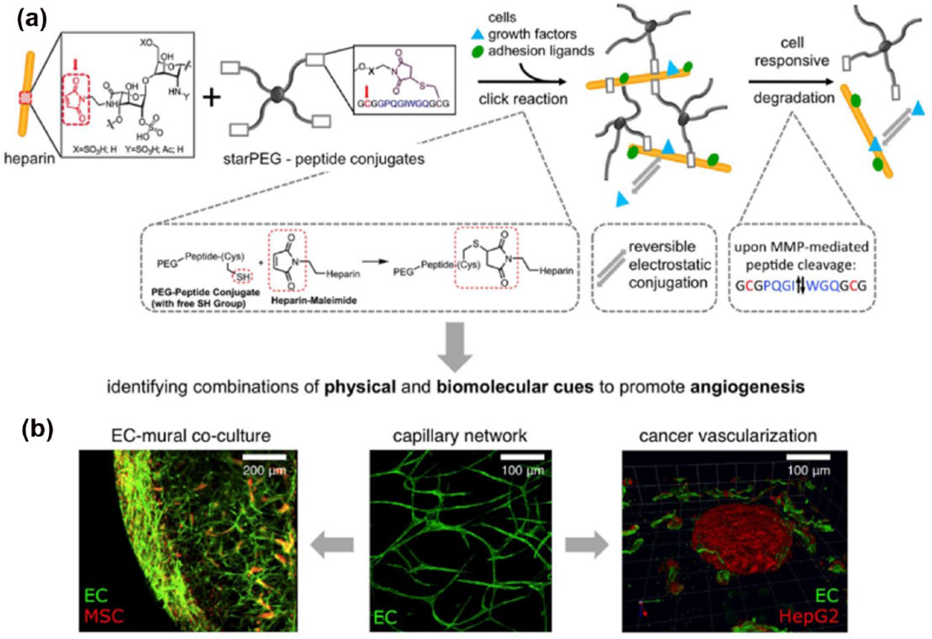
StarPEG-heparin hydrogels as an extracellular milieu to study heterotypic cell–cell interactions during angiogenic events. (a) Maleimide-functionalized heparin units react with terminal thiol groups of starPEG-peptide conjugates during Michael addition reaction resulting in formation of a modular in situ hydrogel platform suitable for displaying multiple adhesion and degradation sites, growth factors, and cell encapsulation. (b) Hydrogels enable studying the role of the extracellular milieu and heterocellular cell–cell interactions during vascularization processes as demonstrated by exploring two distinct processes: vascular capillary stabilization by mural cells and tumor vascularization. (With permission from Chwalek et al. 66 )
Microfluidics
Microfluidics involves the manipulation of the behavior of fluids that flow in channels smaller than 1 mm in at least one dimension. Microfluidic systems offer several advantages, including decreased sample volume, fewer cells, shorter reaction time, and the ability to perform many experiments in parallel.72,73 Microfluidic devices are well suited for biological experiments at the cellular level because microchannels within these devices can mimic the physical size found in vivo. 74 Because of the small size of the microchannels, microfluidic devices allow adequate oxygenation and fast nutrition diffusion. 75 Such an environment helps cells to maintain their local microenvironment better than macroscale cell culture flasks and are subjected to less stress because of the more in vivo-like surroundings, which can lead to more accurate observation on cellular behavior in response to external stimuli. 76 Microfluidic technology allows 3D cell culture with a complex microscale structure and well-controlled conditions to mimic the ECM. Furthermore, microfluidics has great flexibility of design to tailor individual cells, single cell sorting, and co-culture systems. 77
Cell culture using microfluidics has been termed organ-on-a-chip, as it contains a continuous perfusion system populated by a cell arrangement that mimics organ-level physiology. 78 This system allows the study of the organ-level functionality of cells rather than the whole organ. In its simplest from, cells in a microfluidic monoculture system are cultured in a single microfluidic chamber as a monolayer. 79 In a co-culture system or a multiple cell type culture system, two or more microfluidic chambers share a microporous or nanoporous membrane with different cell types on the opposite sides (Figure 4). 80 For example, HUVECs and a breast cancer cell line can be co-cultured in microfluidic chamber as a cancer extravasation model. Similar to the in vivo environment of cancer, the breast cancer cells inside the lumen extravasate through the endothelial barrier. 81

(a) Design of microfluidic co-culture devices, (b) a picture of the microfluidic devices, (c) schematic illustration of co-culture chamber A, (d) a sectional view of the microchannel arrays in C, and (e) an optical image of positions B, C, and D. Scale bar represents 600 μm. (With permission from Mi et al. 80 )
Droplet-based microfluidics are compartmentalized for cell encapsulation using aqueous microdroplets in a water-in-oil (w/o) emulsion, because of their fast, efficient, and high throughput capability compared with conventional cell-based screening process. 82 In this process, a cell-containing medium is injected into microfluidic devices using a syringe pump and oil is also injected vertically to make cell-embedded microdroplets. Using this simplified process, interleukin (IL)-3 secreting MBA2 human leukemia cells and heterogeneous umbilical cord blood (UCB) cells were co-encapsulated in an agarose microgel that allowed the observation of the sub-population responsiveness to the paracrine effect of IL-3. From this co-culture system, the rescue effect of localized IL-3 was determined for hematopoietic cell types in the various sub-populations. 83
Microfluidics are adaptable to custom microenvironments, high-throughput drug screening, cell encapsulation, and co-culture systems. Despite their benefits for tissue engineering, microfluidics have several shortcomings, such as changes in reagent concentration because of diffusion outside of the microfluidic channel. However, microfluidics still shows promise to play an important role in the improvement of personalized medicine.
Patterning
During the construction of new biological tissues, surfaces undergo several developmental processes in living organisms to build elaborate cell patterns, based on intercellular surface tension. This significant surface tension of the biological framework remodels the cell-shape changes and the cell contacts. 84 In addition, pattern formation in a biological architecture organizes the different types of cells into a spatial arrangement. During morphogenesis of the retina epithelium, a hexagonal array arises on the ommatidium. Within the patterned surface, cells expressing different cadherins were spatially localized. 85 Thus, it is important to develop artificially patterned surfaces to investigate how cells cope with the surface during morphogenesis.
Over the past decade, a wide range of strategies for patterning polymers has been developed rapidly. The interest in polymer patterning has arisen from the design of existing natural and synthetic polymers and the ability of these patterned surfaces to acquire functionality that alters the behavior of cells and biomolecules. 86 Previously, patterned surfaces were applied in indirect co-culture systems, because of their convenience for separating different types of cells. For example, patterned elecrospun fiber mats were produced on a silver micropatterned glass surface. Co-culture of hepatocytes, fibroblasts, and ECs was established by stacking each patterned fibrous mat with distinct ridges, grooves, and different cell types. Due to the indirect connections between the cells on the patterned surface, hepatocytes showed higher hepatic activities that correlated with in vivo observed phenomena (Figure 5). 87 Other patterning techniques also permit the spatial arrangement of cells on patterned polymer surfaces. For instance, a polydimethylsiloxane (PDMS) stamp fabricated from a silicon wafer was pressed onto a HA-coated TiOH surface, and subsequently, a homogeneous micropattern of parallel HA stripes was constructed on the TiOH surface. The co-culture of SMC and ECs on this patterned surface better regulated SMC behavior and EC functionality compared with monoculture. 88 Various patterning technologies enable the use of diverse polymer-patterned surfaces for biochemical applications. Engineered patterned surfaces are applicable to biomolecule immobilization, 89 single cell patterning, 90 and co-culture systems. Furthermore, surface patterning can be used for the spatial arrangement of cells that require a specific cellular orientation and alignment, such as cardiomyocytes and neurons, to perform their particular functions.
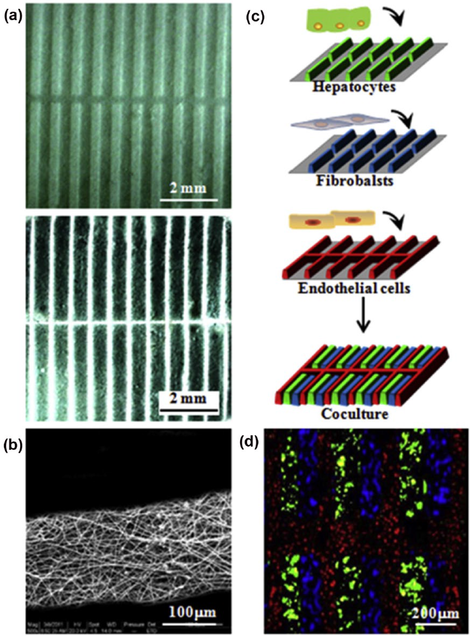
(a) SEM morphologies of patterned fibrous mats with ridge/groove widths of 200/300 and 100/400 μm. (b) Typical SEM morphologies of fibers in the ridges. (c) Schematic illustration of the micropatterned co-culture of hepatocytes with fibroblasts and ECs. (d) Merged CLSM image of immunofluorescent staining of collagen I secretion (blue) by fibroblasts, collagen IV (red) by ECs, and albumin (green) by hepatocytes after micropatterned co-culture for 5 days. (With permission from Liu et al. 87 )
Other strategies
For a long time, researchers have developed new techniques to overcome limitations and improve the properties of existing co-culture systems. Recent advances in the development of more efficient co-culture systems have included the modeling of an ECM-like multicellular environment, comprising 3D structures with tissue-specific morphology aimed at improving the outcome of tissue regeneration.
Layer-by-layer (LBL) fabrication for co-culture systems usually use cell-sheet engineering, which utilizes monolayer cells in a cell sheet and allows control of direct intercellular interactions by stacking different types of cell sheets. For example, EC co-cultured cardiac cell sheets are stacked and overlaid on an engineered vascular bed to promote angiogenesis for anastomoses with existing blood vessels. Co-cultured ECs within piled cell sheets successfully migrated into the vascular bed and linked with existing blood vessels in vitro; implantation of these cell sheets with blood vessel anastomoses enabled maintenance of vascular structure and beating for 2 weeks in vivo. 91 Similarly, a cellular LBL co-culture system of MSCs and chondrocytes layers using biodegradable, nanothin, highly porous (BNTHP) membranes enhanced the chondrogenic differentiation of MSCs. The BNTHP membrane provides 3D geometry for direct or indirect co-culture systems and enhances the cellular interactions than conventional bilayer co-culture systems. 92 This strategy provides more multicellular interactions, enabling the cell behavior that is more similar to that in the natural ECM compared with other approaches.
3D bioprinting technology is also an innovative approach to produce a biomimetic cellular and extracellular environment of tissues or organs. This method allows the distribution of different types of cells and biomaterials, termed as bioink, with high resolution to mimic the elaborate microarchitecture of tissues. For example, when human-induced pluripotent stem cells (hiPSCs) and chondrocytes were 3D bioprinted with a nanofibrillated cellulose (NFC) bioink, hiPSCs were directed into the chondrogenic lineage. In this co-culture system, the appearance of hyaline cartilaginous tissue increased at 5 weeks and the number of cells in the bioink also increased. 93 Furthermore, as patient-tailored cell therapy approaches become more common, 3D bioprinting technology, together with co-culture systems, promises to become a useful technology in tissue engineering and regenerative medicine fields.
Applications of co-culture systems
Co-culture systems can be used in biochemical applications for repair, regeneration, or mimicking of natural tissues. Most co-culture systems involve the cellular interaction of two or more types of cells in biomaterials that strongly influence cell behavior and the extracellular environment. In this section, the combination of co-culture systems with various biomaterials to regenerate several types of tissues is discussed.
Bone tissue regeneration
Bone defects resulting from fracture, trauma, or congenital disorders require bone grafts for the repair and regeneration of bone tissue. However, because bone grafts have significant limitations in successful regeneration, bone tissue engineering with co-culture of various cell types has been explored as an alternative. For example, co-culture of human-induced pluripotent stem cells derived mesenchymal stem cells (hiPSC-MSCs) and macrophages (hiPSC-macrophages) on hydroxyapatite-coated PLGA/PLA (HA-PLGA/PLA) scaffolds promoted in vitro and in vivo mature bone formation. 94 Similarly, when MSCs were co-cultured with ostocytes and osteoblasts in a transwell system, the synergetic relationship between MSCs and osteocytes induced greater alkaline phosphatase (ALP) activity and calcium deposition in MSCs. 95 While stem cell differentiation into osteogenic cell types has shown promising results in bone tissue regeneration, co-culture of bone cells and ECs has also been shown to induce bone regeneration.96,97 For instance, when human osteoprogenitors (HOPs) and HUVECs were co-encapsulated in RGD-grafted alginate microspheres, HOPs with HUVECs showed a higher VEGF expression level in vitro and promoted new bone formation and tissue mineralization in a bone defect site in vivo (Figure 6). 98 Co-culture of MSCs and HUVECs on a nanopatterned surface 99 and biodegradable beta-tricalcium phosphate (β-TCP) scaffolds 100 promoted vascularization and osteogenesis. Because bone is a calcified and peripherally vascularized tissue consisting of various cell types, including osteogenic cells and endothelial cells, co-culture dramatically enhances bone regeneration. Various studies of bone regeneration using co-culture systems have shown meaningful results, suggesting a synergistic effect in osteogenesis and angiogenesis. However, these studies have not demonstrated better bone regeneration or improved physical properties of new bone tissue compared with conventional bone grafts. For successful bone regeneration, co-culture employing appropriate biomaterials should (1) guide the reparative growth of the natural bone, (2) encourage undifferentiated cells to differentiate into active osteogenic cells, (3) promote mineralization of bone tissue, and (4) induce neovascularization. Furthermore, use of a scaffold with physical properties similar to those of the natural bone ECM is important to induce bone regeneration.

(a) Mineralization of immobilized cells after in vitro culture. Human osteoprogenitors (HOPs) or co-cultures of HOPs with human umbilical vein endothelial cells (HUVECs) were immobilized within RGD-alginate microspheres, embedded in paraffin and stained with von Kossa after 1 and 22 days of in vitro dynamic culture conditions. Mineralization of HOPs seemed to slightly decrease after 22 days of culture. However, the co-immobilization of HOPs with HUVECs promoted increased mineralization even after 22 days of culture. (b) X-ray micro-computer tomography (μCT) images of bone regeneration. Bone defects (0.9 mm of diameter) were performed in the femoral metaphysis of nude mice. RGD-alginate microspheres containing no cells, only human osteoprogenitors (HOPs) or HOPs co-immobilized with HUVECs were implanted and bone regeneration was followed by μCT after 3 and 6 weeks. Little or no mineralization was observed when alginate alone was implanted in the bone defect. When only HOPs were implanted, modest mineralization around and inside the microspheres was generated 3 and 6 weeks post-implantation, respectively. However, co-immobilization of HOPs and HUVECs led to an intense mineralization inside the microspheres already 3 weeks after implantation. Higher magnifications of the white squares are shown below (×2). (With permission from Grellier et al. 98 )
Cartilage tissue regeneration
Chondral and osteochondral lesions in weight-bearing joints caused by injury or genetic predisposition can lead to total cartilage destruction. 101 Currently, a wide range of techniques for cartilage tissue regeneration is used, including surgery, re-attachment, microfracture, osteoarticular transfer, grafts, and emerging gene therapy techniques. 102 Commonly, these approaches produce regeneration of fibrocartilage instead of hyaline cartilage that has better compressive, tensile, and frictional properties, which are necessary to bear impact at joints. Recently developed methods including stem cell stimulation, autologous chondrocyte implantation (ACI), and matrix-induced chondrocyte implantation (MACI) provide more promising solutions to restore fully functional cartilage. Co-culture systems also show promise for expanding the regenerative capacity of articular cartilage and to produce a long-term stable chondrogenic phenotype. Co-culturing MSCs with chondrocytes has the potential to catalyze articular cartilage tissue formation and chondrocyte phenotype maintenance. For example, bone marrow–derived MSCs and bovine chondrocyte were co-cultured in nonwoven fibrous PCL scaffolds directly and indirectly. It was found that MSCs provided juxtacrine and paracrine signaling for production of the articular cartilage ECM such as collagen and glycosaminoglycans (GAGs). 103 Similarly, another group co-cultured these cells in bilayered agarose gels separately. A single layer of chondrocytes showed low levels of collagen synthesis, but cells in bilayered scaffolds showed increased levels of collagen synthesis, with no mineralization in the chondrogenic medium for 49 days in vitro. After subcutaneous dorsal implantation of cell-laden scaffolds into mice, production of type II collagen and GAGs was enhanced in bilayered scaffolds and levels of type I collagen and X decreased. However, the opposite results were observed in single-layer MSCs. 104 Other types of stem cells also have the capacity to induce the formation of articular chondrocytes. Adipose-derived mesenchymal stem cells (ADSCs) are an alternative cell source to reduce the number of articular chondrocytes in ACI or MACI. 105 For example, different ratios of human ADSCs and chondrocytes were co-cultured in a fibrin matrix or a type I collagen scaffold. Using histochemical staining, proteoglycan expression was observed in 5% of chondrocytes and in 95% of ADSCs in the co-cultured groups. 106 Similarly, these two cell types were mono- or co-encapsulated in poly(ethylene glycol diacrylate) (PEGDA)—chondroitin sulfate methacrylate (CSMA) scaffolds. The mono-encapsulating scaffolds were separately cultured with conditioned medium from the other cell type or bilayered with an acellular matrix at the midpoint of the two scaffolds to induce a co-culture paracrine effect. To observe direct interactions between the two types of cells, mixed cells were co-cultured in the same scaffolds at different ratios. In the medium-changing and bilayered co-culture groups, ADSCs showed a slight increase in expression of chondrogenic markers and a decrease in expession of fibroblastic markers. However, in the mixed co-culture group, ADSCs showed remarkable enhancement of collagen type 2 levels, which is a hyaline cartilage marker, resulting from cell–cell interactions. Furthermore, replacement of 90% of chondrocytes with ADSCs showed promising results in expressing the chondrogenic phenotype and inducing cartilage regeneration (Figure 7). 107 Articular cartilage is an avascular tissue that contains a single cell type, and chondrocytes are physiologically non-self-restoring. 108 Thus, tissue engineering approaches for articular cartilage regeneration should be accompanied by a biocompatible scaffold and a co-culture system to improve the functionality of chondrocytes. Furthermore, the addition of growth factors such as transforming growth factor beta-1 (TGF-β1), which enhances chondrogenic differentiation in cell culture system, is a promising method for entire cartilage regeneration.
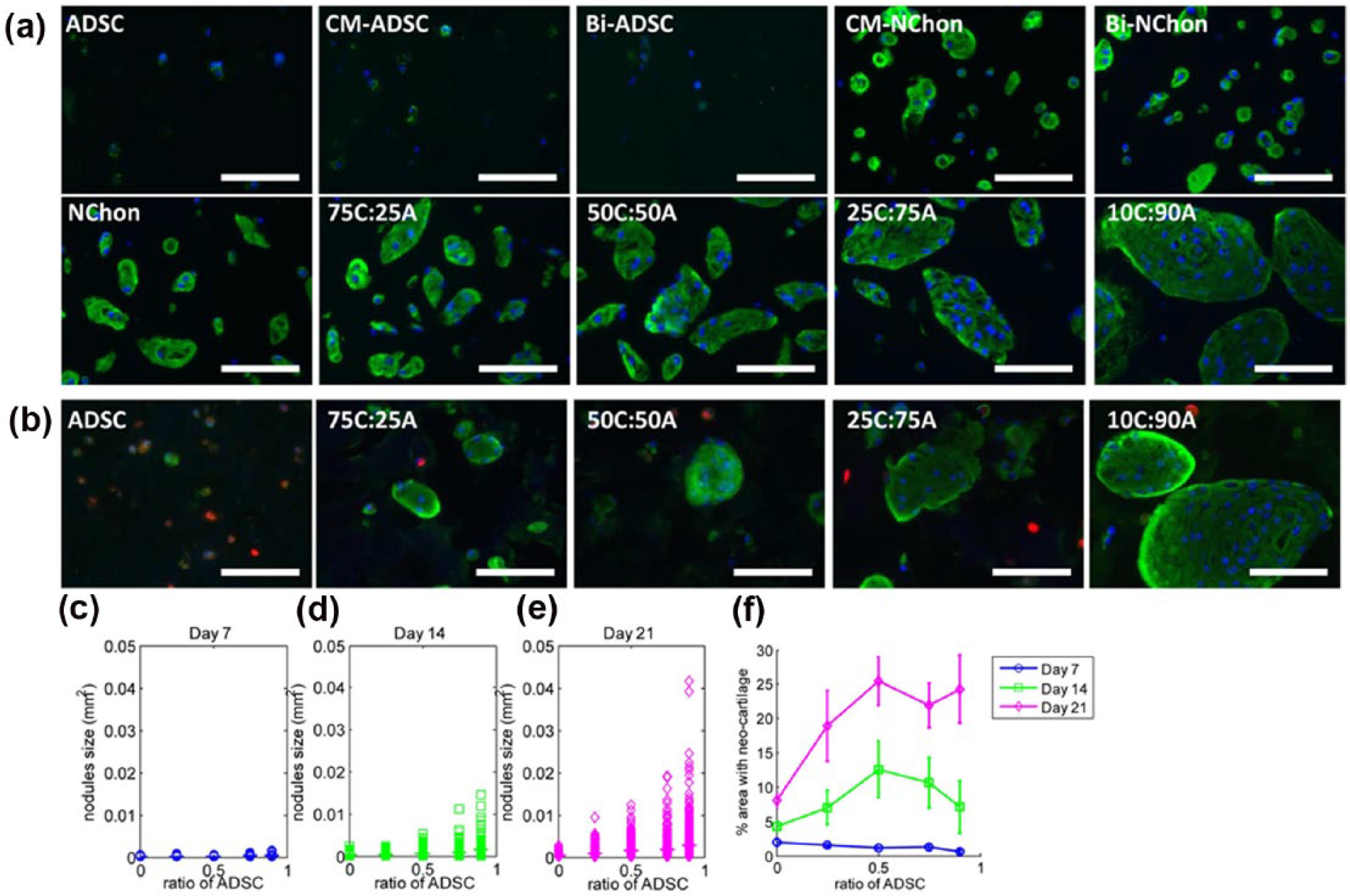
Morphology of newly formed cartilage nodules and cell distribution over 21 days. (a) Newly formed cartilage nodules were visualized by immunostaining of type II collagen at Day 21. Scale bars represent 100 μm. (b) To determine the distribution of the two cell types in the mixed cell culture, ADSCs were membrane-labeled (red) prior to encapsulation in the hydrogels; co-staining with type II collagen (green) revealed that ADSCs always resided outside the neocartilage nodules. Scale bars represent 100 μm. (c–f) Quantification of type II collagen immunostaining images, including cartilage nodule size at different ratios of ADSCs at (c) Day 7, (d) Day 14, and (e) Day 21 (horizontal bars indicate average size), as well as the (f) total percentage of area occupied by cartilage nodules at different cell ratios at Days 7, 14, and 21. Both the cartilage nodule size and the total area of hydrogel being replaced by cartilage nodules increased with an increase in ADSC ratio in the mixed co-culture. (With permission from Lai et al. 107 )
Vascular tissue regeneration
Blood vessels play important roles in the supply of oxygen and nutrients to maintain cell viability, and also enhance matrix production surrounding most natural tissues, except avascular tissues such as cornea or cartilage.7,109 For complete reconstruction of natural tissue, blood vessels should be regenerated during the tissue regeneration process. Recent studies have focused on blood-vessel reconstruction in tissue regeneration and wound-healing models using co-culture of vascular-forming ECs and other cell types. For example, ECs directly co-cultured with myoblasts or triple-cultured with fibroblasts on PLA and PLGA composite porous scaffolds fabricated using a salt leaching process showed notable enhancement of blood vessel and muscle construction in vivo. The pre-existing blood vessels provided physical, as well as biochemical, support for angiogenesis. 110 Similar systems of co-culture of vascular ECs with SMCs also showed angiogenic properties, including vasculature structure formation, and improved EC survival and proliferation.111,112 Stem cells are also involved in vascular tissue formation. For general tissue engineering, stem cells are an alternative source of organotypic cells. Pluripotent embryonic stem cells and adult stem cells differentiate into tissue-specific cell types dependent on various physical and chemical stimuli.113–116 In addition, stem cells secrete large amounts of biomolecules to stimulate biological activity of other cells in vivo. 46 For example, MSCs produce growth factors that are essential for ECs, fibroblasts, immune-related cells, and tissue progenitor cells in situ. Leveraging these properties, several studies have conducted co-culture of MSCs with ECs for vascular tissue formation. For instance, gelatin methacrylate (GelMA) hydrogel–containing MSCs and ECs support vascular morphogenesis and formation of capillary-like networks in vitro (Figure 8). 117 In addition to MSCs, fibroblasts, which are involved in vascular tissue formation, can also facilitate angiogenesis of ECs. Therefore, co-culture of ECs and fibroblasts has the potential to enhance vascularization and angiogenesis. For example, HUVECs were indirectly co-cultured with fibroblasts using conditioned media containing calcium silicate (CS) extracts used for fibroblast culture. Co-cultured HUVECs showed increased expression of VEGF and gap junction protein and capillary network formation. 118 Thus, co-culture of ECs with other cell types is more beneficial to construct vascular tissues compared with monoculture. Angiogenic factors secreted from stem cells or other organotypic cells including VEGF and angiopoietin regulate vascular network formation of ECs. Thus, co-culturing ECs with stimulating cells can enhance the formation of mature vascular and capillary networks. Angiocrine factors produced by capillary-forming ECs maintain organ homeostasis and determine the capacity for organ regeneration.

Stabilization of the ECFC-lined capillaries by perivascular cells. Constructs containing both DsRed-ECFCs and MSCs were cultured for 7 days in GelMA hydrogels with different methacrylation degrees. The ability of the MSCs to differentiate into perivascular cells was analyzed by confocal microscopy after immunofluorescence staining with antibodies against smooth muscle markers. (a, c) Representative confocal images showing the spatial distribution of the DsRed-ECFC-lined capillaries surrounded by αSMA-expressing MSCs. Higher magnification images depicting details of a capillary (top) and a cross-sectional image taken in the direction of the white arrows (bottom) are shown to the right of these images in panels (a) and (c). (b, d) Representative confocal images showing the spatial distribution of both DsRed-ECFC-lined capillaries surrounded by sm-MHC-expressing MSCs. A representative video of a rotating 3D reconstruction of confocal images showing MSC-wrapped capillaries is available. (With permission from Chen et al. 117 )
Liver tissue regeneration
The liver is an important organ that functions in metabolism, blood circulation, detoxification, secretion of proteins, and biosynthesis in the body. Damage of the hepatocytes that constitute the liver can trigger innate immune responses. 119 Several studies have shown the effect of co-culture of hepatocyte with stellate cells to enhance hepatic function. To form engineered liver tissue, hepatocytes and fibroblasts are indirectly co-cultured on double-layered PCL fibrous scaffolds incorporated with PEG hydrogel micropatterns. Because the hydrophilic hydrogel restricts the proliferation of hepatocytes toward the scaffold exterior, hepatocytes form spheroid aggregates and same-size microarrays. Co-culture with fibroblasts enhanced albumin secretion compared with monoculture of hepatocytes (Figure 9). 120 Similarly, hepatocyte spheroids can be cultured on ECM-coated micropatterned PDMS, with fibroblasts cultured in the intervening space around the cell spheroids. This system mimics the actual morphology of liver tissue over multiple weeks and demonstrates the hepatotropic life cycle. 121 In addition to hepatocytes–fibroblasts co-culture, hepatocytes–ECs co-culture has been used to enhance the hepatic function. For example, direct and indirect co-culture of hepatocytes with ECs between type I collagen hydrogel layers promoted better hepatic function of hepatocytes compared with monoculture. 122 The triple culture of hepatocytes with HUVECs and fibroblasts on micropatterned fibrous scaffolds induced the successful construction of functional hepatic tissues and capillary-like structures for drug metabolism. 123 As the proportion of ECM cells in the co-culture increased, cell–cell and cell–ECM interactions increased, resulting in better organization of the reconstructed liver tissue. Co-culture of hepatocytes with macrophages has been used to investigate the hepatotoxic potential of drugs and immune-mediated pathogenesis. For example, hepatocytes and Kupffer cells (KCs) were co-cultured directly on collagen-coated multi-well plates. KCs modulated hepatocyte function and elevated the intracellular Ca2+ level of hepatocytes in an investigation of the cytoplasmic response to drug-induced liver injury. 124 In addition, co-culture of human hepatocytes with macrophages upregulated the immune response following liver cell infection with a zoonotic tularemia-causing bacterium. 125 Hepatocytes are a major component of liver tissue, and improvement of hepatocyte function can be an indicator of the efficacy of liver disease treatment. In particular, hepatocytes in aggregated spheroid form present a versatile and promising approach to investigate liver diseases and drug screening. 126 In addition, incorporating co-culture of hepatocytes with hepatic stellate cells in a microscale culture system 127 shows promise for liver tissue regeneration.

Co-culture of HepG2 and fibroblasts using double-layered fibrous scaffolds incorporated with hydrogel micropatterns. (a) Schematic diagram of the cross-sectional view of the double-layered fibrous scaffolds used for the co-culture studies. (b) Cumulative amount of albumin secreted from HepG2 cells. (c) Fluorescence image (from Cell Tracker Orange) and (d) SEM image of HepG2 spheroids formed within 200 × 200 μm2 patterns after 8 days of culture. (e) Live/dead fluorescence viability assay of HepG2 spheroids after 8 days of culture. (f) Fluorescence image of fibroblasts stained with Cell Tracker Green after 8 days of culture. Scale bars in the figures are 200 μm. (With permission from Lee et al. 120 )
Challenges of co-culture system
This review has discussed the types of co-culture systems and various studies that have used the electrospinning process, hydrogel, microfluidics, and patterning, which are remarkable strategies for the co-culture systems. In contrast to conventional monoculture system, co-culture systems using these approaches present alternative platforms for better tissue regeneration. However, these methodologies for the co-culture system do not mimic the natural ECM precisely. The four methods mentioned above implement the co-culture system by providing a 3D environment; however, it is difficult to imitate the structure or appearance of the actual ECM. Therefore, there remains a need to develop technologies to construct well-organized scaffolds using biocompatible substances with high mechanical strength, porosity, biodegradability, and biocompatibility. These challenges for elaborate scaffolds can be improved by surface modification or synthesis of advanced substances for scaffold fabrication. Recently, 3D printing technologies have been developed to generate ECM-like scaffolds with a high degree of complexity and precision. For a hybrid approach, decellularized ECM can be used as well as natural and synthetic polymers to make ECM-like scaffolds using 3D printing technologies. The availability of functional and well-organized scaffolds for tissue engineering may hold the key for organ transplantation failure and dysfunction caused by diseases or damage.
Summary
Co-culture systems have been shown to improve the functionality of various cells in tissue formation. As a strategy to mimic the structure of natural tissue ECM, the co-culture system more closely mimics cell–cell or cell–ECM interactions compared with monotypic culture for tissue engineering. 128 A number of studies have demonstrated the benefits of co-culture with various cell types. Stimulation by direct or indirect co-culture elicits better functionality of target cells. In addition, the type of biomaterial and the structure of scaffold affect the behavior of cells; therefore, cellular scaffolds can be designed to provide an appropriate 3D environment for co-culture. For these reasons, many studies are currently underway to develop co-culture systems with 3D cellular scaffolds for tissue regeneration. Furthermore, exogenous biomolecular activation of cells in co-culture through scaffold or culture medium may also be a promising source to trigger intracellular responses to enhance cell functionality and tissue regeneration.
Footnotes
Declaration of conflicting interests
The author(s) declared no potential conflicts of interest with respect to the research, authorship, and/or publication of this article.
Funding
This work was supported by the National Research Foundation of Korea (NRF) grant funded by the Korean government (Ministry of Science, ICT & Future Planning, MSIP; 2016M3A9B4919711, 2015R1D1A1A01060444, and 2009-0093823 “Priority Research Centers Programs”).