
Abstract
The development of biomaterial surfaces possessing the topographical cues that can promote mesenchymal stem cell recruitment and, in particular, those capable of subsequently directing osteogenic differentiation is of increasing importance for the advancement of tissue engineering. While it is accepted that it is the interaction with specific nanoscale topography that induces mesenchymal stem cell differentiation, the potential for an attendant bioactive chemistry working in tandem with such nanoscale features to enhance this effect has not been considered to any great extent. This article presents a study of mesenchymal stem cell response to conformal bioactive calcium phosphate thin films sputter deposited onto a polycrystalline titanium nanostructured surface with proven capability to directly induce osteogenic differentiation in human bone marrow–derived mesenchymal stem cells. The sputter deposited surfaces supported high levels of human bone marrow–derived mesenchymal stem cell adherence and proliferation, as determined by DNA quantification. Furthermore, they were also found to be capable of directly promoting significant levels of osteogenic differentiation. Specifically, alkaline phosphatase activity, gene expression and immunocytochemical localisation of key osteogenic markers revealed that the nanostructured titanium surfaces and the bioactive calcium phosphate coatings could direct the differentiation towards an osteogenic lineage. Moreover, the addition of the calcium phosphate chemistry to the topographical profile of the titanium was found to induce increased human bone marrow–derived mesenchymal stem cell differentiation compared to that observed for either the titanium or calcium phosphate coating without an underlying nanostructure. Hence, the results presented here highlight that a clear benefit can be achieved from a surface engineering strategy that combines a defined surface topography with an attendant, conformal bioactive chemistry to enhance the direct osteogenic differentiation of human bone marrow–derived mesenchymal stem cells.
Keywords
Introduction
It is well established that both the chemical and physical properties of a biomaterial surface have a significant effect on cell behaviour thereon, and that by altering specific properties, different cell responses can be induced and controlled. Mesenchymal stem cells (MSCs), which are an increasingly popular cell choice for a range of tissue engineering and regenerative medicine applications, have been shown to be particularly sensitive to the surface cues that they encounter. In particular, it has been shown that subtle changes in topography, chemistry and stiffness can have a significant effect in determining MSC fate1–3 and, as such, present an attractive way by which to manipulate their behaviour for given applications. In the case of bone tissue engineering, there is currently great interest in the development of clinically relevant biomaterials that can directly induce the osteogenic differentiation of MSCs to deliver improvements in orthopaedic therapies.
A number of nanoscale topographies have been shown to be as effective as biochemical media supplements (dexamethasone, etc.) in directing MSCs to an osteogenic lineage.4–7 Additionally, various types of surface chemistry are capable of determining stem cell fate.1,8,9 Hence, an approach that brings together the necessary nanoscale topography with a bioactive surface chemistry could present a superior strategy by which to induce the osteogenic differentiation of stem cells. However, a significant challenge is to retain the topographical features at the same time as providing the necessary chemical stimulus.
Radio frequency (RF) magnetron sputtering is a well-established thin film deposition process whereby high-energy ions created in gaseous plasma bombard a solid target causing atoms and molecular fragments of the material to be ejected and subsequently amalgamate as a thin film on a chosen substrate surface. Sputtering has many advantages over alternative coating techniques, including the provision of uniform coatings, controllable deposition rates and the provision of highly pure layers of a range of materials from metals to ceramics.10,11 The technique has been shown to be particularly useful for the deposition of calcium phosphate (CaP) thin films from hydroxyapatite (HA) and related target materials. Importantly, the process provides good adhesion between the substrate and the coating without the need for surface roughening.12,13 Such sputter deposited CaP thin films have been shown to be bioactive and thereby capable of promoting attachment, proliferation and differentiation of osteoblasts, leading in specific circumstances to mineralisation of attendant extracellular matrix (ECM).10,14–16
In general, as-deposited CaP sputtered thin films have an amorphous structure that conforms to the topography of the substrate onto which they are coated. 16 Therefore, a topographical profile can be provided to a sputter deposited CaP coating by the introduction of underlying substrate topography with predetermined features. A convenient means by which this can be achieved is via the fabrication of a sputter deposited polycrystalline titanium interlayer comprising controllable homogeneous topography in the sub-micro to nanoscale range. This provides a novel combination of bioactive CaP chemistry and nanoscale topography which has the potential to further enhance the nanoscale surface cues known to directly induce osteogenic differentiation in human bone marrow–derived MSCs (hMSCs). In this regard, the titanium and CaP sputter deposited thin films of interest here have been recently shown to promote early stage events in hMSCs that are indicative of commitment to the osteogenic lineage. 17
The work presented here reports the capacity of a combination of CaP bioactivity coupled with nanoscale topography (as supplied by a sputter deposited titanium interlayer) to induce direct osteogenic differentiation in hMSCs without the need for media supplementation or biochemical factors.
Methods and materials
Sputter deposited surfaces
Three types of surface were fabricated by RF magnetron sputter deposition denoted as follows: a titanium thin film on a glass substrate (Ti), a CaP thin film on a glass substrate (CaP) and a CaP thin film on a glass substrate with a Ti interlayer (TiCaP). All of the surfaces were produced using a custom-designed RF magnetron sputter deposition system (Kurt J. Lesker Co. Ltd, UK) comprising a vacuum chamber equipped with three Torus 3M sputtering sources operating at a frequency of 13.65 MHz and attached to individual impedance base matching networks. Samples to be coated are attached to a rotating substrate holder mounted 10 cm vertically above the sources. This system and the process employed to produce the titanium and CaP surfaces have been described in detail previously. 17 Briefly, prior to sputtering, the chamber was evacuated to a base pressure of 5 × 10⁻⁶ mbar. The plasma source gas utilised for all depositions was argon (99.999%; BOC Gases, UK). Titanium layers were deposited from 76 mm diameter, 99.995% pure titanium targets (Kurt J. Lesker Co. Ltd) at 200 W for 2 h at a plasma discharge pressure of 5.0 × 10⁻³ mbar with an argon gas flow rate of 15 sccm. CaP layers were deposited from dry pressed HA (Plasma Biotal, UK) targets at 150 W for 10 h at a discharge pressure of 5.0 × 10⁻³ mbar and an argon gas flow rate of 15 sccm. Prior to deposition, the HA targets were subject to a break in procedure comprising a power ramp-up of 5 W/min in order to avoid thermal damage such as cracking.
The main chemical and topographical characteristics of each of the surfaces of interest here have been reported previously 17 and are summarised in Table 1. The Ti coating comprises a TiO2 chemistry with a primarily rutile crystallographic structure and possesses a nanoscale topographical profile with features in the form of island-shaped grains of 138 ± 15 nm in width and 47 ± 13 nm in height that are homogeneous throughout the surface (as determined by atomic force microscope (AFM) analysis). The CaP thin film coating deposited on a flat glass substrate is highly oriented with a Ca:P ratio of 1.83. Topographically, this CaP coating is relatively smooth with no significant surface features evident. In the case of the CaP deposited onto the sputtered polycrystalline Ti interlayer (TiCaP), the resulting Ca:P ratio of 1.75 is similar to that of the CaP coating on glass. However, the topography of the surface is similar to that of the polycrystalline Ti surface, although the average grain size is larger and they have a greater variability in size.
Summary of the main characteristics of each of the sputter deposited surfaces.
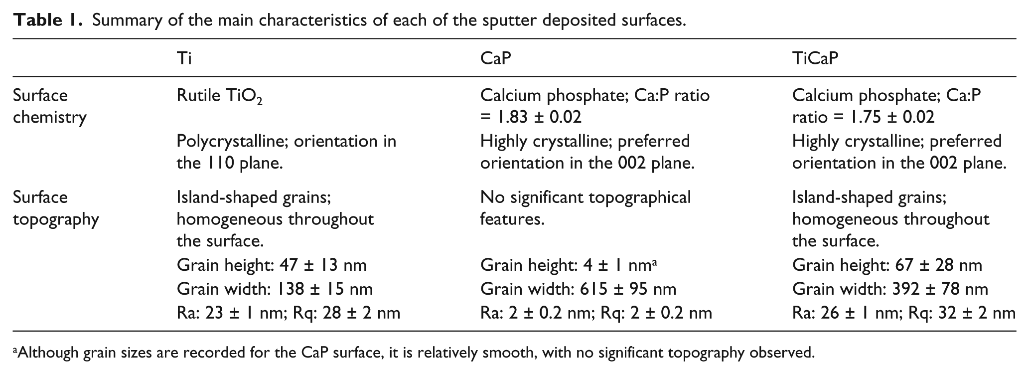
Although grain sizes are recorded for the CaP surface, it is relatively smooth, with no significant topography observed.
MSCs
Commercially obtained, hMSCs (Lonza, UK) derived from the normal bone marrow of a healthy donor were used throughout this study. hMSCs were cultured in Dulbecco’s modified Eagle’s medium (DMEM) high glucose (Invitrogen, UK) supplemented with 10% foetal bovine serum and 0.1% penicillin/streptomycin (Invitrogen). All of the studies were undertaken with hMSCs at passage 5 or earlier. To determine the osteogenic potential of the Ti, CaP and TiCaP sputter deposited thin films, hMSCs were cultured directly on each of the surfaces with a tissue culture plastic as the control, over a period of 28 days. hMSCs were seeded onto the control and sputter deposited surfaces at 4000 cells/cm2. Additionally, as a positive control for the osteogenic differentiation of this source of hMSCs, they were cultured on tissue culture plastic in normal hMSC media supplemented with 10 nM dexamethasone, 50 µM ascorbic acid-2-phosphate and 10 mM β-glycerophosphate (Sigma–Aldrich, UK).
DNA quantification
The DNA content of hMSCs cultured in contact with the relevant surfaces was measured using a PicoGreen® dsDNA quantification assay (Invitrogen) after 1 and 7 days, as per the manufacturer’s instructions. A standard curve was prepared using bacteriophage lambda DNA. Fluorescence was measured at an excitation wavelength of 480 nm and an emission wavelength of 520 nm on a TECAN GENios FL (TECAN, Switzerland). The DNA concentration in each sample was then calculated based on the data from the standard calibration curve.
Alkaline phosphatase activity
Alkaline phosphatase (ALP) activity of the hMSCs cultured on the various substrates was determined at 5, 10 and 15 days in culture, using the SensoLyte® pNPP Alkaline Phosphatase Assay Kit (AnaSpec, USA). The concentration of ALP in each sample was determined from a standard calibration curve (generated with ALP derived from calf intestine) and normalised to the quantity of total protein present which was determined using a bicinchoninic acid (BCA) Protein Assay Kit (Pierce, USA).
Gene expression
The expression of a number of key genes associated with osteogenic differentiation of hMSCs was analysed by quantitative real-time polymerase chain reaction (qPCR) experiments, at days 7, 14, 21 and 28. RNA was extracted from hMSCs using the TriPure Isolation Reagent (Roche Diagnostics, UK), as per the manufacturer’s instructions. The quality of extracted RNA was assessed by analysis on a Bioanalyzer (Agilent, UK), and each sample had an RNA integrity number (RIN) of at least 7.9. First strand complementary DNA (cDNA) was synthesised from the extracted RNA using the Transcriptor First Strand cDNA Synthesis Kit (Roche Diagnostics, Germany) with the use of random hexamer primers, according to the manufacturer’s instructions.
With the exception of osteopontin, RealTime ready assays (Roche Diagnostics, Germany) were used for the qPCR experiments; SYBR Green I (Roche Diagnostics, Germany) based reactions were performed for osteopontin determination. Details of the primers used for the qPCR reactions are listed in Table 2. Thermal cycling conditions for the RealTime ready assays were as follows: 95°C for 10 min then 45 cycles at 95°C for 10 s, 60°C for 30 s and 72°C for 1 s, followed by 40°C for 30 s for the final cooling stage; and for SYBR Green I reactions: 95°C for 5 min then 45 cycles at 95°C for 10 s, 60°C for 10 s and 72°C for 10 s, and then a ramp to 97°C at 0.1°C/s for melt curve analysis followed by 40°C for 10 s for the final cooling stage. Analysis was performed in triplicate using a 96-well Roche LightCycler® 480 instrument (Roche Diagnostics, Germany). Pre-confluent, passage 4 hMSCs were used as a positive calibrator, and messenger RNA (mRNA) expression of target genes was normalised to reference gene expression and then calculated as fold change using the comparative CT (2−ΔΔCT) method.
Primer sequences used in qPCR experiments.

qPCR: quantitative real-time polymerase chain reaction.
Immunocytochemistry
At day 28, cells were fixed and permeabilised in 4% paraformaldehyde (PFA) + 0.1% Triton X-100 for 20 min at room temperature and then incubated with 5% normal goat serum (NGS) (Sigma–Aldrich) for 15 min. Following this, the cells were then incubated with one of the following primary antibodies for 1 h at room temperature: 3 µg/mL polyclonal anti-osteocalcin raised in rabbit, 3 µg/mL polyclonal anti-osteopontin raised in rabbit or 3 µg/mL polyclonal anti-osteonectin raised in rabbit. Cells were then incubated with 10 µg/mL goat anti-rabbit IgG (Invitrogen) secondary antibody conjugated to Alexa Fluor® 488 for 1 h at room temperature. The cells were subsequently counterstained with 600 nM 4′,6-diamidino-2-phenylindole (DAPI) (Invitrogen) for 2 min at room temperature before 10 µL of mounting media was added to each sample and then sealed with a glass cover slip and clear varnish. Fluorescent images were acquired using an LSM 5 Pascal Confocal Laser Scanning Microscope (CLSM) (Carl Zeiss, Germany).
Results
hMSC viability
By the 7-day culture point, all three sputter deposited surfaces were found to support significant levels of hMSC viability, with the level of viable cells in all cases greater (p < 0.001) than that for the control (Figure 1). Of the sputter deposited surfaces, the highest rate of proliferation was observed on the (polycrystalline) Ti surface over the culture period, whereas the TiCaP surface engendered the lowest level compared to both the Ti and CaP surfaces.

(a) DNA quantification of hMSCs cultured on the different sputter deposited surfaces over 7 days. The difference between each value is statistically significant (p < 0.05) unless otherwise indicated by NS (not significant). (b) CLSM images of hMSCs cultured on control, Ti, CaP and TiCaP surfaces for 7 days. The cytoskeleton (actin) and nuclei are immunofluorescently labelled with Alexa-Flour® phalloidin-488 (green) and DAPI (blue), respectively (scale bar = 50 µm). An hMSC monolayer formed on each of the surfaces, with the Ti surface supporting the highest level of viability.
Protein and ALP activity
Protein production, an increase in which can be indicative of matrix deposition, 18 was measured in hMSCs cultured on the sputter deposited surfaces across 15 days of culture (Figure 2(a)). Consistent with the DNA quantification data, a higher amount of protein was measured on the Ti surface after 5 and 10 days in culture, as compared to that on the CaP and TiCaP surfaces (Figure 2(a)). However, by day 15, the protein content had increased significantly on each of the sputter deposited surfaces to values in all cases that were similar to the levels for the osteogenic control (hMSCs cultured with 10 nM dexamethasone, 50 µM ascorbic acid-2-phosphate and 10 mM β-glycerophosphate).

(a) BCA whole cell protein estimation of hMSCs cultured on the various surfaces over 15 days. Protein content was increased significantly on each of the sputter deposited surfaces compared to the control. (b) ALP activity of hMSCs cultured on the various surfaces over 15 days with data normalised to total protein concentration. ALP activity increased on all sputter deposited surfaces; however, Ti induced the greatest level compared to either CaP or TiCaP.
ALP activity increased on each of the sputter deposited surfaces by day 15, as compared to that for the normal control (p < 0.001) (Figure 2(b)). The CaP and TiCaP surfaces induced similar levels of ALP by the end of the culture period; however, the Ti surface induced a higher level than both of these samples at day 10. Contrary to the descending pattern of ALP activity observed for the osteogenic control, the hMSCs cultured on the three sputter deposited surfaces exhibited a trend of increasing ALP activity throughout the 15-day culture period.
Gene expression
The expression of several genes that are integral to the process of osteogenic differentiation was analysed by qPCR for the hMSCs cultured on the sputter deposited surfaces over a period of 28 days, at 7-day intervals. The Ti and CaP surfaces both demonstrated patterns of increasing COL1A1 expression over the culture period with similar levels of maximal expression observed by day 28 (Figure 3). The TiCaP surface, however, induced a considerable increase in COL1A1 expression from the hMSCs after day 21 of the culture period, with values that were significantly greater (p < 0.001) than either the Ti or CaP surface and comparable to that of the osteogenic control.

mRNA expression of COL1A1 in hMSCs cultured on control and sputter deposited surfaces over 28 days. The TiCaP surface induced the greatest increase in COL1A1 expression compared to either the Ti or CaP surface.
The pattern of expression of ALP mRNA observed in the hMSCs cultured on the sputter deposited surfaces (Figure 4) varies throughout the culture period. This is in contrast to the osteogenic control which demonstrates maximal levels in the early period followed by a steady decrease thereafter. ALP expression in the hMSCs cultured on the sputter deposited surfaces peaks at day 14, and while this then decreases on the CaP surface, this level is maintained on the Ti and TiCaP surfaces throughout the duration of the culture period.

mRNA expression of ALP in hMSCs cultured on control and sputter deposited surfaces over 28 days. During the culture period, each of the surfaces induced ALP expression at levels greater than that for the normal control.
Expression of osteopontin increased significantly by day 28 in hMSCs cultured on the various sputter deposited surfaces (Figure 5) with the peak expression levels being significantly higher (p < 0.001) than that for the normal control. Of the three sputter deposited surfaces, the TiCaP samples induced the highest level of peak osteopontin expression, although the difference between the TiCaP and the CaP surfaces was not significantly different.

mRNA expression of osteopontin in hMSCs cultured on control and sputter deposited surfaces over 28 days. Osteopontin was up-regulated late in the culture period in hMSCs on each of the sputter deposited surfaces, with a greater increase observed for TiCaP compared to Ti.
The CaP and TiCaP surfaces induced osteocalcin expression patterns similar to that for the osteogenic control, with the highest levels observed at day 21 (Figure 6). Again, all of the sputter deposited surfaces induced significantly higher (p < 0.001) levels of osteocalcin expression than the normal control, with the CaP and TiCaP surfaces inducing higher peak expression levels compared to that for the Ti surface.

mRNA expression of osteocalcin in hMSCs cultured on control and sputter deposited surfaces over 28 days. Significant increases in osteocalcin expression were observed for each of the sputter deposited surfaces; however, CaP and TiCaP induced higher peak expression levels compared to that for Ti.
Immunocytochemistry
The immunocytochemical localisation of osteopontin, osteocalcin and osteonectin in hMSCs cultured on the sputter deposited surfaces for a period of up to 28 days has been used to highlight their relative effect on the later stages of osteogenic differentiation (Figure 7). A limited amount of osteocalcin is detected for hMSCs cultured on the Ti surface, with higher levels observed for those cultured on the CaP and particularly the TiCaP surface with areas observed throughout the monolayer that indicate the formation of discrete areas of mineralisation. Osteonectin immunolabeling can be observed for the hMSCs on the Ti surface, while larger and more frequent areas were detected on both the CaP and TiCaP surfaces. A limited amount of osteopontin immunolabeling is present in the hMSCs cultured on the Ti surface with only minimal areas of fluorescence detected. Again, there are more significant areas of positive staining observed in the hMSCs cultured on both the CaP and TiCaP surfaces, highlighting the formation of mineral deposits throughout the cells.
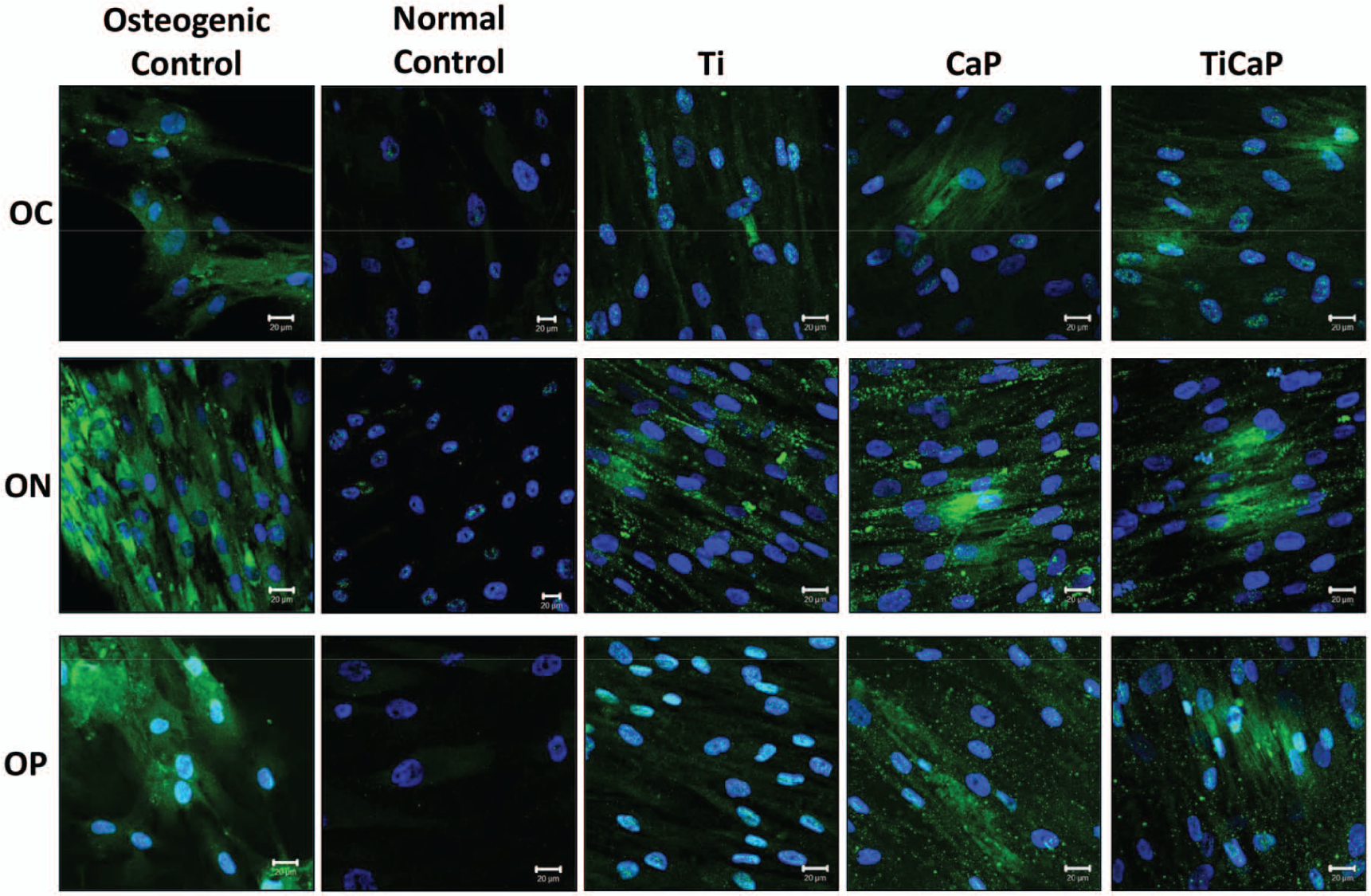
Immunocytochemical localisation of osteocalcin (OC), osteonectin (ON) and osteopontin (OP) (each labelled green) in hMSCs cultured on the various surfaces for 28 days. The nucleus is counterstained with DAPI (blue) (scale bar = 20 µm).
Discussion
The ability to induce substrate-mediated osteogenic differentiation in hMSCs is key to the development of improved implants, scaffolds and cell delivery systems. RF sputter deposited thin films of titanium and CaP are known to promote a favourable osteoblast-like response in cells cultured thereon and, as such, hold promise for being able to direct the osteogenic differentiation of hMSCs for the purposes of bone repair and regeneration. Hence, the response of hMSCs to sputter deposited surfaces that possess distinct surface properties comprising both topography and bioactive chemistry offers the potential to induce osteogenesis without the addition of biochemical stimulants in the culture media.
All three of the sputter deposited surfaces investigated here were found to support high levels of hMSCs viability, as determined by DNA quantification. The highest levels of adhesion and proliferation were observed on the polycrystalline titanium surface (Ti). This is likely to be due to the hydrophilic nature of its TiO2 surface, 17 as it has been previously shown that osteoblast cells attach and proliferate more readily on such surfaces. 19 Furthermore, it has been shown that roughened titanium surfaces promote increased absorption of fibronectin, 20 a major protein known to support cell adhesion. 21 Therefore, the topography and surface chemistry of the sputter deposited Ti engenders a high level of hMSC adhesion and proliferation.
Previous studies have shown higher levels of osteoblast attachment and proliferation on CaP surfaces deposited onto a sputtered Ti interlayer similar to that used here. 16 However, the DNA content measured for hMSCs cultured on the TiCaP surface at both days 1 and 7 is lower than that for the CaP surface. These results are similar to those obtained by Dulgar-Tulloch et al. 22 who reported decreased adhesion and proliferation of hMSCs to HA surfaces with grain sizes of approximately 50 nm, compared to those cultured on smoother surfaces. Furthermore, initial low numbers of hMSCs attached to biomaterial surfaces have been associated with greater levels of subsequent osteogenic differentiation.5,6,23
Total protein estimation of hMSCs in contact with the various types of surface over 15 days in culture indicated higher protein levels on the sputtered Ti surface early in the culture period compared to the CaP or TiCaP surface. This can be attributed to the higher rate of hMSC adherence and proliferation on the Ti surface; however, by the end of the 15-day culture period, the three sputter deposited surfaces had similar levels of protein content which were considerably greater than that for the normal control. This behaviour is known to be associated with matrix production and mineralisation during osteogenic differentiation. 18
ALP is a key early marker of osteogenic differentiation and plays an important role in the regulation of mineralisation. 24 ALP activity levels were considerably larger in cells cultured on the sputter deposited surfaces than for the normal control, indicating that all are capable of directing hMSC differentiation towards an osteogenic lineage. The Ti surface promoted the highest level of ALP activity after 15 days in culture with the values recorded significantly greater than the CaP or TiCaP surface. However, it is noted that while the osteogenic control exhibited peak expression followed by a gradual decrease, a contrasting pattern of increasing activity is observed for the MSCs cultured on the sputter deposited surfaces. This is suggestive of a more subtle osteogenic effect being induced by the sputtered surfaces compared to that of the biochemical supplements used in the osteogenic media.
Increased ALP activity on both the CaP and TiCaP surfaces supports the findings from previous studies on similar sputter deposited CaP thin films with a comparable chemistry. 25 However, as there is no significant difference between the levels recorded for the two surfaces, this suggests that at early time points, the topography of the TiCaP coating does not induce additional levels of osteogenic differentiation in hMSCs compared to the effect of the CaP chemistry.
All sputter deposited surfaces were found to induce significant up-regulations in expression of key genes associated with osteogenic differentiation when analysed over a 28-day period. A summary comparison of the peak expression levels of each of the genes examined for the three sputter deposited surfaces is presented in Table 3. The highest levels of gene expression were recorded for the hMSCs cultured on the TiCaP surface with the Ti surface generally inducing lower levels of expression by comparison. The TiCaP surface was also found to have higher levels of gene expression than the CaP surface for most, but not all, genes of interest. Whereas the Ti surface induced higher levels of ALP activity over the shorter 15-day culture period, the TiCaP surface supports higher levels of gene expression. It would therefore seem that the early increase in ALP activity on the Ti surface was due to the increased MSC proliferation supported by this surface, which reflects the well-established inverse relationship between proliferation and differentiation during osteogenesis. 26
A comparison of the peak level of gene expression (fold change) observed in hMSCs cultured on the Ti, CaP and TiCaP surfaces where yellow = highest, orange = intermediate and red = lowest level of expression out of the three sputter deposited surfaces.

hMSCs: mesenchymal stem cells.
Value is statistically different (p < 0.005) to TiCaP.
Value is statistically different (p < 0.005) to CaP.
Value is statistically different (p < 0.005) to Ti.
There is a considerable up-regulation of COL1A1 in hMSCs cultured on the TiCaP surface compared to that for either the Ti or CaP surface. COL1A1 encodes the main component of type 1 collagen present in most connective tissues and is abundant in bone. It therefore plays an important role in ECM maturation and mineralisation in hMSCs. Although expression of this gene in the hMSCs cultured on the Ti and CaP surfaces was significantly greater compared to the normal control, the combination of the bioactive nature of the CaP surface and a nanoscale topography, as provided by the polycrystalline sputter deposited Ti interlayer, is evidently much more effective in promoting COL1A1 than either of the sputtered CaP or Ti coatings alone. Hence, the production, maturation and mineralisation of a collagen I–rich ECM, which is crucial in osteogenic differentiation,26,27 are greater in hMSCs cultured on the TiCaP surface, compared to those of the Ti or CaP surface.
The CaP and TiCaP surfaces were both effective in inducing the expression of the relatively late-stage markers of osteogenic differentiation, osteopontin and osteocalcin, which suggests the formation of a mature osteoblast phenotype. Immunocytochemical localisation of these proteins shows discrete areas of osteopontin and osteocalcin expression, indicative of mineralisation, for hMSCs cultured on the CaP and TiCaP surfaces; however, this effect is less distinct for the Ti surface. Areas of positive staining for osteonectin in the hMSCs cultured on the CaP and TiCaP surfaces, and to a lesser extent the Ti surface, also suggest that areas of mineralisation are beginning to form within the monolayer. Observations made from immunocytochemistry strengthen the proposition that the hMSCs cultured on the CaP and TiCaP sputter deposited surfaces are undergoing significant levels of osteogenic differentiation and may be transitioning to a mature osteoblast phenotype. This enhanced expression of late-stage markers of differentiation, induced by the presence of the CaP chemistry, can be attributed to the known affinity of non-collagenous matrix proteins to bind to HA. 28
The ALP activity, gene expression and immunocytochemical protein localisation results clearly show that each of the sputter deposited surfaces can induce osteogenic differentiation in hMSCs to different levels, and that this behaviour is attributable to the different physical and chemical cues they possess. The TiO2 (rutile) surface chemistry of the sputtered Ti coating promotes higher levels of hMSC attachment and proliferation compared to either the CaP or TiCaP surface and is also capable of directing MSC differentiation towards an osteogenic lineage. However, although ALP activity was initially greatest on this surface, longer term gene expression and protein analysis indicated that the Ti surface was not as effective for promoting a mature osteoblast phenotype as compared to either the CaP or TiCaP surface. Several studies have highlighted the utility of micro- to nanostructured titanium surfaces in directing osteogenic differentiation.5,6,29–31 For example, Sjöström et al. 31 and McNamara et al. 29 have shown that although levels of osteogenic differentiation could be induced by titanium oxide pillars of 50 nm in height, increased differentiation was observed by those that were 15 nm in height. The Ti surfaces here have grain heights of 47 ± 13 nm, and although they are effective in inducing levels of osteogenic differentiation, it would seem that the feature size may limit the effectiveness at later time points.
By sputtering a CaP thin film directly onto a flat substrate such as glass, the effects of topography are negated, and therefore, the induction of osteogenic differentiation can be directly attributed to the chemistry of the coating. Up-regulations of ALP, COL1A1, osteopontin and osteonectin over a 28-day culture period indicate that the CaP surface chemistry can indeed direct differentiation of hMSCs to a mature osteoblast phenotype. Additionally, the data presented here demonstrate that the CaP surface induces higher levels of hMSC differentiation than the Ti surface.
The TiCaP coating possesses a surface chemistry similar to that of the CaP sample, and surface topography of the sputtered Ti interlayer. Although the CaP and TiCaP surfaces induced similar levels of osteopontin and osteocalcin mRNA expression, expression of ALP and COL1A1 was significantly greater for hMSCs cultured on the TiCaP surface. The grain sizes in this sample (height: 67 ± 28 nm) are larger than for the CaP or Ti coatings, and as discussed previously, smaller features of less than 20 nm in height have been shown to have a greater effect in inducing osteogenic differentiation.29–31 Hence, in order to fully exploit the benefits of topography and CaP chemistry combined, smaller grain sizes could be useful. This might be achieved by reducing the thickness of the Ti and CaP coatings, but needs to be balanced with maintaining the favourable chemistry of the CaP coating since reducing the coating thickness can have a detrimental effect in that regard.
Conclusion
The sputter deposited surfaces produced for this study have all been proven to be effective in inducing the osteogenic differentiation of hMSCs in vitro, without the addition of biochemical stimulants in the culture media. The polycrystalline Ti coating engendered the highest levels of hMSC adherence and proliferation, but yielded a lower long-term level of osteogenesis compared to the CaP and TiCaP surfaces. The bioactive nature of the sputter deposited CaP surface chemistry, which has previously been shown to promote a favourable osteoblast response, can direct the differentiation of hMSCs towards a mature osteoblast phenotype. However, the combination of the CaP chemistry and topography, as supplied by depositing a CaP thin film coating onto a Ti interlayer, produced a type of surface that has an enhanced capability for inducing osteogenic differentiation of hMSCs compared to the effects of either the Ti or CaP coatings when used separately. These results reaffirm the importance of topography in substrate-mediated hMSC differentiation, but importantly, they also show that although the inherent bioactive nature of the CaP promotes osteogenic differentiation of hMSCs, its effectiveness can be increased with the addition of an appropriate topographical profile.
Footnotes
Declaration of conflicting interests
The authors declare that there is no conflict of interest.
Funding
This research received no specific grant from any funding agency in the public, commercial or not-for-profit sectors.