
Abstract
Background
Direct comparison of enzymatic and original blue cell-counting detections with the multinuclear activation of an indicator (MAGI) cells, so far, remains to be performed in parallel. Although inhibitors for reverse transcription solely inhibit the reverse transcription step, those for HIV-1 entry block syncytium formation of HIV-1-infected MAGI cells in addition to the entry (dual inhibition). It raises a concern that reduction of enzymatic activity is artificially influenced by syncytium-blocking activity of inhibitors for entry.
Methods
The MAGI cells with a syncytium inducible strain, HIV-1IIIB, were used for anti-HIV activity determination both with conventional counting with X-Gal staining and measurement of chlorophenol red β-d-galactopyranoside conversion with a plate reader.
Results
Infectivity of HIV-1 in the MAGI cells was highly correlated with both methods. In microscopic observation, small blue cells with single or a couple of nuclei were dominantly observed in the presence of inhibitors for entry, but not in the presence of those for reverse transcription. Actual anti-HIV-1 activities were comparable or moderately sensitive in the chlorophenol red β-d-galactopyranoside method.
Conclusions
Antiviral activities of inhibitors for entry obtained from both enzymatic and counting methods appear to be comparable, even in infection of a highly syncytia inducible HIV-1IIIB strain.
Introduction
The MAGI assay providing accurate infectivity of HIV-1 is established as a simple and rapid system to screen compounds blocking HIV-1 replication.1–4 However, to evaluate HIV infectivity, it requires tedious counting, which hampers wide application of the MAGI assay to high throughput screenings. Therefore, simple and automated enzymatic assay systems have been developed, such as an assay with TZM-bl cells, which are HeLa-derived cells expressing high expression of CD4 and CCR5 by amphotropic retroviral transduction and containing an HIV-1 transactivator protein, Tat-responsive two reporter genes, firefly luciferase and β-galactosidase, for a sensitive quantitation of HIV-1 infection.5,6 For instance, Maeda et al. evaluated antiviral activity of CCR5 antagonists by enzymatic activity of β-galactosidase with the MAGI cells. 7 In general, these assays provide relatively high throughput screenings with convenient protocols.
There is a concern on correlation whether counting and enzymatic methods for the evaluation of antiviral activities are comparable. For instance, inhibitors for the reverse transcription (RT), they solely inhibit the RT step, while those for entry can block two steps, syncytium formation at the late replication step in addition to the entry step. The single-round MAGI assay indeed detects only antiviral activity of early steps, since it utilizes 5-bromo-4-chloro-3-indolyl-β-D-galactopyranoside (X-Gal) conversion by expression of LTR-driven β-galactosidase through Tat 2 . In this regard, syncytium formation that takes place in the late phase of HIV replication is also blocked by inhibitors for entry, resulting in production of small blue cells. In the counting method, either small or large infective focus will be counted as one infection focus, while in enzymatic assay, small syncytia produce less enzymatic activity, led to possible artificially influenced antiviral activity. To our best knowledge, there are no comparison in antiviral activity obtained from methods by microscopic counting and enzymatic activity. Here, we compared both methods with HIV-1IIIB and HIV-1NL4-3, which produce high-level syncytia with its infection, to investigate inhibitory effect of syncytia formation on the antiviral activity.
Methods
Cells and viruses
HeLa-CD4-LTR-β-gal cells 2 were kindly provided by Dr. M. Emerman through the AIDS Research and Reference Reagent Program, Division of AIDS, National Institute of Allergy and Infectious Disease (Bethesda, MD), and used for the antiviral assay as previously described.8,9 A laboratory wild-type stain, HIV-1IIIB was propagated in MT-2 cells, harvested and stored at –80°C as described previously. 3 Recombinant infectious clones of HIV-1 carrying various mutations in the pol gene were generated using pNL101 as previously described. 1 Each molecular clone (2 µg/mL as DNA) was transfected into 293 T cells using the TransIT-LT1 Transfection Reagent (Mirus Bio, Madison, WI). After 24 h incubation, MT-2 cells (106 cells/well) were added and co-cultured with 293 T cells for an additional 24 h. When an extensive cytopathic effect was observed, the cell supernatants were harvested and stored at −80°C until further use.
Antiviral agents
An adsorption inhibitor, dextran sulfate MW 5000 (DS5000) and an HIV-1 nucleoside reverse-transcriptase inhibitor, 3′-azido-3′-deoxythymidine (AZT) was purchased from Sigma-Aldrich Japan (Tokyo, Japan). A non-nucleoside reverse-transcriptase inhibitor, efavirenz, was obtained through the NIH AIDS Reagent Program. A CXCR4 antagonist, AMD3100 was kindly provided from Prof. Shiro Shigeta, Fukushima Medical University (Fukushima, Japan). Peptide-based HIV-1 fusion inhibitors were chemically synthesized using standard Fmoc-based solid-phase techniques, as previously described.10,11
Determination of drug susceptibility
Drug susceptibility with counting was determined as previously described.8,9 Briefly, HeLa-CD4-LTR-β-gal cells were plated in flat 96-well culture plates (104 cells/well). On the following day, the cells were inoculated with HIV-1IIIB (60 blue cell-forming units (BFU)/well, resulting into 60 blue cells after 48 h incubation) and cultured in the presence of drugs. Forty-eight hours after virus inoculation, the cells were fixed with phosphate-buffered saline (PBS) containing 1% formaldehyde and 0.2% glutaldehide for 5 min, washed with PBS three times, and incubated with 0.4 mg/ml X-Gal, 4 mM potassium ferricyanide, 4 mM potassium ferrocyanide, and 2 mM of MgCl2 in PBS for 1 h at 37°C. All the blue cells stained with X-gal were counted in each well. The activity of compounds was determined as the concentration that reduced HIV-1 infection by 50% (EC50).
Drug susceptibility with enzymatic activity was also determined as described 7 with some modifications. Briefly, the assay was performed as identical to the counting method, except for the amount of HIV-1IIIB inoculation (300 BFU/well) due to low color development in the spectrometer. After 48 h of HIV-1 inoculation, cells incubated for were lysed with PBS containing 1% Triton-X100 (100 µL) and incubated at 37°C for 1 h with 10 mM chlorophenol red β-D-galactopyranoside (CPRG) in 2 mM MgCl2, and 100 mM KH2PO4 (100 µL). To terminate the reaction, 80 μl of 0.5 M Na2CO3 was added. The optical density (wavelength at 570 nm) was measured in a microplate reader (GloMax®-Multi+ Microplate Multimode Reader, Promega Corporation, Madison, WI). Drug concentrations that brought about 50% inhibition of the β-galactosidase activity were determined.
The amount of HIV-1 p24 gag antigen level representing viral particle was determined on day 2 with a commercially available ELISA kit (RETRO Tek HIV-1 p24 antigen ELISA 2.0, ZeptoMtrix Co., Buffalo, NY) and compared with infectivity examined by a counting method.
All assays were performed in triplicate.
Results
Microscopic observation
A representative syncytium-inducible stain, HIV-1IIIB, infected MAGI cells in the presence of inhibitors for entry and RT (non-entry) just after 48 h post infection were stained with X-Gal and shown in Figure 1. Two RT inhibitors, AZT and EFV, that are incapable of inhibition of syncytium formation, showed large and apparent syncytia as shown in control but the number of foci was apparently reduced. In contrast, infected foci under entry inhibitors, DS5000, AMD3100, and C34, were apparently small and sometimes only consisted of single cells. These results suggest that, depending on the mechanism of action, antiviral activity obtained from counting and enzymatic methods might be artificially influenced between two methods.
Syncytia in the presence of inhibitors.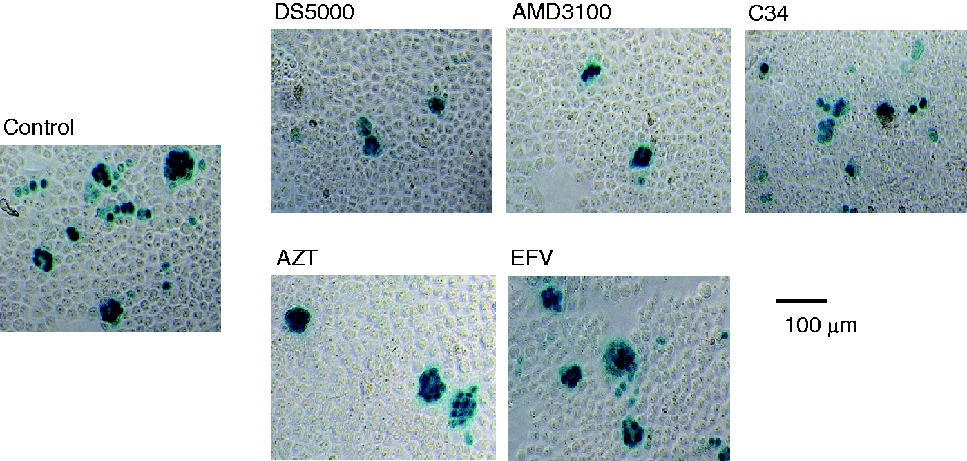
Correlation of infectivity
First, we examined the correlation of the infectivity obtained from counting and enzymatic methods. In the counting method, in a 96-well plate, blue cells indicating HIV-1 infected cells are adjusted to 60 BFU/well to count conveniently with enough accuracy,3,4 since too low viral input makes difficult to obtain accurate data but too many makes difficult to count. However, in the enzymatic assay, it is somewhat not sufficient to generate enough optical density with the CPRG substrate. As shown in Figure 2(a), to produce efficient optical density with CPRG conversion (approximately 1.0) over 300 BFU/well appeared to be required. At least up to 320 BFU/well, correlation of both methods was well maintained (R2 = 0.9839). Therefore, in the absence of inhibitors, both methods are highly correlated with each other. For further confirmation, we examined correlation between the counting and amount of p24 antigen level. Infectivity by the counting and ELISA methods was well correlated (R2 = 0.9999, Figure 2(b)).
Comparison of counting and enzymatic methods.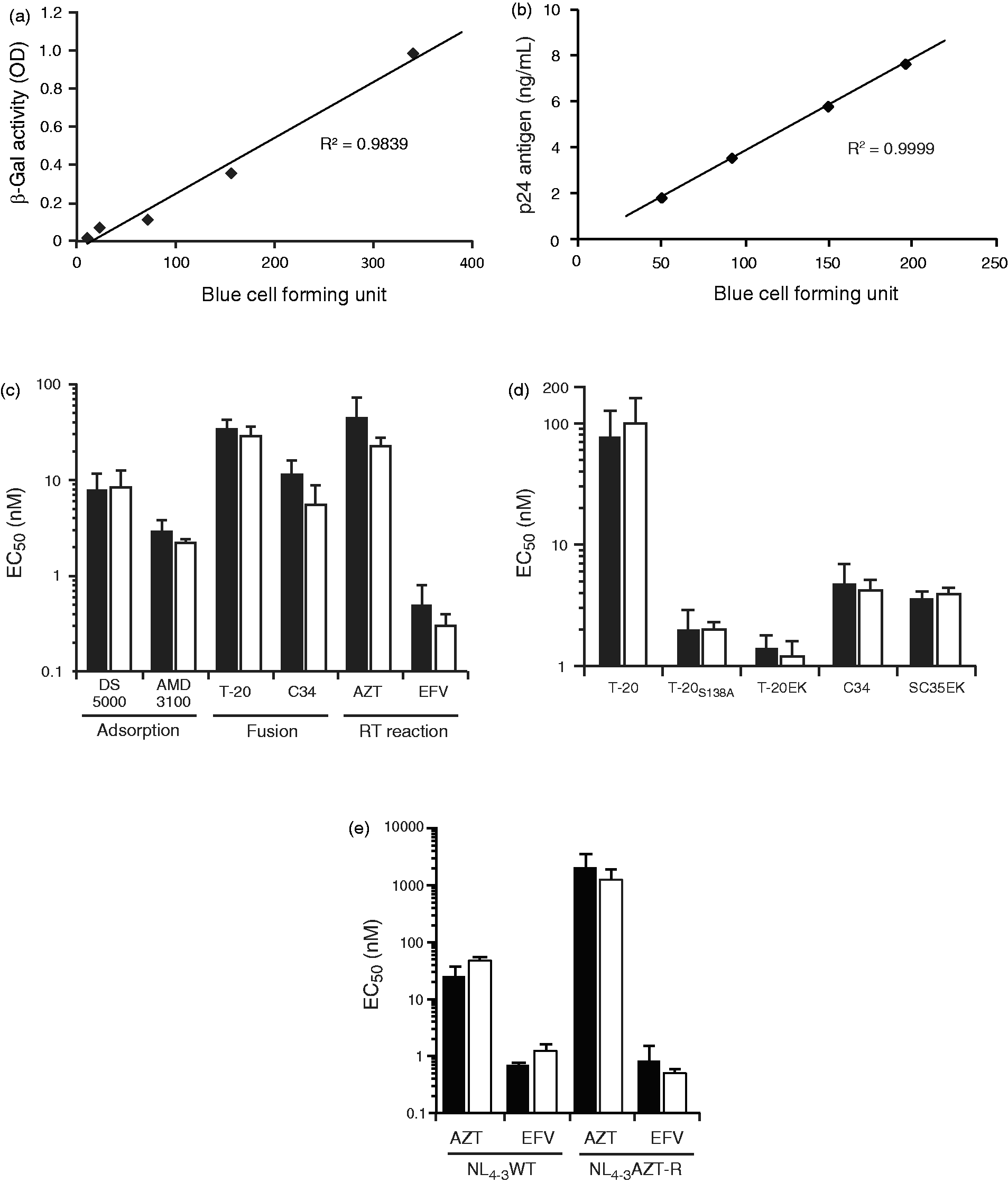
Comparison of antiviral activity
Antiviral activities of inhibitors for entry and RT (non-entry) obtained from both methods were compared in parallel. Activities of AZT, and EFV were relatively enhanced in the enzymatic method without statistical significance (Student’s t-test; p > 0.05), compared with entry inhibitors that block syncytia formation (Figure 2(c)). A fusion inhibitor, C34 that blocks entry and syncytium formation, seems to show relative enhanced activity in the enzymatic method. Therefore, we further examined other fusion inhibitors, a clinically approved T-20 (Fuzeon) and its derivatives, T-20S138A and T-20EK12,13 and a C34 derivative SC35EK. 9 Activities of all fusion inhibitors determined by the both methods were comparable (Figure 2(d)). We further examined with two HIV-1NL4-3-based viruses, wild- and AZT-resistant clones. AZT resistant HIV-1NL4-3 virus containing the resistance mutations in the RT coding region (M41L/D67N/K70E/V75M/L210W/T215Y) and shows high-level resistance to AZT but not to EFV. As shown in Figure 2(e), activities of AZT and EFV were comparable in NL4-3 based HIV-1 viruses. Taken together, the antiviral activity of various compounds obtained from both methods is well comparable.
Discussion
In this study, we compared two representative methods for the detection of HIV-1 infectivity in the MAGI cells in parallel. In the counting method, either small or large syncytia will be counted as one infective focus, while in the enzymatic method, small and large syncytia may produce low and high enzymatic activity, respectively. It is likely that inhibitors for HIV-1 entry might show artificially enhanced activity due to dual mechanisms of action, inhibition of syncytium formation in addition to entry per se. It will make it difficult to compare or evaluate antiviral activity obtained from the two distinct methods. However, anti-HIV activity was, surprisingly, comparable in both methods. It is likely that relatively high input of HIV-1 in 96-well plate (300 MU/well) may reduce or interfere with formation of large syncytia, since adjacent cells may also be infected with HIV-1.
Since β-galactosidase system is well established and can be visualized with X-Gal staining, it will be convenient to standardize HIV infectivity, or titer and suitable to compare characteristics of both methods. Moreover, the β-galactosidase activity can be measured by various commercial available systems. Here, we applied CPRG to detect enzyme activity and successfully detect their activity with a standard spectrometer. This enables to detect even in a resource-limited laboratory. Probably, luciferase system with TZM-bl cells must be more sensitive but it needs luminometer and relatively expensive luciferase substrate, 14 while a CRPG/β-galactosidase system is simple and widely useful for various applications.
In conclusion, we compared counting and enzymatic methods with the MAGI cells and revealed that both systems will work and detect comparable antiviral activity.
Footnotes
Acknowledgements
We are grateful to the Network Joint Research Center for Materials and Devises and the Biomedical Research Core (Tohoku University Institute of Multidisciplinary Research for Advanced Materials, and Graduate School of Medicine, respectively) for technical support.
Declaration of conflicting interests
The author(s) declared no potential conflicts of interest with respect to the research, authorship, and/or publication of this article.
Funding
The author(s) disclosed receipt of the following financial support for the research, authorship, and/or publication of this article: This work was supported by a grant from the Ministry of Education, Culture, Sports, Science, and Technology of Japan, a grant for the Promotion of AIDS Research from the Ministry of Health, Labour and Welfare.