
Abstract
Background
Because guanine-based herpes simplex virus thymidine kinase inhibitors are not orally available, we synthesized various 6-deoxy prodrugs of these compounds and evaluated them with regard to solubility in water, oral bioavailability, and efficacy to prevent herpes simplex virus-1 reactivation from latency in a mouse model.
Methods
Organic synthesis was used to prepare compounds, High Performance Liquid Chromatography (HPLC) to analyze hydrolytic conversion, Mass Spectrometry (MS) to measure oral bioavailability, and mouse latent infection and induced reactivation to evaluate the efficacy of a specific prodrug.
Results
Aqueous solubilities of prodrugs were improved, oxidation of prodrugs by animal cytosols occurred in vitro, and oral absorption of the optimal prodrug sacrovir™ (6-deoxy-mCF3PG) in the presence of the aqueous adjuvant Soluplus® and conversion to active compound N2-[3-(trifluoromethyl)pheny])guanine (mCF3PG) were accomplished in mice. Treatment of herpes simplex virus-1 latent mice with sacrovir™ in 1% Soluplus in drinking water significantly suppressed herpes simplex virus-1 reactivation and viral genomic replication.
Conclusions
Ad libitum oral delivery of sacrovir™ was effective in suppressing herpes simplex virus-1 reactivation in ocularly infected latent mice as measured by the numbers of mice shedding infectious virus at the ocular surface, numbers of trigeminal ganglia positive for infectious virus, number of corneas that had detectable infectious virus, and herpes simplex virus-1 genome copy numbers in trigeminal ganglia following reactivation. These results demonstrate the statistically significant effect of the prodrug on suppressing herpes simplex virus-1 reactivation in vivo.
Introduction
Drugs currently used for human herpes simplex virus (HSV) infections are nucleoside (“acycloguanosine”) analogs, such as acyclovir, its prodrug valcyclovir (Valtrex®), the prodrug famciclovir (Famvir®), as well as the pyrophosphate analog foscarnet (Figure 1). Although these drugs are effective in treating HSV infections during the symptomatic and infectious replicative stages, they are only partially effective in suppressing reactivation of the viruses from the latent state in neurons.1,2 In fact, there is no “cure” for herpes virus latency, and despite treatment with current therapeutics both symptomatic and asymptomatic recurrent shedding and infection occur. This represents a significant weakness of current therapies as infectious virus is unwittingly transmitted via mucosal membranes to partners. This is problematic with ocular infections by HSV-1 and genital infections with HSV-2. The latter infects the genitalia of both sexes and can be transmitted to sex partners even in the absence of active disease.
Antiherpes drugs.

Thus, in order to control the reactivation of the virus, especially in immunocompromised patients where development of drug resistant variants to acyclonucleoside drugs has become common, we3–5 and others6,7 have developed several families of inhibitors of HSV thymidine kinase (TK) whose expression is required for reactivation of virus from the latent state in neurons. Interestingly, we showed that both 2-(phenylamino)-6-oxopurines such as the 9-(4-hydroxybutyl) derivative HBPG and N2-[3-(trifluoromethyl)phenyl]guanine, mCF3PG (Figure 2) are potent inhibitors of HSV types 1 and 2 TKs,4,5 block HSV reactivation from latently infected nerve ganglia in cell cultures,
8
and that HBPG, given intraperitoneally, reduced recurrent HSV disease in mice
9
and monkeys,
10
as well as HSV encephalitis in mice.
11
Although the in vivo results are promising, the low water solubility and poor oral absorption of lead analogs HBPG and mCF3PG have limited further testing of these compounds. Based on the success of famciclovir, a 6-deoxy prodrug of the active penciclovir, we reasoned that analogous prodrugs of TK inhibitors may better penetrate cells, and, in appropriate formulations, may have increased oral bioavailability of the ultimate inhibitors.
HSV thymidine kinase inhibitors and prodrugs.
The marketed drug famciclovir (Figure 3) undergoes both ester cleavage and oxidation to provide effective plasma levels of the drug penciclovir.
12
In addition, various nontoxic adjuvants (facilitators) have been developed to increase water solubility of poorly soluble compounds and, in some cases, enhance their oral absorption. In this paper we, therefore, describe synthesis of 6-deoxyguanines corresponding to the active TK inhibitors, their oxidative conversion to the ultimate drugs by incubation in animal cytosols, their water solubility, and their oral absorption and conversion, alone and in combination with various facilitators, by mice. While preliminary results
13
indicated that 6-deoxyHBPG was not oxidized by cytosols, 6-deoxy-mCF3PG (aka sacrovir™, Figure 2) was, although the latter was converted to both the active inhibitor and a regioisomeric 8-oxo form in varying ratios depending on the source of cytosol. This led us to synthesize certain 8-substituted analogs of mCF3PG in order to block the 8 position of the corresponding 6-deoxy compound, viz. the 8-methyl, 8-aza, and 7-deaza-8-aza derivatives, and test them as TK inhibitors. Only 8-aza-mCF3PG was found to inhibit TK, and its prodrug form, i.e. 8-azasacrovir™, was oxidized by all cytosols. However, although orally absorbed by mice, 8-azasacrovir was not converted rapidly to the 6-oxo compound. Sacrovir itself, given in drinking water containing 1% Soluplus, suppressed HSV-1 reactivation in a mouse model. This finding indicates that sacrovir™ may be a useful prodrug for treatment of HSV recurrences.
Metabolism of famciclovir.
Materials and methods
General
All new compounds were characterized by 1H NMR, 13 C NMR, LC–MS, and HPLC analyses (see Supplementary Material for details of syntheses). HBPG and mCF3PG were prepared as previously described.3,4 NMR spectra were measured with a Bruker Avance 300 instrument; chemical shifts of 1H are relative to internal TMS and of 13 C relative to solvent at 39.51 ppm. HLPC analyses were done with a Agilent ProStar dual pump system. Standard method 1 was from 100% A (water containing 0.1%TFA) to 100% B (acetonitrile containing 0.1% TFA) for 12 min with a 2 min hold at 100% B and 2 min equilibration at 100% A. The flow rate was 1 ml/min, and acquisition was from 0 to 12 min. Standard method 2 was identical except that the gradient was run over 8 min. The column was Waters Xbridge C18, 150 mm × 4.6 mm, 5 µm. Injection was 2 µl of ca. 1 mg/ml in DMSO with detection by UV at 265 or 275 nm. LC–MS analyses employed a LCQ Advantage (Thermo-Fisher) MS detector interfaced with a Surveyor LC system and PDA detector. LC generally employed an XBridge C18 column, 150 × 2 mm, 3 µm, with a guard column. Mobile phases were A, 0.1% acetic acid in water. B, acetonitrile. Flow rate was 200 µl/min. and column temperature was 45°C. Injection volume was 3 µl via partial loop mode. UV detection by PDA was 200–420 nm. HR–MS data were acquired on a Thermo Orbitrap velos instrument at UMass Medical School in the electrospray positive mode. For solubility measurements, solubilizing agents included Solutol® HS 15 and Soluplus®, gifts from BASF Inc., and Povidone (Kollidon®) (Sigma-Aldrich). Mice (Swiss Webster, males, ca. 28 g) were from Taconic Farms or Charles River Laboratories. Animal work at LSU and at GLS was performed according to guidelines of the NIH, and euthanasia was consistent with standards of AVMA.
Cytosol incubations
Animal cytosols were pooled samples from liver (Bioreclamation Inc., Westbury, NY). Test compounds (50 µM) were incubated at 37°C in mixtures containing 1 mg/ml cytosolic proteins in phosphate buffer (pH 7.4) in glass vials. At various times, aliquots of the incubation mixture were removed and mixed with 2 volumes of acetonitrile, centrifuged at 13,000 × g, the supernatants filtered through 10 k cutoff centrifugation filters, and analyzed by LC–MS. Standard curves were generated from authentic prodrugs and oxidation products.
Solubility
Aqueous solubility was determined at 25°C by stirring an excess of compounds in water or water containing various adjuvants overnight, filtering and analysis by HPLC with UV quantitation of the filtrate. Comparison with a standard curve in each case allowed calculation of concentration. Adjuvants included variable percentages of Soluplus®, a polyvinyl caprolactam–polyvinyl acetate–polyethylene glycol graft copolymer, 10% Solutol® HS 15, a polyoxy 15-hydroxystearate, and 10% Kollidon® (Povidone), polyvinylpyrrolidone of avg. MW 10,000.
Oral bioavailability studies
Bolus
Sacrovir (12 mg) was dissolved with sonication in solutions of 1, 5, and 10% Soluplus in deionized water (2 ml). Swiss Webster mice (avg wt 28.7 g) were given these solutions at 10 ml/kg orally via gavage. Animals were anesthetized with isoflurane at 20 min postdosing, and blood was removed by cardiac puncture and placed in heparinized tubes on ice. Animals were then sacrificed by CO2 inhalation and double pneumothorax. Blood samples were centrifuged at 1500 × g for 5 min at rt, and plasma was drawn off and frozen at −70°C until processed.
In drinking water
Sacrovir (186 mg) was dissolved in 10 ml of 10% aqueous Soluplus with stirring, and this suspension was added to 90 ml of deionized water. This solution containing 1.86 mg/ml of sacrovir in 1% aqueous Soluplus and 1% Soluplus alone were provided as the sole source of drinking water to groups of five mice ad libitum. Fresh solutions were prepared each day. Animal weights and water consumption volumes were measured each day. After the end of the experiment, animals were anesthetized with isoflurane and exsanguinated by cardiac puncture. Blood was processed as above, and LC–MS was used to quantitate prodrug or drug concentrations in plasma.
Effects of sacrovir on HSV-1 reactivation
Corneas of ketamine/xylazine-anesthetized seven-week-old female BALB/c mice (Charles River Laboratories) were scarified bilaterally in a cross-hatch pattern and infected topically with a 3 µl drop containing 25,000 PFU/eye of wild-type HSV-1 McKrae strain. Animals were injected intraperitoneally with 200 µl normal human serum to increase survivability. At days 3 and 5 postinfection, the efficiency of infection was assessed in each eye by fluorescent slit-lamp biomicroscopy and by detection of infectious virus in ocular swabs. To assess complete HSV-1 latency, at both 4 and 5 weeks postinfection, ocular swabs were cultured on Vero cells and tested negative for infectious virus. At greater than 40 days postinfection, surviving mice were randomly assigned into treatment groups: (1) control; normal drinking water; (2) 1% Soluplus; or (3) sacrovir (1.86 mg/ml) in 1% Soluplus. Fresh solutions were prepared each day and provided ad libitum as the sole source of drinking water for each group for two days prior to initiating HSV-1 reactivation by hyperthermic stress as described previously. 14 Following hyperthermic stress-induced reactivation, mice were dried, returned to their cages, and provided food and water containing freshly prepared treatments. At 24 h postreactivation, animals were humanely euthanized and assessed for the presence of infectious virus in paired ocular swabs, paired eviscerated trigeminal ganglia (TG), and individually excised cornealscleral buttons. The presence of infectious virus in each sample was determined by coculture on CV-1 cells. Samples that showed no HSV-1-mediated cytopathic effect at day 7 were scored as negative. In a parallel study, individual TGs were removed, eviscerated in ATL buffer (Qiagen), and frozen until processed for DNA extraction.
Quantification of HSV-1 genomic viral loads in TG
HSV-1 genome copy number in TG was determined by quantitative real-time PCR of the UL30 DNA polymerase gene. UL30 primers and PCR reaction conditions were as previously published. 14 Total DNA was extracted from individually isolated TG using DNeasy columns (Qiagen) and quantified spectrophotometrically. Target genomes in 150 ng of DNA were amplified and detected using BioRad IQ SYBR Green Supermix. Samples were assayed on a BioRad CFX96 in 96-well plates relative to a standard curve with a plasmid containing the HSV-1 UL30 gene. The number of UL30 gene copies in each sample was determined against the standard curve, and the number of viral genomes per TG was calculated based on the total DNA extracted from each TG. Data were plotted in Graphpad and statistical significance between all groups was determined by one-way ANOVA with subsequent Tukey’s multiple comparisons posttest.
Results
Syntheses
Prodrugs
6-DeoxyHBPG was prepared as shown in Figure 4. Details of this and other new syntheses are in Supplementary material.
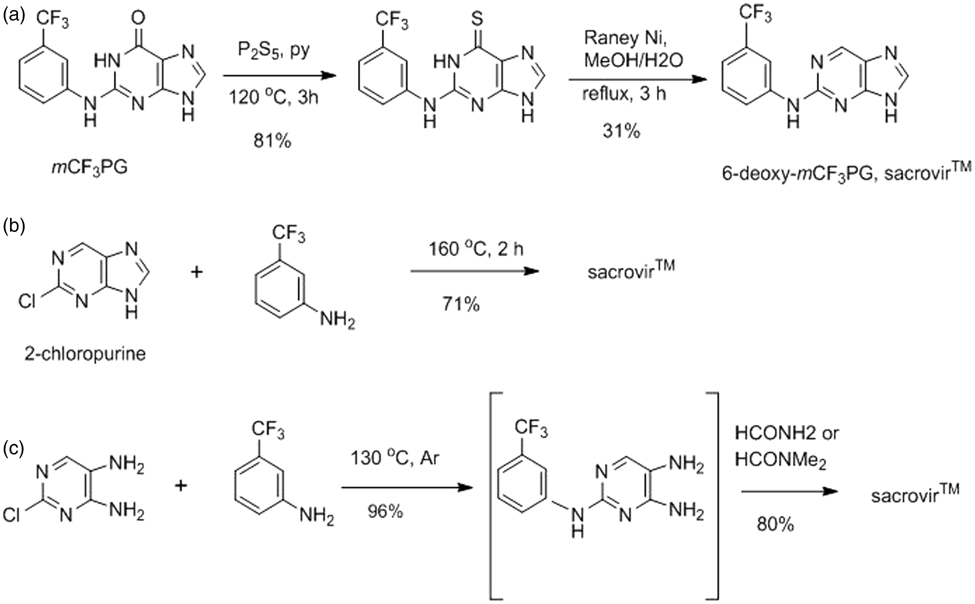
6-Deoxy-mCF3PG (sacrovir™) was prepared from mCF3PG via its 6-thio analog (Figure 5(a)) and from 2-chloropurine (Figure 5(b)) by fusion with 3-(trifluoromethyl)aniline. The 2-chloropurine used in 3(b) was made by selective reduction of the available 2,6-dichloropurine (not shown). An alternate and preferred method involves fusion of the commercially available 2-chloro-5,6-diaminopyrimidine with 3-(trifluoromethyl)aniline in 96% yield, followed by purine ring closure with formamide or dimethylformamide (Figure 5(c)).
In order to determine the identity of the second oxidation product of sacrovir™, the authentic 8-oxo analog was prepared from the pyrimidine intermediate in 96% yield as summarized:
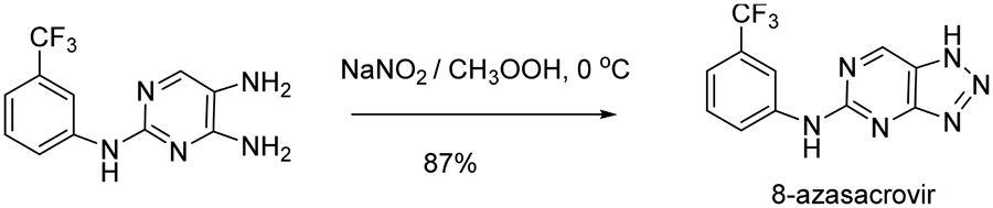
Because of the uncertainty in in vivo conversion of sacrovir, we chose to evaluate cellular oxidation of several 8-blocked forms of this compound as potential prodrugs and the corresponding guanines as TK inhibitors. Synthesis of 6-deoxy-8-aza-mCF3PG (8-azasacrovir™) was straightforward. This compound was made by cyclization of the pyrimidine intermediate with nitrous acid and isolated in 87% yield:
TK inhibitors
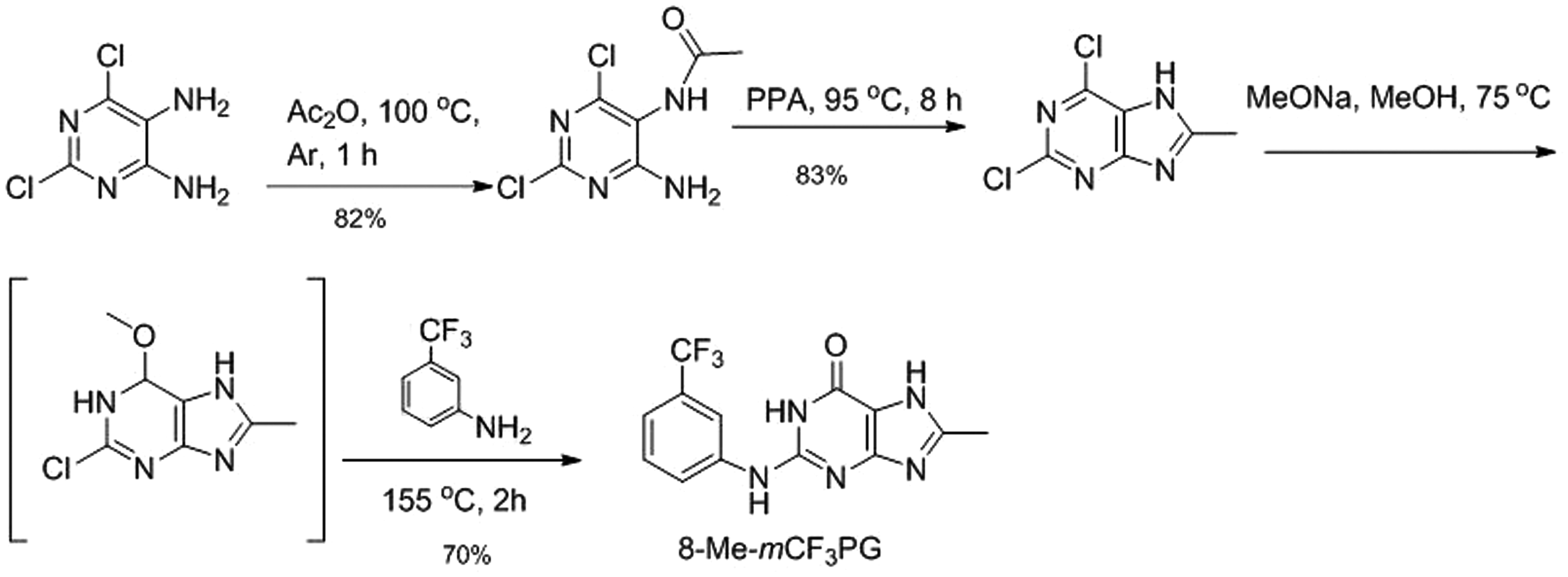
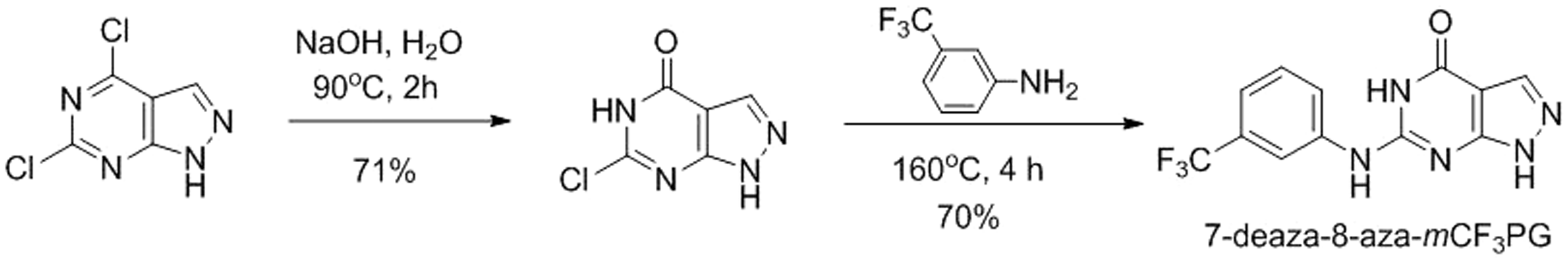
HBPG and mCF3PG, were prepared as described.3,4 As candidate 8-substituted TK inhibitors, 8-methyl-mCF3PG was prepared as summarized in Figure 6, and 7-deaza-8-aza-mCF
3
PG was prepared as summarized in Figure 7.
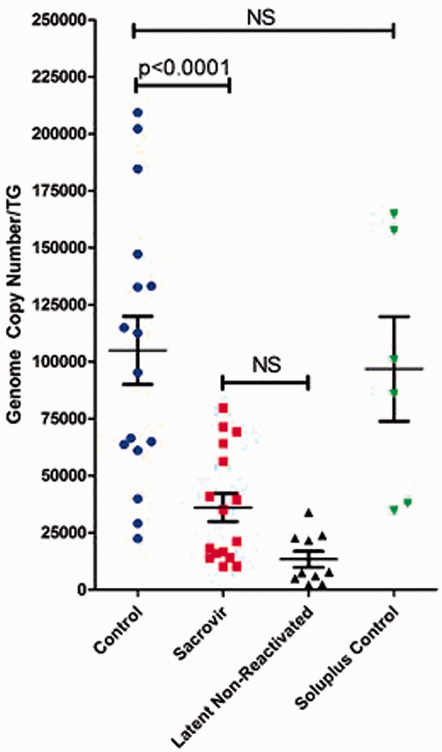
Synthesis of 6-deoxy-HBPG. Synthesis methods for 6-deoxy-mCF3PG, sacrovir™ Synthesis of 8-methyl-mCF3PG. Synthesis of 7-deaza-8-aza-mCF3PG. HSV-1-specific genome copies in trigeminal ganglia. NS: not significant.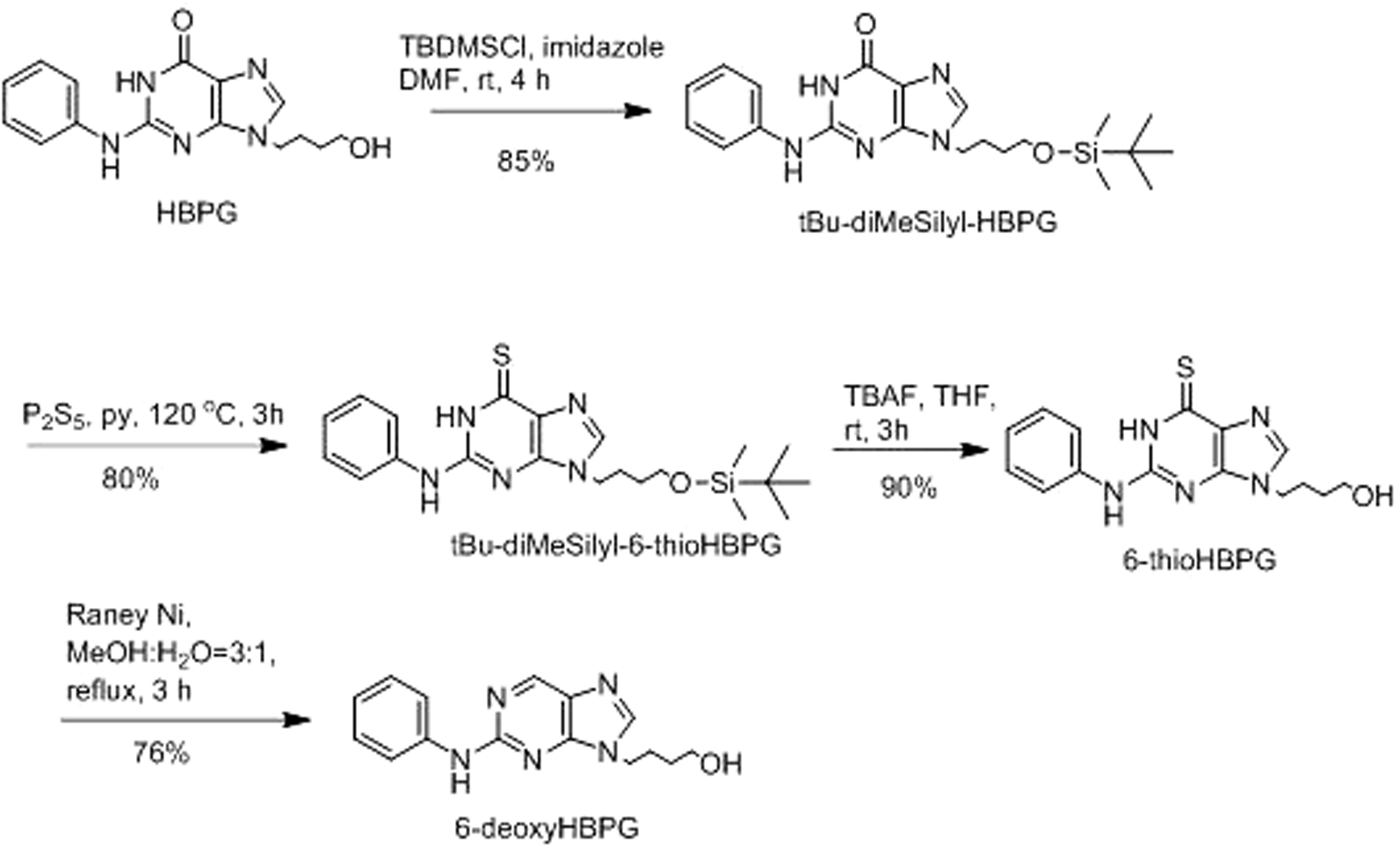
8-Aza-mCF3PG could not be synthesized chemically. It was first isolated and identified by LC–MS and 1H NMR from incubation mixtures of 8-azasacrovir™ in guinea pig cytosol. Mobile phase was evaporated on a Speed-Vac®, and the solid, identified by LC–MS, was dissolved in DMSO to make a 10 mM stock solution. It was isolated by preparative HPLC and had the appropriate exact mass in HR–MS. Subsequently, commercial xanthine oxidase was found to give this oxidation product quantitatively. 15
Enzyme inhibition
Recombinant HSV TKs were cloned, expressed, purified, and assayed as described. 5 Briefly, the enzymes with and without test compounds were assayed in the presence of Km concentrations of substrate (thymidine, 1 and 2 µM for HSV-1 and HSV-2 TKs, respectively). 5 8-Methyl-mCF3PG and 9-deaza-8-aza-mCF3PG were inactive at 50 µM, as was sacrovir itself, but 8-aza-mCF3PG gave IC50 values close to 1 µM versus both HSV-1 and HSV-2 TKs. For these inhibitors, double reciprocal “Lineweaver-Burk” plots revealed that, like mCF3PG, 8-aza-mCF3PG was competitive with the substrate (Ki = 0.6 µM) for HSV-1 TK, but, in contrast with mCF3PG (competitive Ki = 0.65 µM), it appeared to be uncompetitive with substrate for HSV-2 TK (Ki = 0.11 µM).
Solubility of sacrovir and 8-azasacrovir
Aqueous solubilities of prodrugs (mg/ml). a
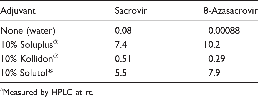
Measured by HPLC at rt.
Oxidation by animal cytosols
Oxidation of prodrugs by animal cytosols. a
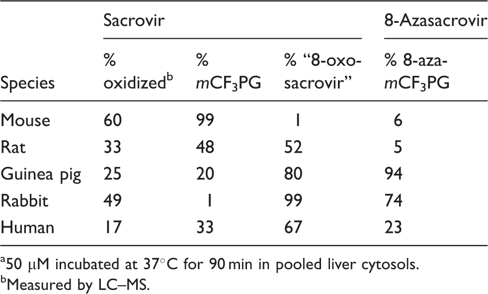
50 µM incubated at 37°C for 90 min in pooled liver cytosols.
Measured by LC–MS.
Absorption and bioavailability of oral sacrovir in mice
Bolus dosing by gavage
Plasma concentration of mCF3PG after bolus gavage dosing of sacrovir to mice. a
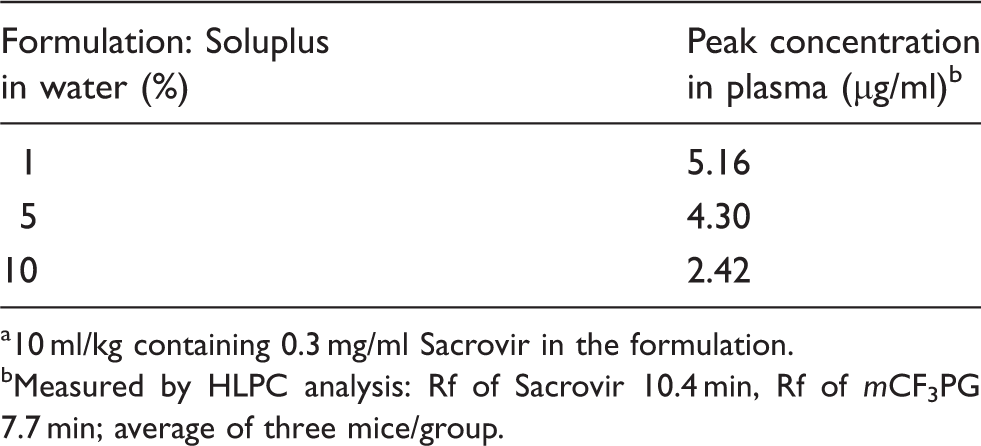
10 ml/kg containing 0.3 mg/ml Sacrovir in the formulation.
Measured by HLPC analysis: Rf of Sacrovir 10.4 min, Rf of mCF3PG 7.7 min; average of three mice/group.
In drinking water
Consumption of sacrovir in drinking water by mice. a
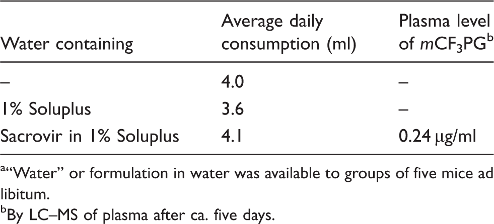
“Water” or formulation in water was available to groups of five mice ad libitum.
By LC–MS of plasma after ca. five days.
Effect of sacrovir on HSV-1 reactivation from latently infected mice
Effect of treatments on HSV-1 reactivation in mice.
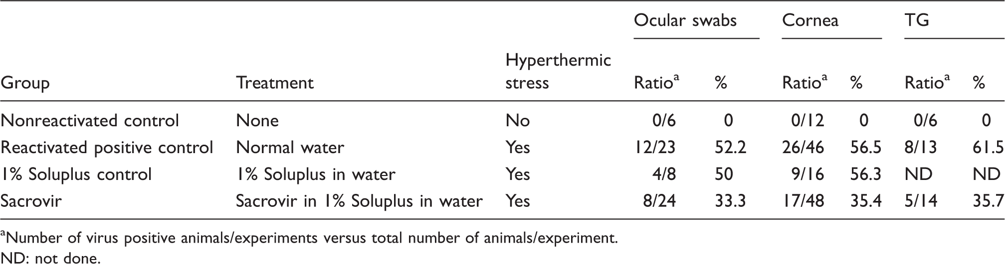
Number of virus positive animals/experiments versus total number of animals/experiment.
ND: not done.
In a parallel study, HSV-1 genomic viral loads within the TG were quantitatively determined by RT-PCR of individually isolated TGs from reactivated and nonreactivated animals under the various treatment conditions. 14 The HSV-1 genomic viral load from individual TG isolates and the mean of each treatment group are shown in Figure 8. In nonreactivated TGs, HSV-1 genomic viral loads ranged from 2103 to 23,777 copies per TG with an average of 13,282. In reactivated positive controls, the TG averaged 104,983 copies per TG and ranged from 22,386 to 184,617 copies per TG. Control treatment with the 1% Soluplus adjuvant alone did not differ significantly from positive controls: a viral load range from 34,556 to 164,712 copies per TG, resulting in an average genomic viral load 96,853. By contrast, sacrovir treatment reduced the overall average genomic viral load in heat-stress reactivated animals to 36,012 copies per TG with a range of 10,150–79,690 copies per TG.
These results demonstrate that sacrovir is absorbed chronically by mice and converted to mCF3PG, and effectively blocks HSV-1 reactivation in mice. However, the average consumption of sacrovir in 1% Soluplus was lower (ca. 2.4 ml/day/mouse) than that observed in experiments in uninfected mice (Table 4), leading to a lower “dose” of ca. 208 mg/kg/day/mouse and thus to lower plasma levels of mCF3PG than expected.
Discussion
6-DeoxyHBPG, i.e. 2-phenylamino-9-(4-hydroxybutyl)purine, was not oxidized to HBPG by animal cytosols nor was it orally absorbed in mice (results not shown) and was not studied further. 6-Deoxy-mCF3PG (sacrovir™), in contrast, was readily oxidized to the active 6-oxo compound (mCF3PG) and an additional form (8-oxo) in cytosols, but exclusively to mCF3PG in vivo in mice. The possibility that, like famciclovir, sacrovir is oxidized in vivo predominantly by systemic aldehyde oxidase 17 suggests that the cytosol results may be artifactual. An analog of sacrovir meant to block oxidation at C-8, i.e. 8-aza-6-deoxy-mCF3PG (8-azasacrovir), was oxidized by cytosols to give a single product, 8-aza-mCF3PG, although the rate of conversion was minimal in mouse and rat cytosols. Although 8-azasacrovir was more water soluble than sacrovir (Table 2) and was readily absorbed orally by mice, it was not oxidized to give substantial plasma levels of 8-aza-mCF3PG in vivo (results not shown). Among 8-substituted analogs of mCF3PG, only the 8-aza analog was active as an inhibitor of HSV TKs. Other substituted analogs, i.e. 8-methyl-mCF3PG and 7-deaza-8-aza-mCF 3 PG were inactive as was sacrovir itself, up to 50 µM. Thus, sacrovir became the focus of our attention as a prodrug.
Sacrovir was soluble in 1–10% Soluplus (Table 3) and readily absorbed orally in 1% Soluplus in water. It was also important, in order to deliver drug consistently to animals, to measure the acceptability of sacrovir and Soluplus by mice. Both sacrovir in 1% aqueous Soluplus and 1% aqueous Soluplus alone were accepted by mice in the drinking water for five days, the length of the reactivation experiment (Table 4). The “dose” of sacrovir was ca. 368 mg/kg/day. Soluplus® is the graft copolymer polyvinyl caprolactam–polyvinyl acetate–polyethylene glycol, and it is used either in aqueous solution or by hot-melt extrusion to enhance solubility of poorly soluble compounds. 18 It often increases oral absorption of drugs at pharmacologically acceptable levels. It is stated by the manufacturer to be “virtually non-toxic after a single ingestion.” It also did not affect the consumption of drinking water by mice, both in the absence and presence of sacrovir (Table 4). Soluplus represents the best adjuvant to increase the oral absorption of sacrovir and related compounds, at least among those reported herein (and others unpublished).
Ad libitum oral delivery of sacrovir albeit at a lower “dose” than expected was effective in suppressing HSV-1 reactivation in ocularly infected latent mice as measured by four parameters: (1) percentage of mice shedding infectious virus at the ocular surface, (2) percentage of TG positive for infectious virus, (3) number of corneas that had detectable infectious virus, and (4) HSV-1 genome copy numbers in TG following reactivation. The first three are summarized in Table 5, and the genome copy number data are summarized in Figure 8, demonstrating the statistically significant effect of the prodrug on suppressing HSV-1 reactivation in vivo.
Footnotes
Acknowledgements
This work is dedicated to the memory of Dr James Hill, LSU Eye Center, New Orleans, LA.
Declaration of conflicting interest
The author(s) declared no potential conflicts of interest with respect to the research, authorship, and/or publication of this article.
Funding
The author(s) disclosed receipt of the following financial support for the research, authorship, and/or publication of this article: The authors were supported, in part, by SBIR grant 1 R43 AI100300-01 from the National Institutes of Health (to GEW) and by FIRB grant 2BAU011.SR4.001 (to FF). LS was supported by a fellowship provided by Regione Lombardia, Project ID 13810040.
Supplementary Material
Supplementary material includes details of synthesis and characterization of all new compounds.