
Abstract
Objective
Osteoarthritis (OA) is a degenerative joint disease that has no cure, and current therapies are intended to minimize pain. There is, therefore, a need for effective pharmacologic agents that reverse or slow the progression of joint damage. We report herein on an investigation of the effects of intra-articular injections of ganglioside sugars on the progression of OA in an experimental rabbit model.
Design
Knee OA was induced Japanese in White rabbits by anterior cruciate ligament transection (ACLT). Ganglioside sugars at concentrations of 0.1, 0.3, and 0.9 mg/ml were then intra-articularly injected as a possible treatment for OA. Controls received intra-articular injections of saline. Knees were assessed macroscopically, histologically, and mechanically at 13 weeks after ACLT induction.
Results
Macroscopically, knees of the groups that received ganglioside sugars at concentrations of 0.3 and 0.9 mg/ml exhibited milder cartilage degradation compared to the controls. Consistent with these results, histological scores for these knees were significantly higher than the corresponding values for the control knees. Lectin histochemistry staining revealed that the treatment with ganglioside sugars at concentrations of 0.3 and 0.9 mg/ml was associated with a remarkable increase in the levels of GalNAc-positive chondrocytes in cartilage. Coefficient of friction testing also demonstrated that cartilages treated with ganglioside sugars had a lower coefficient of frictions than the values for the control group.
Conclusions
Intra-articular injections of ganglioside sugars prevented cartilage degeneration in an OA-instability model. These results highlight the promising therapeutic potential for using ganglioside sugars in the treatment of progressive OA.
Introduction
Osteoarthritis (OA) is the most prevalent chronic joint disease that affects 70% of the world population with ages ranging between 55 and 70 years. The disease is characterized by the degradation of articular cartilage accompanied by synovial inflammation, subchondral bone sclerosis, and the formation of osteophytes.1-3 The destruction of cartilage occurs when chondrocytes fail to maintain a homeostatic process in the cartilage resulting in a reduction in the synthesis of extracellular matrix (ECM) molecules. To date, there are no effective pharmacologic agents that reverse or slow the progression of OA joint damage. 4
Articular cartilage is an aneural and avascular tissue that has limited healing capacity. It is composed of a specialized matrix composed of collagen molecules, proteoglycans, and noncollagen proteins that form a fibrillar network. Cartilage homeostasis is mediated by chondrocytes that function to maintain the synthesis and degradation of the ECM. However, a decrease in the deposition of newly synthesized collagen and proteoglycan molecules by chondrocytes in the matrix is generally thought to be the primary factor leading to cartilage destruction in OA.5,6 Gangliosides, a class of glycosphingolipids (GSLs) containing sialic acid, are biologically active molecules that are found in eukaryotic cell membranes and play an essential role in cellular physiology. GSLs can be classified into several groups depending on their carbohydrate structure, including the o-, a-, and b-series.7-9 GSLs are involved in the attachment of cells to matrices, cell-to-cell interactions, and the recognition of pathogens.7,8 Glucosylceramide synthase (Ugcg) is the enzyme that is involved in first step in GSL synthesis and has been documented to be essential in organ development including cartilage. 9 There is a growing body of evidence to indicate that GSLs are critical for the maintenance of chondrocyte function and for cartilage homeostasis.9-12 In fact, the deletion of NeuAcα(2-3)Galβ(1-4)Glc-Cer (GM3) synthase (GM3S) enhances cartilage degradation in mice. 10 Moreover, cartilage degeneration due to OA has been reported to be correlated with a 40% decrease in total ganglioside content.10,11 Supplementation of gangliosides sugars (GA2, GM2, GD2), which are modulated by the β1, 4-N-acetylgalactosaminyl transferase (GalNacT) to cultured chondrocytes that had been stimulated with IL-1α was reported to suppress the expression of catabolic factors and the action of cartilage degradative enzymes. 12 These collective findings strongly indicate that GSLs are involved in the regulation of chondrocyte function and cartilage metabolism.
Given the essential role of GSLs in maintaining chondrocyte function and cartilage metabolism, we evaluated the antiarthritic activity of ganglioside sugars in an experimental model of OA. The therapeutic effects of intra-articular administrations of the ganglioside sugars were evaluated in the knees of a rabbit model in which OA was induced by anterior cruciate ligament transection (ACLT).
Methods
Ethics Statement
All procedures for animal experiments were performed consistent with the ethical guidelines approved by the Animal Care Committee of Hokkaido University. (Approval ID:17-0085). All procedures were performed in accordance with ARRIVE guidelines (https://arriveguidelines.org).
Animals and Treatment With Ganglioside Sugars
Japanese white rabbits (7-month old) with an average weight of 3.3 kg (Japan SLC Inc., Hamamatsu, Japan) were used in this study. Animals were appropriately anesthetized by intravenous injection of 0.05 mg/kg of pentobarbital, followed by isoflurane in oxygen gas anesthesia. Their bilateral legs were shaved, prepared, and draped in a sterile fashion. ACLT was performed through a medial parapatellar approach to induce OA in the knees of the animals.
13
The patella was everted, and the anterior cruciate ligament was then transected. The knee joints were irrigated, and the capsule and skin were closed with running 3-0 nylon sutures (Suprylon, Vomel, Germany). The instability of the knee was confirmed by Lachman testing. The rabbits were allowed to move freely in their individual cages. Animals were monitored regularly for signs of pain, distress, or abnormal behavior. Ganglioside sugars, including GA2, GM2, GD2, (Elicityl, Crolles, France) (Molecular weight GA2:545.49, GM2:858.73.49, GD2:1171.96) (
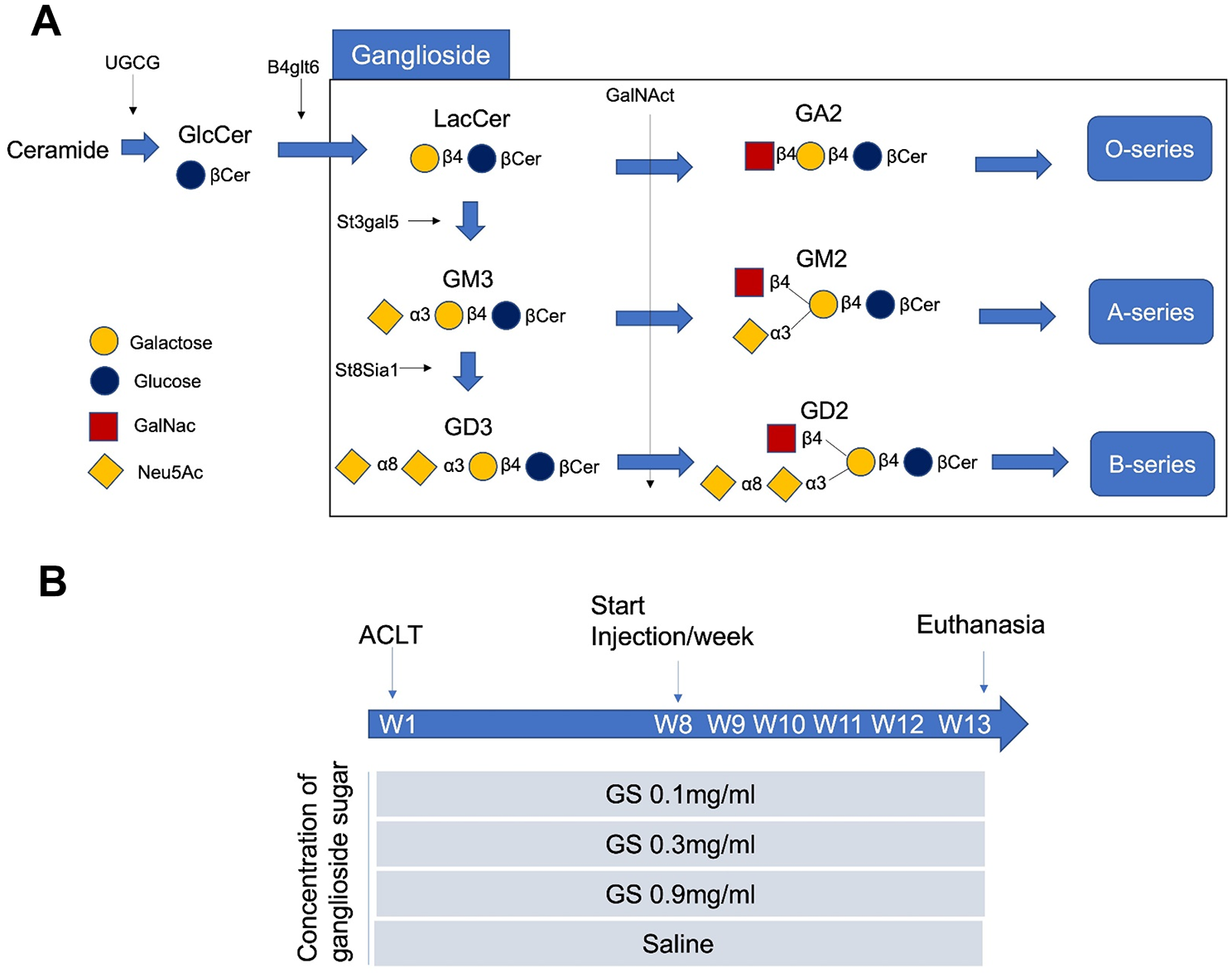
GSLs types and treatment procedure. (
The injections were performed weekly for 5 weeks post-surgery, and the joints were evaluated after 13 weeks (
Gross Morphological Assessment
All specimens exhibited complete transection of the ACLT at the time of sacrifice. Gross morphological assessment of the knee (9 knees in each group) was performed as the scoring system of the modified version of the Osteoarthritis Research Society International (OARSI) scoring system (Supplementary Table S1). 14 Briefly, India ink diluted with phosphate-buffered saline (PBS) (1:5) was used to stain the articular cartilage. The stained surfaces were photographed with a high-resolution digital camera (Nikon, Tokyo, Japan). The assessment was conducted in a blind manner.
Histological Assessment
The distal femurs from each knee (9 knees for each group) were fixed with 4% phosphate-buffered paraformaldehyde (Wako, Japan) for 24 h, decalcified in 10% ethylenediamine tetra-acetic acid solution (EDTA, pH 7.4) for 1 week, and then embedded in paraffin. After making 5-μm thickness sagittal sections from the center of the MFC in each sample, the sections were stained with Safranin-O. All sections were randomly examined in a blind manner and histological gradings were scored as recommended by the OARSI (Supplementary Table S2). 14
Lectin Histochemistry Assessment
Paraffin-embedded sections were deparaffinized in xylene, rehydrated through a series of graded alcohol solutions, and washed twice with PBS for 5 minutes. Sections were exposed to 30% H2O2 solution for 3 minutes to block endogenous peroxidase activity, washed twice with PBS and then incubated with Wisteria floribunda Agglutinin biotinylated probes (WFA, Vector Labs, Burlingame, CA, USA) overnight at 4°C. Sections were counterstained with hematoxylin, rinsed and mounted for microscopy. Sections of human quadriceps muscle were used as internal controls for optimizing the reaction and conditions. 15 Sections of 6 normal control rabbits’ knees were preliminary stained.
Friction Testing
A total 20 knees (5 knees in each group) were subjected to friction testing using a pendulum friction tester that was designed in our laboratory.
16
Soft tissues, except for tendons, ligaments, and joint capsules around the knee, were removed from the joint. After resection at the proximal end of the femoral shaft and at the distal end of the tibia, the knees were secured to polyethylene tubes with bone cement. During the testing, the joints were kept moist with an NS injection, and the tubes were attached to the pendulum friction tester. The friction testing was performed in a temperature-controlled environment to prevent variability that could affect the behavior of lubricants or tissue samples and the testing chamber was equipped with a humidity control system to ensure consistent moisture levels, which can affect the friction of biological materials. The total mass of the frame of the pendulum and the pendulum weight was 40 N.
17
After allowing 1 minute for preloading, the displacement was measured between Laser displacement sensor 1 (LDS 1: LK-G30, Keyence Corp., Osaka, Japan) and Laser displacement sensor 2 (LDS 2: LK-G80, Keyence Corp., Osaka, Japan) mounted on the upper plate of the frame (
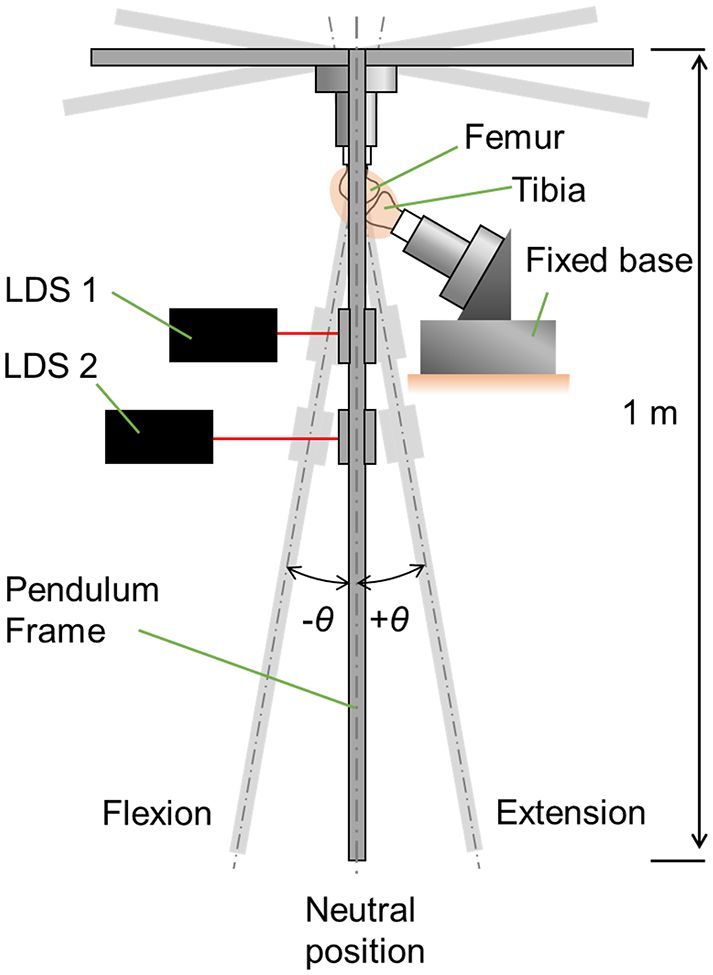
Pendulum friction tests. Schematic illustration of the pendulum friction tester used in this study.
Statistical Analysis
A power analysis was performed to determine the sample size based on previous studies13,20 (data not shown), and the sample size was considered sufficient for each statistical analysis. The statical analysis was performed using Kruskal-Wallis test for gross morphological assessment and histological assessments and one-way analysis of variance for lectin histochemistry assessment and friction testing followed by Tukey’s multiple-comparison procedure (GraphPad Prism, San Diego, USA). A P value of less than 0.05 was considered statistically significant. The histological assessments and scoring were performed by blinded observers to prevent bias. Histological slides were randomized during evaluation to avoid bias related to processing order or evaluator fatigue. Intra- and interobserver reliability was assessed on 3 randomly selected knees from each group. Two observers (M.H. and T.E.) independently assessed 12 knees were measured twice in a blinded manner at 1-month intervals. The intraclass correlation coefficients for intraobserver reliability were 0.93 (M.H.) and 0.91 (T.E.), and the intraclass correlation coefficient for inter-observer reproducibility was 0.92.
Results
Macroscopical Observations of Knees After Treatment by Different Dosages of Ganglioside Sugars
All knees exhibited joint effusion with synovial fluid and mild to severe degenerative changes in the cartilage surfaces. The degenerative changes in the cartilages were more severe in the femoral condyle compared to the surfaces in the tibial plateau. Of note, Saline and 0.1 mg/ml groups showed greater cartilage erosion mainly at the medial femoral condyle (MFC) (
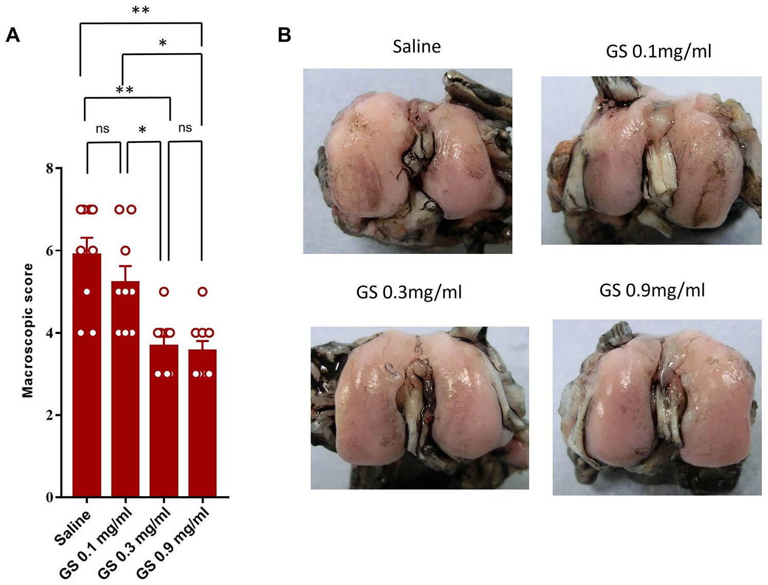
Macroscopic assessment of the medial condyles from each group 13 weeks after surgery. (
Histological Observations of Knees After Treatment by Different Dosages of Ganglioside Sugars
The rabbits that had been treated with concentrations of 0.3 and 0.9 mg/ml solutions of ganglioside sugar exhibited significantly lesser erosions in cartilage than these observed in animals that received 0.1 mg/ml and saline (
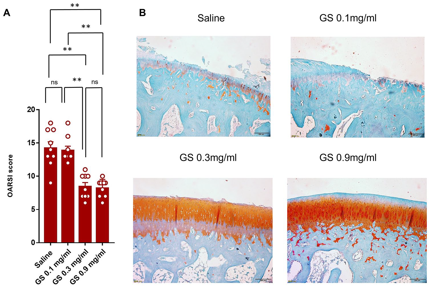
Histological observations of stained sections of the medial femoral condyles by Safranin-O. (
Lectin Histochemistry Staining for Detecting GalNAc-Positive Chondrocytes in Cartilage After Treatment
Furthermore, sections of cartilage were subjected to WFA staining for the detection of GalNAc. The WFA staining of chondrocytes and inter-territorial matrix was positive for the majority of intact cartilage areas (mainly superficial zone of cartilage) (
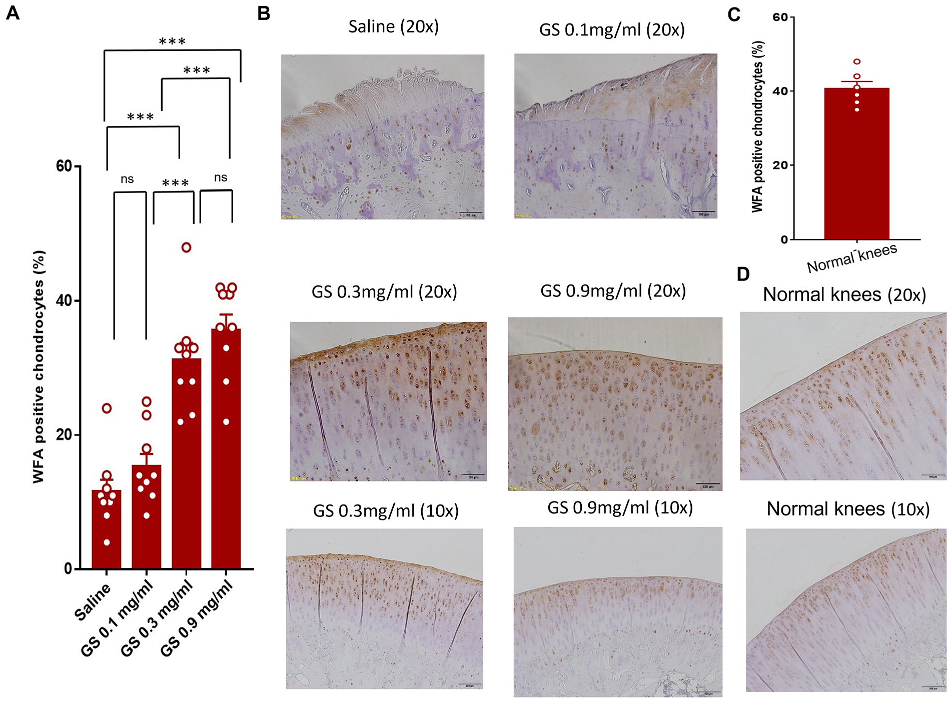
WFA-histochemical staining of medial femoral condyles from each group. (
Friction Testing of Knees After Treatment by Different Dosages of Ganglioside Sugars
To gain further insights into the frictional behavior of the articular cartilages under stress, frictional testing was performed. Importantly, the coefficients of the knees from rabbits that received 0.3 and 0.9 mg/ml solutions of ganglioside sugars were significantly lower than these for the saline-treated rabbits (
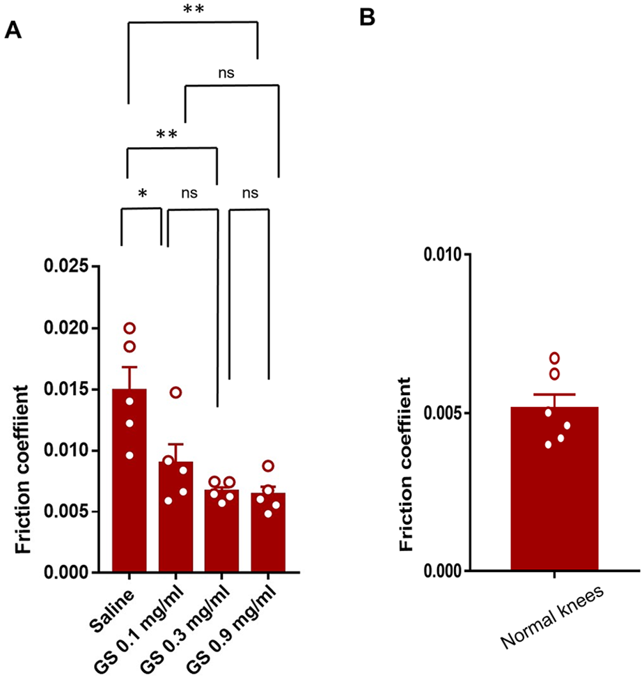
Assessment of pendulum friction tests. (
Discussion
The OA is a degenerative disease of the joint that remains the most prevalent type of arthritis worldwide with no treatment available to prevent the progression of the disease. Given the importance of developing a new treatment, in this study, we explored the antiarthritic effects of the intra-articular administration of ganglioside sugars in an ACLT-induced OA model. Our results demonstrated that this treatment significantly alleviated the progression of OA by suppressing the degenerative changes in articular cartilage.
In our study, OA was induced in rabbits by ACLT, which is the most common model for evaluating the effects of treatment,21,22 characterized by the occurrence of severe cartilage degeneration in the medial femoral condyle and lateral condyle.23,24 Bilateral surgeries of knees were performed and severe degeneration in cartilage of the ACLT rabbits were observed in medial femoral condyle followed by lateral condyle. Despite the use of this severe model, macroscopical and histological observations as well as friction tests of cartilage revealed that the intra-articular administration of ganglioside sugars has the potential for exerting a preventive effect on the progression of OA. It is noteworthy that in a joint lubrication test, the average values for the friction coefficient of rabbit ACLT-induced knee OA were increased to 0.015 after the treatment, and the recorded values were comparable to values that were previously reported for intact knees.20,25 Importantly, the ganglioside sugar treatment resulted in a remarkable increase in WFA staining, which is indicative of an increased level of GalNAc on chondrocytes. In fact, a weak reaction to GalNAc is associated with the severity of cartilage damage in OA.15,25-27 These results suggest that the therapeutic effects of ganglioside sugars could arise from the anabolic effects of GalNAc on chondrocytes, since this treatment suppresses the expression of genes for matrix metalloproteinases in chondrocytes and cartilage catabolism induced by interleukin-1.12,28 Likewise, the pharmacological potentials of gangliosides have been documented in several experimental models. For example, it has been reported that exogenous gangliosides rescue neuronal cells from death after trophic factor deprivation 29 and promote the regeneration of sciatic nerve defects after repair with cryopreserved peripheral nerve allografts. 30 We used a mixture of GA2, GM2, and GD2 in this study based on previous research. 12 in which 50μM concentration was used in vitro. Taking consideration of molecular weight (ganglioside concentrations in biological tissues are typically much lower) and range intended to test both low, medium, and high doses to observe the potential dose-dependent effects of the ganglioside sugars on the experimental outcome, as well as past studies using the rabbit OA mode, 20 we employed concentrations of (0.1, 0.3, 0.9 mg/ml) in our study. Our study highlights potential beneficial effect of GalNac supplementation in OA. A more complete understanding of this mechanism would help in establishing an adequate treatment protocol, including the interval and frequency of injections needed. We plan to explore a broader concentration range in follow-up studies to better define the upper and lower limits of the therapeutic window and dose response relationships.
The major limitations of this study include that only one postoperative time point was examined, the sham surgery group is absent from the experimental groups, and results were obtained using a rabbit model of 7 months. In fact, there is possibility of the incomplete skeletal maturity of the 7-month-old Japanese white rabbits used in this study as Masoud et al. 31 reported that 7- to 8-month-old New Zealand white rabbit exhibit incomplete matured skeletal maturity. The possibility of incomplete skeletal maturity might affect this study’s outcomes. Therefore, considering clinical applications, examining the therapeutic effects of ganglioside sugars in a large and older and other animal OA models and comparison with existing OA treatments would be of great interest in terms of gaining additional evidence of the effects. Moreover, in this study, we used bilateral surgeries model of ACLT, which is usually more severe than unilateral ACLT model. In contrast to the findings reported by Gushue and colleagues that higher joint contact forces pass through the lateral compartment in rabbits’ knee, 32 our model led to severe damage in the medial compartment of the cartilage. Finally, biological effects of ganglioside sugars on synovium and pharmacodynamics of ganglioside sugars were not clarified. This emphasizes the need for future study aims at clarifying metabolism and the fate of ganglioside sugars in the joint as well as their pharmacodynamics for treatment of OA. Future research should focus on addressing the limitations of this study while expanding the scope to more clinically relevant models. Studies with sham surgery, long-term follow-up, mechanistic insights, and human trials will be critical to confirm the therapeutic potential of ganglioside sugars. This will help bridge the gap between preclinical research and clinical application, ensuring that the findings translate into meaningful benefits for OA patients.
In conclusion, our results indicate that the intra-articular administration of ganglioside sugars is an effective strategy for preventing the degeneration of articular cartilage and improving joint lubrication of OA knees in ACLT model. These findings suggest that ganglioside sugars have promising potential as an effective agent for the treatment of OA.
Supplemental Material
sj-docx-1-car-10.1177_19476035241311542 – Supplemental material for Intra-Articular Administration of Ganglioside Sugars Protects Cartilage from Progressive Degeneration in an Instability OA Rabbit Model
Supplemental material, sj-docx-1-car-10.1177_19476035241311542 for Intra-Articular Administration of Ganglioside Sugars Protects Cartilage from Progressive Degeneration in an Instability OA Rabbit Model by Masanari Hamasaki, Tomohiro Onodera, Junichi Furukawa, Masahiro Todoh, Yuma Sakai, Taku Ebata, Mohamad Alaa Terkawi, Kentaro Homan and Norimasa Iwasaki in CARTILAGE
Footnotes
Acknowledgements and Funding
Authors thank Mrs. Eriko Tagishi for their kind help and technical support in our laboratory. This study was supported by a Challenging Exploratory Research Grant from the Japan Society for the Promotion of Science (grant no. AI24659661). The sponsors of the study had no roles in study design, data collection, data analysis, data interpretation, or writing of the report. All authors had full access to all data of this study and had final responsibility for the decision to submit for publication.
Author Contributions
Study conception and design: M.A.T., T.O., and N.I. Acquisition of data: M.H., T.E., K.H., and Y.S. Analysis and interpretation of data: M.H., T.O., J.F., and M.T. Drafting: M.H., M.A.T., T.O., and N.I. All authors contributed significantly to this research and have seen and approved the contents of the manuscript.
Declaration of Conflicting Interests
The author(s) declared no potential conflicts of interest with respect to the research, authorship, and/or publication of this article.
Ethical Approval
Data Availability
The datasets used and analyzed during the current study are available from the corresponding author on reasonable request.
References
Supplementary Material
Please find the following supplemental material available below.
For Open Access articles published under a Creative Commons License, all supplemental material carries the same license as the article it is associated with.
For non-Open Access articles published, all supplemental material carries a non-exclusive license, and permission requests for re-use of supplemental material or any part of supplemental material shall be sent directly to the copyright owner as specified in the copyright notice associated with the article.