
Abstract
Objective
Meniscus progenitor cells (MPCs) have been identified as promising candidates for meniscus regeneration, and it is crucial for us to understand meniscus injury repair mechanism at the cellular level. In this study, we investigate the biological properties of MPCs isolated from different species using the differential adhesion to fibronectin (DAF) technique. We aim to characterize MPCs in different species and evaluate the feasibility of these models for future meniscal investigation.
Design
MPCs were isolated from freshly digested meniscus from rat, rabbit, goat, and human cells using DAF. Biological properties, including proliferation, colony-forming, multilineage differentiation, and migration abilities, were compared in MPCs and their corresponding mixed meniscus cell (MCs) population in each species.
Results
MPCs were successfully isolated by the DAF technique in all species. Rat MPCs appeared cobblestone-like, rabbit MPCs were more polygonal, goat MPCs had a spindle-shaped morphology, human MPCs appear more fibroblast-like. Compared with MCs, isolated MPCs showed progenitor cell characteristics, including multilineage differentiation ability and MSC (mesenchymal stem cells) markers (CD166, CD90, CD44, Stro-1) expression. They also highly expressed fibronectin receptors CD49e and CD49c. MPCs also showed greater proliferation capacity and retained colony-forming ability. Except for goat MPCs showed greater migration abilities than MCs, no significant differences were found in the migration ability between MPCs and MCs in other species.
Conclusion
Our study shows that DAF is an effective method for isolating MPCs from rat, rabbit, goat, and human. MPCs in these species demonstrated similar characteristics, including greater proliferation ability and better chondrogenic potential.
Introduction
The meniscus is a fibrocartilage situated within the knee joint. It is crucial for maintaining joint stability, facilitating load transmission, and providing lubrication. 1 Injury to the meniscus significantly affects its normal functioning.2-4 Although meniscal repair is often the preferred therapeutic option, the rate of successful healing remains variable, ranging from 42% to 88%. 5 In scenarios where the injury happened in the avascular area or repair efforts prove unsuccessful, meniscectomy becomes an ultimate resort, with the potential consequence of early-onset knee osteoarthritis (OA). 6 Novel approaches, including meniscus allograft transplantation and tissue engineering reconstruction, have attracted increasing attention. However, they remain in the early stages of development,4,7-9 and more pre-clinical assessment and long-term observation are needed to ensure the safety and efficacy before broader clinical application.
Tissue-resident stem/progenitor cells have emerged as key players in tissue regeneration.10-12 Over recent years, an increasing interest has been directed toward meniscus progenitor cells (MPCs). Researchers have successfully isolated MPCs according to their colony-forming capacity,13-17 migration potential,18-21 marker expression,22,23 and differential adhesion to fibronectin (DAF).24-27 DAF was initially used in the investigation of cartilage progenitor cells (CPCs), 28 and its recent adaptation in meniscal studies has proven to be a facile and practical approach in MPCs isolation.24-27
However, human MPCs were mostly isolated from meniscus from patients with OA,13,15,24,26,27 while the animal MPCs were from normal meniscus.14,16,17 Since researchers also observed a significant shift in meniscus cell types in OA meniscus compared with normal meniscus in recent single-cell studies,29,30 MPCs derived from OA meniscus may fail to represent the cellular characteristics in other conditions. Apart from that, animal and human meniscus were reported to have different regeneration abilities, while meniscus could heal automatically in rat and rabbit, this was not reported in caprine and human.14,31,32 Therefore, characteristics of MPCs from different species is still not clear. Since the establishment of an appropriate animal model holds unique value in facilitating in-depth research on certain diseases and their treatment,16,20,21,33 it can not only enable researchers to elucidate the underlying mechanisms of tissue-resident stem/progenitor cells’ response to meniscus regeneration or degeneration but can also validate novel therapies and monitor their effectiveness. To clarify whether findings in DAF isolated MPCs from OA human still hold in healthy, acute-injured and surgery-induced OA meniscus or vice versa is the prerequisite for future in-depth study. Besides, existing studies evaluate the properties of MPCs using various methods and perspectives, making it hard to draw a comprehensive conclusion, thus adding to the uncertainty of the effective translation from preclinical to clinical application.
In this study, we isolated MPCs from acutely injured human meniscus and the most commonly employed animal models in meniscal research, including rat, rabbit, and goat using the DAF technique, and characterized their biological properties. Through this, we hope to verify the existence of MPCs in different animal model and further prepare their applications in future investigations.
Methods
Meniscus Specimen Acquisition
The study was approved by the Institutional Review Board of Sun Yat-sen Memorial Hospital (Approval number: SYSKY-2023-1115-01). The human meniscus was harvested during arthroscopic meniscectomy from 3 donors (age 26-31) diagnosed with acutely torn meniscus. Written informed consents were provided by the participants.
The following protocol for the use of the animals was approved by the IACUC of the Sun Yat-sen University. Menisci from 6 male Sprague-Dawley (SD) rats (180-220 g), 6 male New Zealand white rabbits (1.8-2.5 kg), and 3 male goats (20 ± 3 kg) were used in all experiments. After sacrifice, the knee joints were separated, and menisci were extracted for experiment use.
MPCs Isolation
MPCs were isolated from menisci by DAF, according to previous reports (Fig.1).24,25,27 Briefly, 6-well plates were coated overnight at 4°C with 10 μg/ml fibronectin (Sigma-Aldrich, United Kingdom) in phosphate-buffered saline (PBS) with 1 mM MgCl2 and 1 mM CaCl2. Plates were washed in serum-free medium before use.
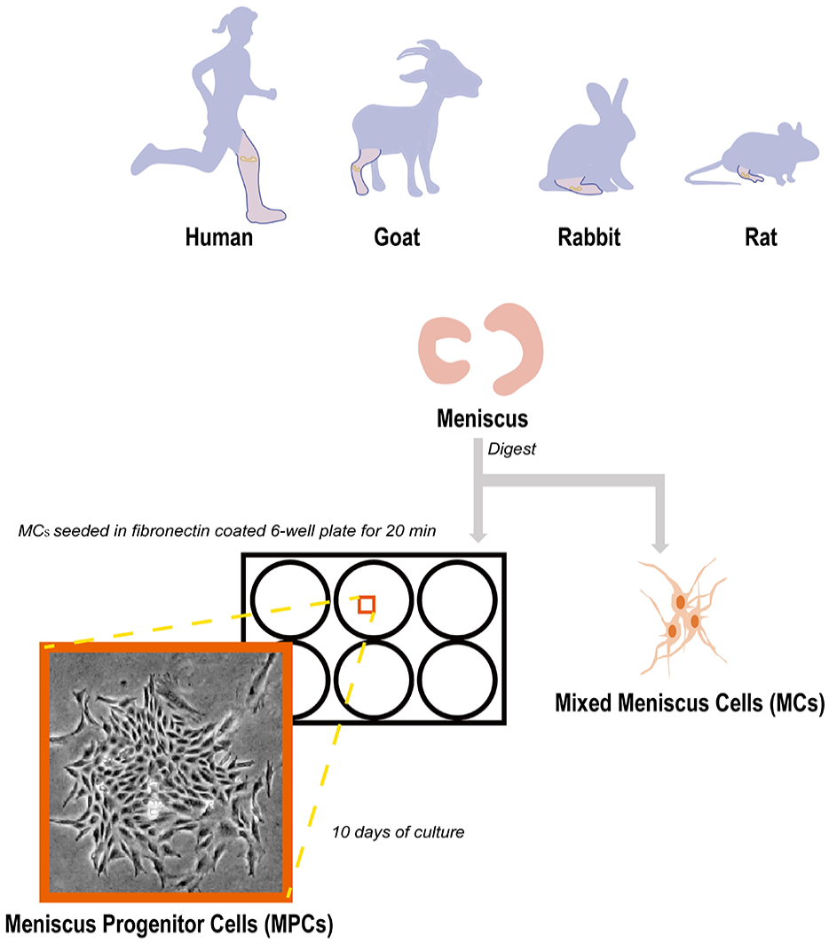
Flow diagram schematic of the experimental design.
Menisci were minced into 1 mm3 pieces, sequential digested in 2 mg/ml pronase (70 U/ml, 1 hour at 37°C) in Dulbecco’s Modified Eagle Medium (DMEM) (Gibco-BRL, Inc.) for 1 hour at 37°C, followed by 2 mg/ml collagenase type II (245 U/ml, 12 hours at 37°C; Worthington, United States) in DMEM at 37°C overnight.
For MPCs isolation, cells were seeded onto the coated plates at a low density of 200 cells/cm2 (2,000 cells/ml) for 20 minutes at 37°C in DMEM with 1% P/S allowing for adhesion. After 20 minutes, supernatant containing non-adherent cells was discarded. Two milliliters of fresh DMEM supplemented with 10% FBS and 1% P/S were added into each well. After 10 days of culture, colonies containing more than 32 cells were considered MPC colonies, and photos of colonies in each species were taken by Olympus IX73 inverted Microscope (Olympus, IX73; Japan). Progenitor colonies from each species were trypsinized by 0.25% trypsin/EDTA (Gibco-Thermo Fisher Scientific, United States) and reseeded; MPCs at passage 1 to passage 4 were used in this study.
After tissue digestion, a mixed population of meniscus cells (MCs) was directly transferred to monolayer culture in DMEM with 10% fetal bovine serum (FBS) (Gibco, United States) and 1% penicillin–streptomycin (P/S) (ThermoFisher Scientific, United States). The complete culture medium was changed every 3 days. Cell morphology Images were taken by Olympus IX73 inverted Microscope. MCs at passage 1 to 4 were used.
Multilineage Differentiation Potential of MPCs
For osteogenic differentiation, cells were seeded at a density of 1.5 × 10⁵ per well in a 6-well plate with osteogenic induction medium (Procell Life Science & Technology Co., Ltd., PD-014). The induction medium was changed every 3 days. After 21 days of differentiation, cells were washed with PBS, fixed in 4% Paraformaldehyde Fix Solution (PFA) for 3 minutes, rinsed gently with PBS, and stained with 0.2% Alizarin Red S solution (Solarbio, G1450) for 30 minutes.
For adipogenic differentiation, cells were seeded at a density of 1.5 × 10⁵ per well in a 6-well plate in the adipogenesis induction medium (Cyagen Biosciences Inc., GUXMX-90031) and incubated for 21 days following the manufacturer’s instruction. Cells were washed with PBS, fixed in 4% PFA for 3 minutes, rinsed gently with PBS, and stained with Oli Red O solution (Cyagen) for 30 minutes. Images were taken by Olympus IX73 inverted Microscope.
For chondrogenic differentiation, 4 × 10⁵ cells were centrifuged into a cell pellet with chondrogenic induction medium and cultured for 28 days according to the manufacturer’s instruction (Cyagen Biosciences Inc., HUXMA-90042). Three pellets from each cell type and species were used for biochemical quantitation of GAG/DNA and historical analysis.
GAG/DNA Analysis
Each pellet was digested in 200 µl of papain digestion solution containing 125 µg/ml papain (BioFroxx, 1364GR005), 20 mM EDTA (BioFroxx, 1340GR500), and 20 mM cysteine-HCl (Macklin, L805178) at 60°C for 3 hours. After papain digestion, all samples were centrifuged at 200 g for 5 minutes, and supernatants were stored at −30°C before use. The total GAG content was measured by Blyscan Sulfated Glycosaminoglycan assay kit (Biocolor, UK). The DNA content was measured using PicoGreen dsDNA Assay kit (Invitrogen). The GAG content was normalized to the corresponding DNA content per pellet for analysis.
Histology
Three pellets from each sample were processed for cryosections at 8 μm thickness. Cryosections were fixed in 4% PFA for 30 minutes and stained in 1% toluidine blue O solution (Leagene, DA0052) for 5 minutes.
Cell Counting Kit-8 (CCK-8) Assay
MPCs and MCs from each species were seeded onto a 96-well plate at 3 × 10³ cells/well and cultured in DMEM supplemented with 10% FBS and 1% P/S in a humidified incubator. After 1, 3, and 5 days of culture, 10 μl of the CCK-8 solution (Dojindo, Kumamoto, Japan) was added to each well of the plate; after incubated for 3 hours, the absorbance at 450 nm was measured using a microplate reader.
Cell Cycle Analysis
MPCs (1 x 10⁶) and MCs from each species were fixed in ice-cold 70% ethanol overnight and washed with PBS. Cells were incubated in FxCycle™ PI/RNase Staining Solution (Invitrogen, USA) for 20 minutes at room temperature in dark and analyzed using a BD FACSVerse™ flow cytometer.
Colony-Forming Assay
For the Colony-forming assay, 104 MPCs and MCs from each species were seeded in a 6 cm2 plate. After 7 days of culture, colonies were fixed with 4% PFA and stained with 1% crystal violet. Images were taken by Nikon Ni-U Microscope.
Flow Cytometry
Cells (5 × 10⁵) were harvested for each sample and incubated in PBS with 5% (w/v) bovine serum albumin (BSA; BioFroxx) at 4°C for 30 minutes for non-specific antibody-binding blockade. The cells were then stained with antibodies against stem cell markers (CD166, CD105, STRO-1, CD29, CD44) or adhesion molecules (CD49b, CD49c, CD49e) at 4°C for 30 minutes. Cytometric analysis was performed using a BD FACSVerse™ flow cytometer. Results were further analyzed using FlowJo software.
Migration Assay
MPCs (1 x 10⁵) and MCs from each species were seeded in 12-well plates. The monolayer cells were scratched by a 1,000 µl sterile pipette tip after reaching confluency and cultured in an FBS-free culture medium. Wound closure was monitored by imaging at 0 and 24 hours later. Each experiment was performed in triplicate. The wound area was quantified by a Wound healing size tool plugin 34 using ImageJ/Fiji Software. 35
Statistical Analysis
GraphPad Prism (Version 9.3.0, San Diego, California, USA) was used for statistical analysis. Unpaired t-test was used to analyze the GAG/DNA assay, CCK-8 proliferation assay, cell cycle analysis, and cell migration assay of MPCs and MCs within each species. Data were presented as mean ± standard deviation (SD) in the graphs and text; P < 0.05 indicated a significant difference.
Results
Morphology of MPCs and MCs in Different Passages
After seeding onto the fibronectin-coated 6-well plates, MPCs were able to adhere within 20 minutes and form colonies 10 days late in all species (
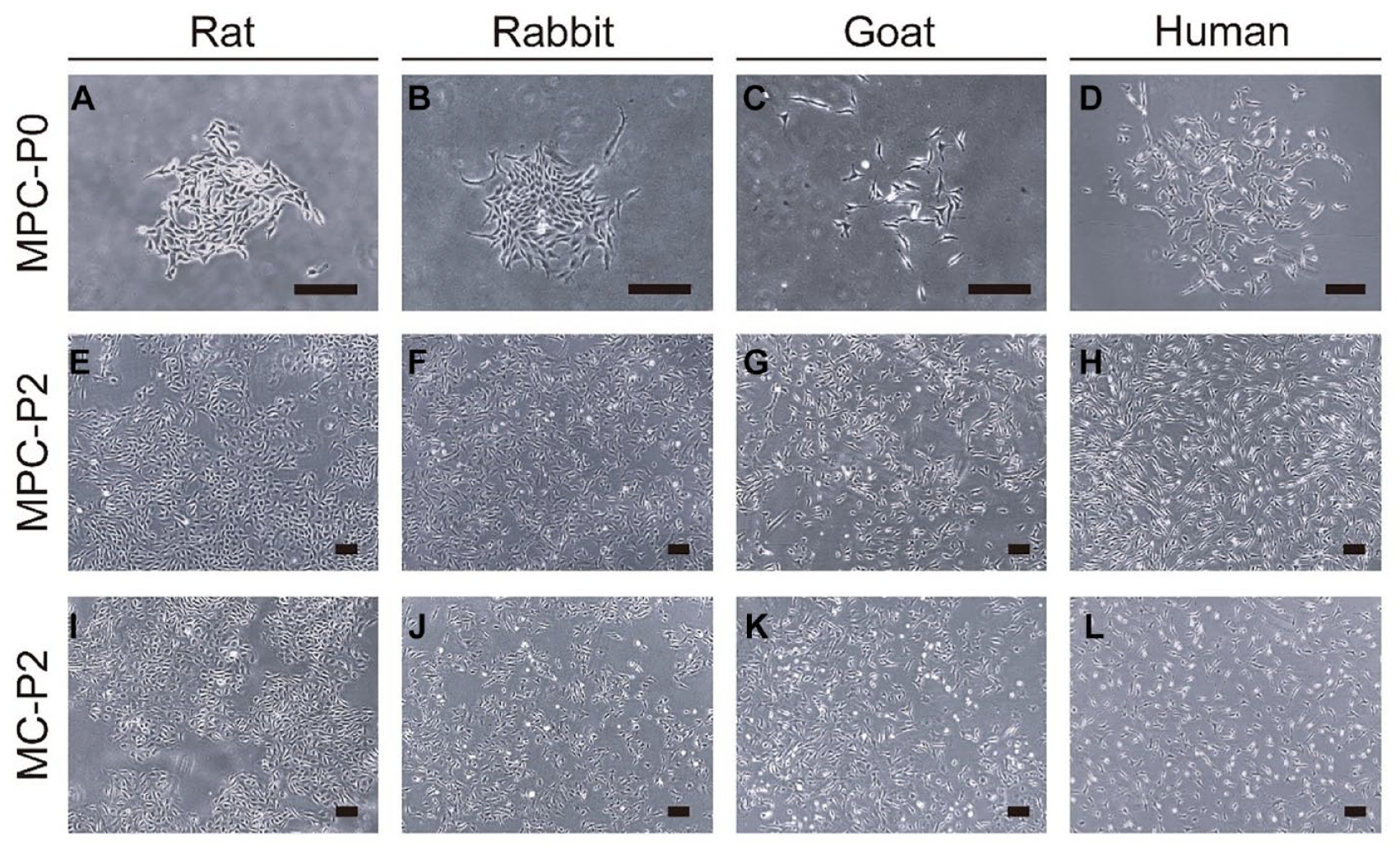
Representative images of meniscus progenitor cells (MPCs) and meniscus cells (MCs) isolated from rat (
While MCs and MPCs had similar morphology within each species (
MPCs Showed Enhanced Chondrogenic, Adipogenic, and Osteogenic Differentiation Abilities Than MCs
After 21 days of inducing culture in 6-well plate, MPCs showed more oil red (
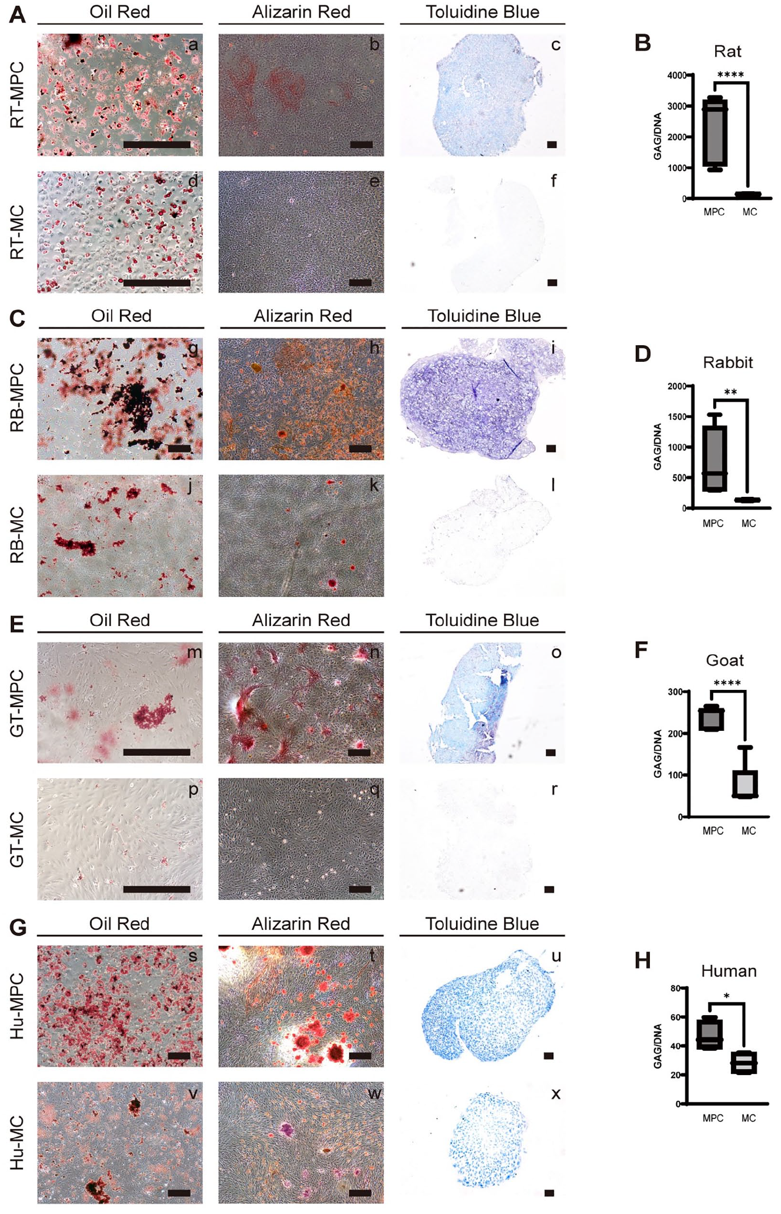
Adipogenic, osteogenic, and chondrogenic differentiation of MPCs and MCs in rat (
The Expression Profile of MPCs and MCs
Surface molecular expressions of MPCs and MCs in each species were measured by flow cytometry (
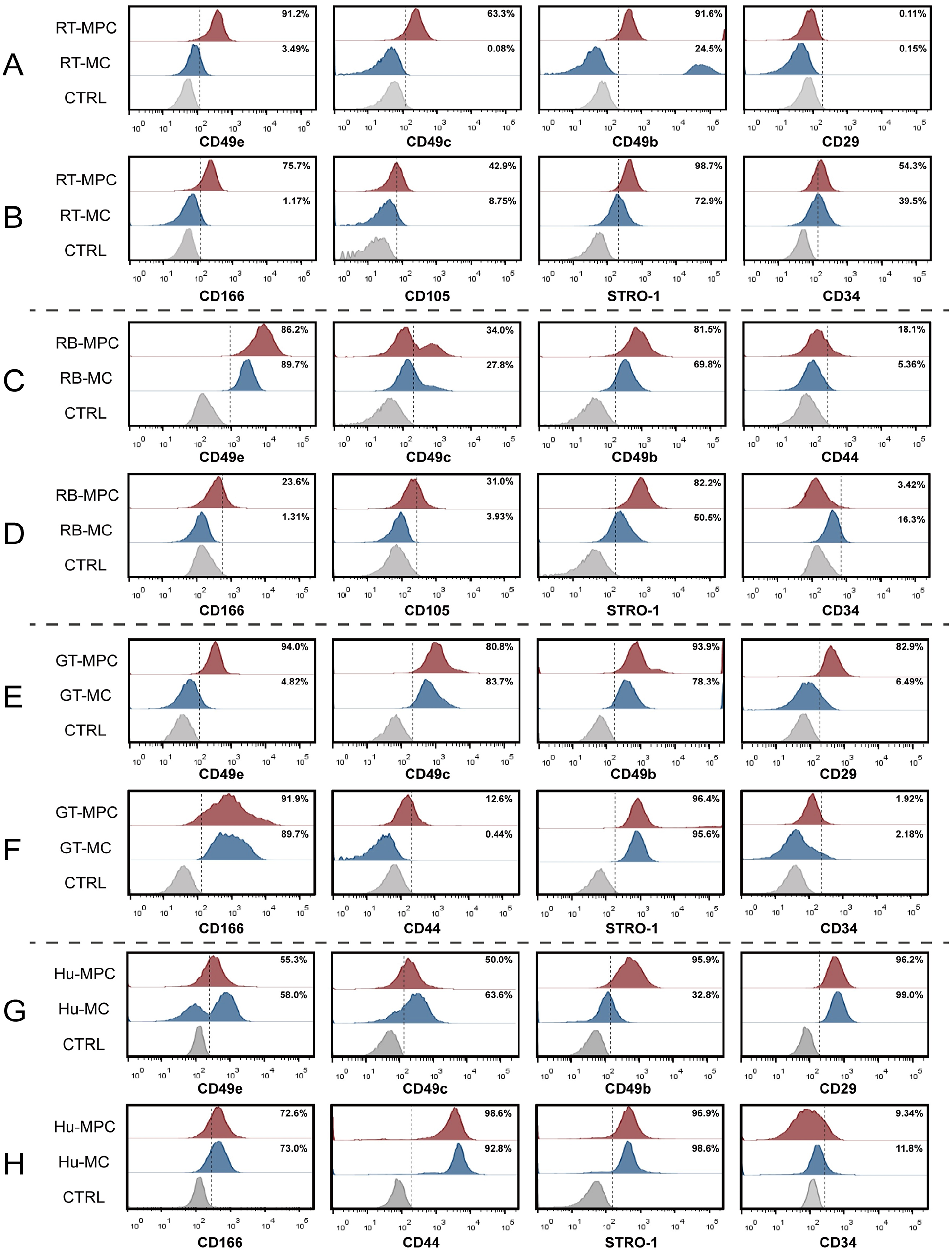
Flow cytometry plots of typical adhesion molecules expression and MSC markers expression in rat (
MPCs Exhibited Greater Proliferation Ability Than MCs
The CCK-8 proliferation assay showed that MPCs grew faster than MCs from day 1 of culture; this difference was maintained on day 5 in rat (P = 0.0018), rabbit (P < 0.0001), goat (P = 0.0005), and human (P = 0.0006) (

(
Colony Formation Ability and Migration Ability Analysis Between MPCs and MCs
The colony-forming assay showed the MPCs retained great colony-forming ability (
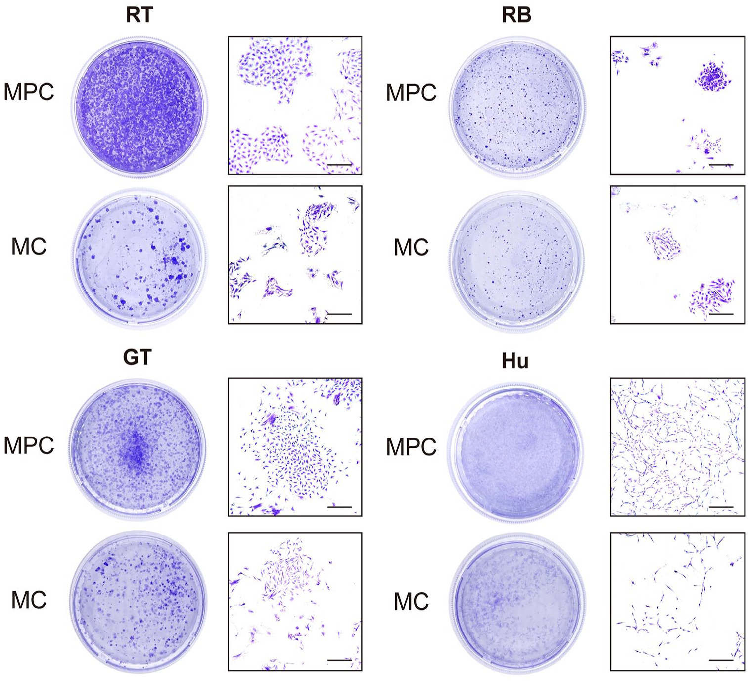
Representative images of colony-forming assay of MPCs and MCs in different species. Scale bar represents 5,000 µm. RT: rat; RB: rabbit; GT: goat; Hu: human.

(
Discussion
DAF method has been used in MPCs isolation from OA human meniscus in previous studies.24-27 However, while investigating the role of MPC in the meniscus and evaluating its therapeutic application largely rely on pre-clinical animal models, no studies have verified its presence and compared animal MPCs with human MPCs yet. In this study, we used DAF to isolate MPCs from rat, rabbit, and goat and studied their biological properties.
Fibronectin was present in developing cartilage 28 and OA cartilage. 36 It also played an important role in the initial reparative stage by facilitating cell migration and proliferation.37-41 Therefore, DAF was used to identify CPCs first by Dowthwaite et al. 28 and then gradually used by more studies.42,43 It was not until recently used in MPCs isolation.24-27 These studies mostly used samples from patients with OA who underwent total knee replacement (TKR).24-27 Whether MPCs can be isolated from healthy meniscus or acutely injured meniscus in human and other animal models was not clear. We showed that DAF is a facile method in extracting MPCs, not only from healthy meniscus of different animals but also from non-OA human. This suggested that MPCs are a group of innate resident cells in normal meniscus, which may respond to injury, degeneration, or whenever the normal static environment of the meniscus is disturbed.
CD49c and CD49e were fibronectin receptors,
44
Williams et al.
42
reported a distinct population of chondrocytes expressing CD49e within the viable full-depth chondrocyte population. We showed an increased proportion of CD49c positive subset in rabbit MPCs compared with MCs (
Although some animal non-separated MCs stained positive for MSC marker (
The white zone of the meniscus had more chondrocyte-like cells and was made up of 60% type II collagen.1,4 The lack of vascularity made it hard to initialize the repairing procedure. Even though some regeneration could be achieved, it was prone to form a fibrous or scar-like tissue, with more type I collagen and lack of GAG component,
4
which cannot provide ideal support to fully perform its load-transmission role. Studies in cartilage cells showed that even chondrocytes gradually lost their phenotype and produced more type I collagen than type II collagen after several passages during in vitro culture,48,49 and the enhanced chondrogenic ability of MPCs (
It was reported that meniscus structural features were more similar in sheep and human but different in rabbit.
54
In addition, spontaneous healing of meniscus was reported in rat and rabbit but limited in caprine and human.32,55 Our results showed that MPCs isolated from healthy menisci in rat, rabbit, goat, and injured menisci from non-OA donors had equivalent characteristics, suggesting that these animals could be candidate models in meniscus investigation concerning stem/progenitor cells. However, it is important to note that although MPCs from different species showed improved biological characteristics compared with MCs, some differences still existed across species. For instance, MPCs from different species have different morphologies (
Some limitations are worth noting. Since it was not eligible to obtain normal human meniscus tissue, we used surgical debris from young acute injury meniscus which was not repairable with meniscectomy (donor age range, 26-31). These teared tissues were mainly generated by knee violent force within young donors, and the majority of the samples were still under almost “healthy condition.” Therefore, we considered human meniscus tissue used in this study as “normal tissue.” In addition, the meniscus was a heterogeneous tissue,
1
and MPCs from different regions may also demonstrate features with slight difference.
27
Human sample used in our study did not cover the whole cellular heterogeneities of meniscus (mainly from white-white region). Besides, injury may also cause microenvironment changes of meniscus which might alter the biological characteristics of human meniscal cells in this study. Altogether, these might be a possible explanation for the differences in multilineage differentiation ability (
In conclusion, DAF was an effective method for isolating MPCs from different species. Isolated MPCs showed stem/progenitor-cell-like properties: the ability to undergo multilineage differentiation and the expression of MSC surface markers. MPCs from these species demonstrated similar biological characteristics; they showed greater proliferation ability and outstanding chondrogenic potential compared with MCs.
Footnotes
Acknowledgments and Funding
The author(s) disclosed receipt of the following financial support for the research, authorship, and/or publication of this article: This study was financially supported by grants from the Natural Science Foundation of Guangdong Province for Distinguished Young Scholars (2020B1515020014) and National Outstanding Youth Science Fund Project of National Natural Science Foundation of China (82022046).
Declaration of Conflicting Interests
The author(s) declared no potential conflicts of interest with respect to the research, authorship, and/or publication of this article.
Ethical Approval
The study was approved by the Institutional Review Board of Sun Yat-sen Memorial Hospital (Approval number: SYSKY-2023-1115-01). Written informed consent for inclusion in this research was obtained from the patients prior to surgery.