
Abstract
Study Design
Retrospective, descriptive observational study.
Objective
The need for revision surgery after mandibular fractures is an indicator for severe postoperative complications. This study aimed to characterise this patient cohort, describe solutions to deal with complications and evaluate treatment quality as a risk variable for complications.
Methods
Patients with revision surgery with refixation after open reduction and internal fixation (ORIF) of a mandible fracture were included. Patient- and therapy-specific information were assessed together with postoperative complications. The quality of fixation was evaluated individually by 6 specialists. Interobserver agreement was analysed using Fleiss’ kappa.
Results
Out of 630 patients, inclusion criteria were met by 17 patients (14 male, 3 female) with an average age of 43.3 (±15.5) years. Complications at the mandible body/angle/symphysis led to refixation in all cases. Main indications for refixation were osteomyelitis (52.9%) or pseudarthrosis (41.2%). Risk factors were drug-related immune suppression, local infection or substance abuse (nicotine, alcohol or drugs). Six patients did not present any of these predictors. Of these, treatment of 4 patients was rated as not in accordance to the AO principles. The interrater reliability of treatment quality assessments was .239.
Conclusions
Patients with risk factors need to be carefully observed perioperatively after ORIF of mandibular fractures and treatments need to be adapted to these patients. Discrepancies of treatments to common guidelines may also be an independent predictor for treatment failure in patients without risk factors. Current treatment guidelines should be re-evaluated concerning additional treatment strategies for patients with specific risk factors.
Introduction
The mandible is one of the most common trauma sites of the face with young male assault victims being most frequently affected. 1 Depending on the type of mandibular fracture, treatment options include non-surgical techniques, closed reduction involving intermaxillary fixation (IMF) and open reduction and internal fixation (ORIF). 2 Despite the high incidence of mandibular fractures, there is still a lack of evidence to support treatments using a single approach. 2 Thus, treatment is usually based on the clinician’s prior experience and individual circumstances. Nevertheless, different recommendations/principles exist concerning fracture types and possible surgical or non-surgical treatment options given by the ‘Arbeitsgemeinschaft für Osteosynthesefragen’ (AO) or Champy.3-5
Independent of the chosen treatment, postoperative complication rates after mandibular fractures have been described as around 6.6%–21.2%.6,7 Complications include numbness, malocclusion, abscess formation, infected hardware, osseous mal/nonunion leading to pseudarthrosis and hardware exposure/extrusion or even fracture.6,7 Independent risk factors for postoperative complications have been identified, particularly: time from injury to treatment, tobacco use and dental extraction. 7 Other studies also discussed the use of osteosynthesis plates, alcohol abuse, angular fracture location, complex fractures, type of antibiotic used and comorbid illnesses (human immunodeficiency virus, diabetes mellitus), but evidence of these results is limited due to missing multivariate analyses.6,8,9
A clear indicator of a major failure of initial fracture treatment is the performance of revision surgery with refixation. This procedure has previously been identified as a primary outcome measure in a study by Scolozzi et al. 10 In that study, 63 patients with severe mandibular fractures and fixation using 2.4 mm titanium reconstruction plates for fixation were analysed and a refixation rate of 3% was detected. 10 Since complications like numbness, malocclusion or wound healing disorders may also be temporary, the rate of specifically major complications indicated by the need for refixation has not been analysed by other studies.
Therefore, the purpose of this study was to 1) define predictors and indications for revision surgery with refixation after mandibular fractures, 2) verify the practicability of the AO-guidelines for mandibular fracture treatments and 3) evaluate the treatment quality as a further risk factor for major complications. The investigators hypothesise that previously defined risk factors for complications will be confirmed and that patients without presentation of known risk factors have been treated in disparity to the AO-guidelines. The specific aims of this study were to: 1) define the patient collective with refixation revision surgery after mandibular fracture treatments more clearly to increase alertness to surgeons and describe solutions to deal with complications 2) evaluate the need for an update of current treatment guidelines
Patients and Methods
Study Design/Sample
A single-center, retrospective, descriptive observational study was performed. All patients with primary ORIF of a mandibular fracture at the department of Oral and Maxillofacial Surgery, Charité – Universitätsmedizin Berlin, between April 2017 and December 2021 were screened. Inclusion criteria were the need for revision surgery with refixation in the postoperative course. Patients <18 years, with their primary treatment at a different clinic and primary treatment other than osteosynthesis (e.g. mandibulo-maxillary fixation) were defined as exclusion criteria. Ethical approval was given by the local ethics committee (EA2/189/21).
Variables
Predictors to analyze were substance abuse (yes/no) including alcohol, nicotine, drug abuse, immune suppression, local infection at the mandible fracture site and quality of treatment. The main outcome variable was the revision surgery with refixation after mandibular fracture treatment. Covariates were patient- and therapy-specific information and postoperative complications.
Patient-specific information included gender, occlusion status, pre-existing illnesses and relevant medication.
Therapy-specific information included the date of initial and revision surgery and mandibular fracture site, accident mechanism (assault, traffic or sports accident, fall, pathological fracture), fracture site (corpus, angle, ramus, condyle) and pattern (simple/complex and single/multiple with regard to AO criteria 11 ), soft tissue defect at the fracture site (yes/no), information on primary and revision treatment including type of refixation (miniplates, conventional reconstruction plate, patient-specific reconstruction plate) and surgical approach (intra-/extraoral), information on iliac crest bone grafting (yes/no), antibiotic treatments after surgeries (yes/no) and histopathological results.
Information on postoperative complications included the indication (pseudarthrosis, osteomyelitis and material failure) and mandibular site causing revision, wound healing disorder (yes/no), abscess formation (yes/no), fistula (yes/no), plate exposure (yes/no), pain (yes/no), malocclusion (yes/no) and material failure (yes/no).
Data Collection Methods
Data collection was performed using clinical records from patient files. Data were recorded using Microsoft Excel (v.16.6, Microsoft Corporation, Redmond, USA). Observation of follow-up ended on October 15th, 2022. Occlusion was defined as any dental contact between the upper and lower dental arch. For the evaluation of treatment quality, 6 senior consultants/specialists in Oral and Maxillofacial Surgery (MG, CD, SK, KK, MH, CR) were asked to evaluate if treatment was performed according to the AO principles by answering a decision question (yes/no). 4 Pre- and initial postoperative radiographs (computed tomography scans, panoramic radiographs and cone-beam computed tomography scans) of all included patients were presented with the same number of randomly chosen patients, which did not meet the inclusion criteria (no revision surgery with refixation). Demonstration was performed in a blinded and random manner. Thus, the identification of cases with complications should be avoided. Insufficient treatment was defined as a minimum of 50% agreement of specialists. Critical treatment was defined as 33.3% of specialists evaluating treatment as insufficient.
Data Analyses
Descriptive statistics were performed using SPSS (SPSS (version 27.0.1.0, IBM Corp, Armonk, New York, USA). Interrater reliability was evaluated using Fleiss’ kappa coefficient.
Results
630 patients were screened initially. 17 patients (2.7%) underwent revision surgery with refixation in the postoperative course and met the inclusion criteria.
Patient Characteristics and Primary Treatment Information.
(SD = Standard deviation; BMI = Body mass index; IMF = Intermaxillary fixation, * both patients were edentulous).
After primary surgery, all patients received antibiotic treatment, on average for 4.8 (±2.2) days. 13 patients (75.5%) received amoxicillin/clavulanic acid, and 4 patients (23.5%) clindamycin due to allergies. Apart from 1 patient with an infected fracture, primary antibiotic application was prophylactic.
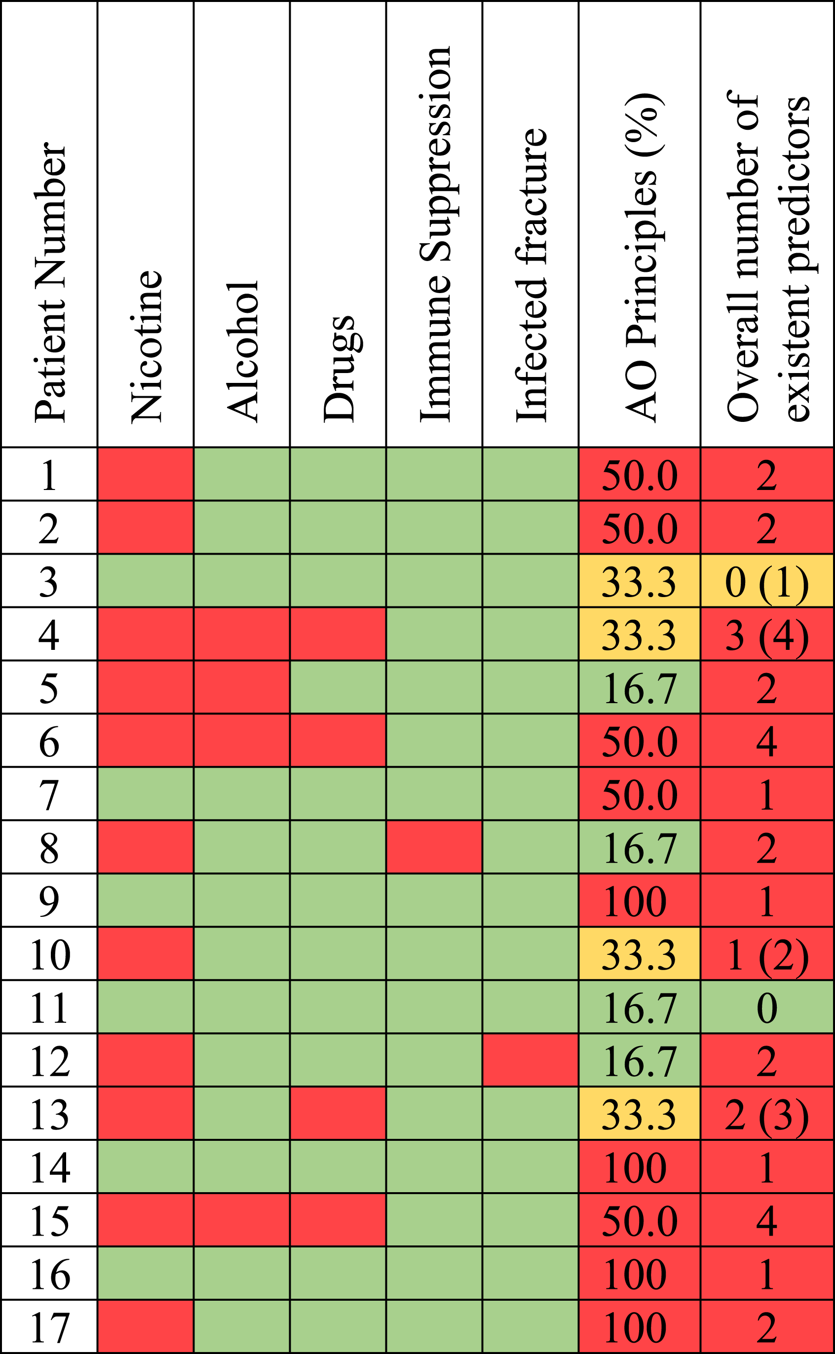
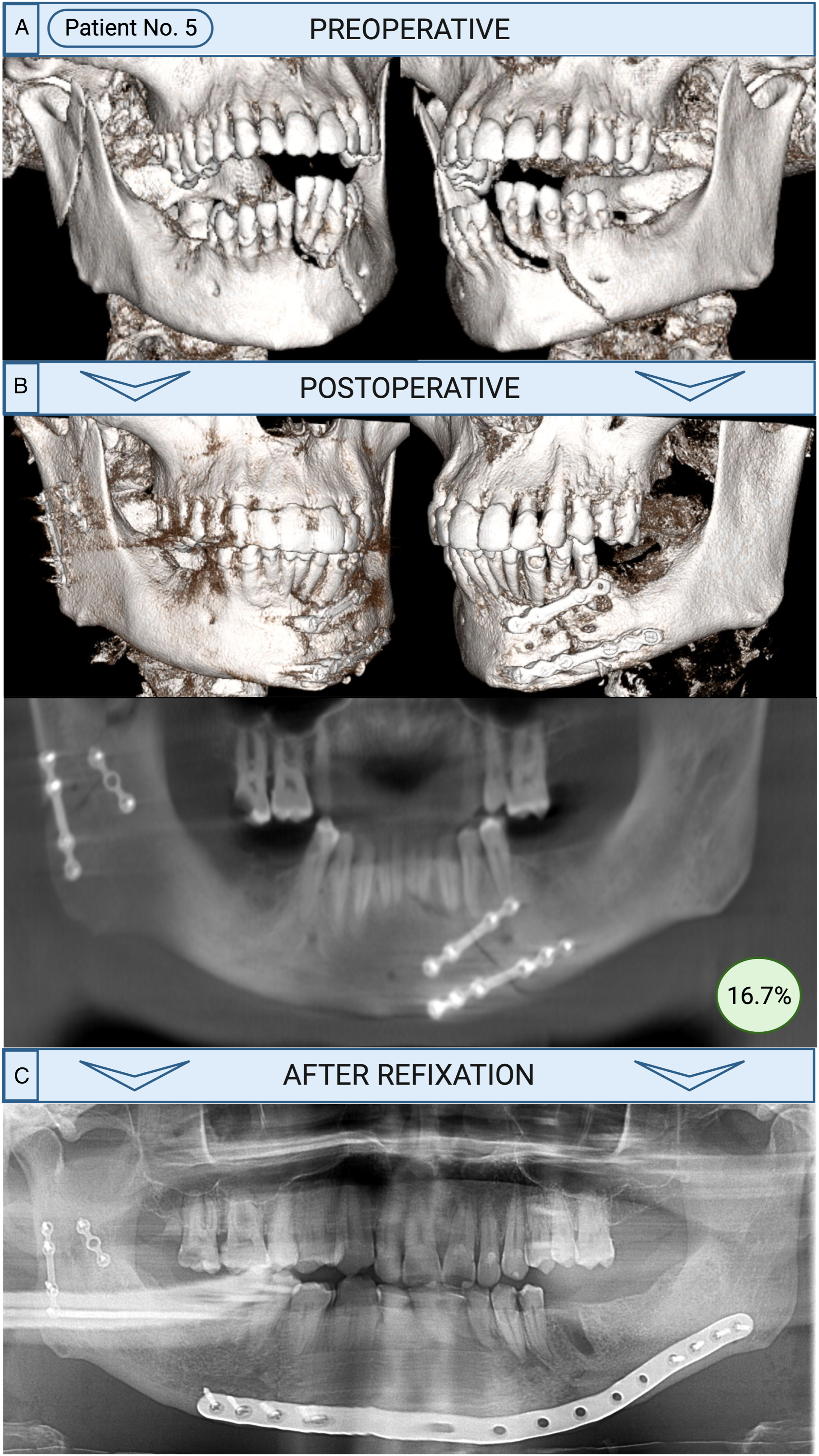
Risk factors were presented by 64.7% including immune suppression, infected fractures or nicotine, alcohol or drug abuse. Six patients (35.3%) did not present any of these risk factors. Of these, 4 patients were not treated according to the AO principles (rated by 50.0% or more specialists as insufficient treatment) and 1 (5.9%) patient (patient No. 3) had critical treatment according to the AO principles (rated by 33.3% of specialists as insufficient treatment). Examples of critical and insufficient treatments are shown in Figure 1. One patient (5.9%) did not present any risk factors (patient No. 11) and only 1 of the observers rated this ORIF as insufficient (Figure 2). This patient demonstrated a fracture of the mandibular angle. A non-retained tooth 48 was not removed and this patient developed an acute osteomyelitis with clinical signs of abscess formation and pain after 1 month. Examples of different insufficient fracture treatments in panoramic radiographs. Revision surgery with refixation after development of complications following initial fracture treatment. 
There were 8 patients (47.1%) with combination AO treatment failure and the presence of any patient risk factors Table 2. In 4 patients (patient 9, 14, 16 and 17), all specialists (100%) rated fracture treatment as insufficient according to AO principles. In all other 13 patients, evaluation differed between specialists, but all initial fracture treatments were rated as a failure of AO principles by at least 1 specialist. The interrater reliability was .239.
Predictors for Refixation. Presence of nicotine, alcohol and drug abuse, immune suppression, infected fractures and failure of treatment according to AO principles (50.0% or more ratings as insufficient treatment) is analysed and marked in red if present. Critical treatment (33.3% ratings as insufficient treatment) according to AO principles is marked in yellow if present. Green indicates the absence of a predictor.
Complications After Initial ORIF and Revision With Refixation.
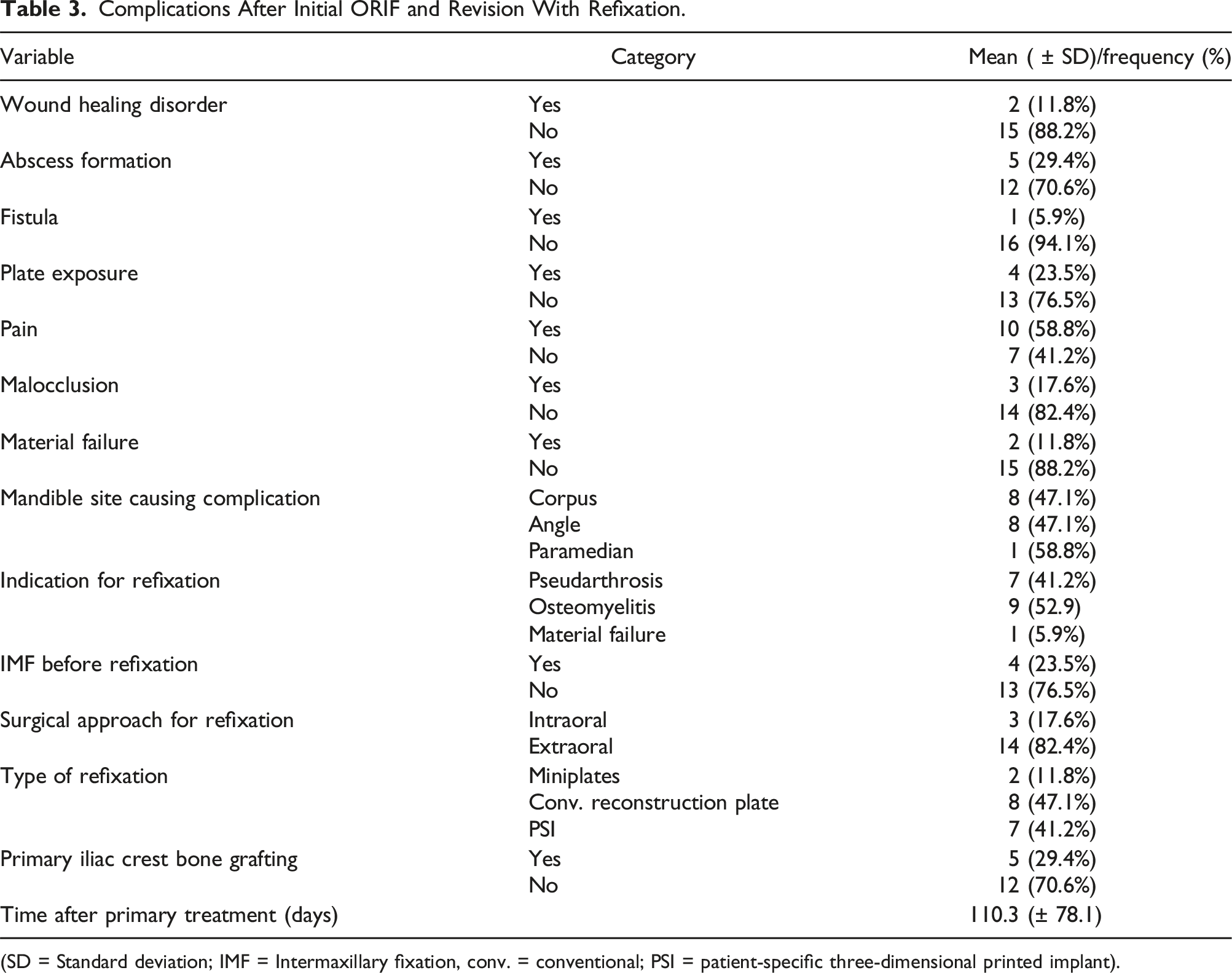
(SD = Standard deviation; IMF = Intermaxillary fixation, conv. = conventional; PSI = patient-specific three-dimensional printed implant).
Refixation after development of complications following initial fracture treatment.
Discussion
The purpose of this study was to 1) define predictors and indications for revision surgery with refixation after mandibular fractures, 2) verify the practicability of the AO-guidelines for mandibular fracture treatments and 3) evaluate the treatment quality as a further risk factor for major complications. The results of this study demonstrate that the patient cohort with re-fixation revision surgeries mostly presents previously identified risk factors for complications and exclusively fractures at the mandibular angle, body or parasymphysis. Furthermore, treatment quality is suggested as a further risk variable for postoperative complications, and current treatment guidelines should be re-evaluated concerning further support tools for surgeons and more specificities.
Generally, with only 2.7% of refixation after mandibular fractures, the patient cohort with severe complications after mandibular fractures is small. The rate of refixation is in accordance with a study by Scolozzi et al, who found a refixation rate of 3%. However, in that study, ORIF with 2.4 mm titanium reconstruction plates exclusively was analysed in 63 patients. 10 Other studies have not reported any refixation rates, although this procedure is an indicator of severe treatment complications.
The results of our study show that patients who undergo a refixation are predominantly male victims of assaults, with open single or multiple mandibular fractures, which always exhibit 1 fracture site at the mandibular angle, corpus or paramedian region. In all patients, these fracture sites were identified as the region of complication and 88.2% were rated as open fractures due to the presence of a tooth in the fracture gap. According to Ellis et al., the presence of a tooth in line of the fracture does not significantly increase the risk of postoperative complications. 12 However, there are still recommendations addressing the question of tooth removals in cases of impaired teeth including fractured roots, partially impacted teeth with pericoronitis or extensive periapical lesions. 13 In our study, all patients with refixations received a prophylactic antibiotic treatment following the initial fracture reduction but osteomyelitis was the reason for treatment failure in every second patient. Given the high number of patients with open fractures due to presence of teeth, a causal relationship of osteomyelitis and presence of teeth can be suspected. However, if the indication for teeth removals to reduce major complications in this specific patient cohort should be given more generously, needs to be assessed by case-control studies.
With 64.7% of patients presenting at least 1 previously identified risk factor for the development of complications after mandible fractures including immune suppression, infected fractures due to delayed treatment or nicotine, alcohol or drug abuse, our findings are in accordance with other studies.6-9 In our cohort, almost every second patient (41.2%) presented multiple factors (Table 3). However, there were 6 patients (35.3%) without the presence of any classical risk factors for postoperative complications. The blinded evaluation by the specialists indicates that 5 of these patients were evaluated with an insufficient treatment quality. In 3 of these 5 patients, there was even a complete agreement (100%) by the specialists. There was only 1 patient (5.9%) who did not present any known risk factor or insufficient osteosynthesis rated by the specialists (Figure 2). The data indicate that the quality of osteosynthesis, which is a component of general treatment quality, 14 is a major factor for long-term success in the treatment of mandibular fractures.
Generally, there is still no consensus concerning a specific approach to mandibular fracture treatment. 2 The AO-guidelines give evidence-based directions concerning favoured treatments and therefore treatment quality.3,4,11 Mittermiller et al. have previously evaluated the interobserver reliability of the AO CMF classification and found weaknesses concerning the level 3 variables (fracture morphology). 15 The assessments of anatomic areas (level 1) and the location of the fractures within specific areas (level 2) demonstrated a higher agreement between observers. Similar to the lower agreement of level 3 variables, the interobserver reliability (.239) in our study is only at a fair level according to Landis and Koch. 16 Apart from 4 patients, there was always at least 1 different rating. Therefore, our study and the results of Mittermiller indicate, that some fractures are difficult to classify, and treatments are sometimes debatable, making general compliance with treatment guidelines difficult. The presence of 5 patients without classical risk factors but complications due to insufficient treatment quality in this study, is an indicator of the need for further support tools for surgeons or the necessity for an update of the current AO-guidelines. For example, the quality of fracture reduction or plate adaption is currently not assessed by the guidelines. Because only 1 patient received Schuchardt arch bars initially, fixation stability may have been underestimated. Furthermore, the guidelines lack specific advises concerning the length of plates or the distance of the screw holes from the facture line. In addition, adaptions to patients’ risks should be introduced, because patients with risk factors might benefit from the prolonged use of intermaxillary fixation or preferential use of load-bearing osteosynthesis plates. The low interrater reliability furthermore shows that the current guidelines may not be easy to apply for every case.
In contrast to the quality of osteosynthesis, postoperative antibiotics and radiographs, as further possible factors of treatment quality, seem to be negligible. A prospective, randomised clinical trial including 144 patients could not find evidence for the need for a prolonged antibiotic treatment after ORIF of mandibular fractures. 17 Also, the results from a meta-analysis including 3285 patients did not reveal any need for prophylactic antibiotics. 18 There are no data in the current literature concerning the number needed to treat in order to prevent infections. 19 Also in our study, all patients received antibiotics postoperatively, but complications could not be prevented. Previous studies also concluded that postoperative panoramic radiographs in order to control fracture reduction add little to the physical examination and history in the detection of complications. 20
Since immediate postoperative occlusion is considered to be a good indicator of treatment quality, many surgeons do not recommend routine postoperative radiographs.21,22 In our clinic, all patients received a postoperative radiograph, but nevertheless, refixation was needed. Standardised intraoperative radiographs allow the optimisation of surgical outcomes of mandibular fractures and therefore spare patients from repeated interventions, especially in complex fractures.23,24 Radiographic examinations are also helpful to confirm osseous union before the removal of fixation materials. 25 Therefore, the correct indication for radiographs should be carefully chosen.
Furthermore, individualised treatment solutions may also increase treatment quality in challenging cases. Our study included 2 patients suffering from atrophic mandibular fractures. El-Gengehi et al found evidence for the beneficial use of computer-based surgical guides for fracture reductions in edentulous mandibles. 26 The combination of patient-specific crib-shaped implants combined with autogenous free bone grafts has been introduced as another promising solution.27,28
Patient-specific implants (PSI) have been used in more than 40% of patients for revision surgeries, indicating the benefits of these plates in terms of mechanical integrity and computerised workflow. 29 The treatment of infected mandibular fractures has previously been recommended to include decortication and primary bone grafting. 30 While decortication was part of every refixation surgery in this study, primary bone grafting was only performed in about 30%. Of all cases in this study, in which removal of the refixation plate was performed at a later stage due to successful bone healing, the rate of primary bone grafting was only 16.7%. This is an indicator, that even in cases receiving revision surgery, bone grafting is not necessary for a positive outcome in every case. Surgeons are advised to evaluate the need for bone grafting by assessing the quality of bony buttressing. Furthermore, in patients with infectious situations, Schuchardt arch bars or IMF screws should be considered in advance of refixation, as performed in 4 patients in this study.
There were several limitations in this study resulting from its retrospective design. Postoperative patient compliance concerning chewing and oral hygiene was not assessed. Furthermore, the experience of the surgeon at the time point of surgery was not assessable and radiographic demonstration of pre- and postoperative fracture situations was performed using image sections instead of entire 3D-visualisations. Nevertheless, image sections were chosen to allow proper assessments.
Conclusion
The patient cohort with revision surgeries with refixation is mainly characterised by fractures at the mandibular angle, body or parasymphysis in patients with risk factors including smoking, alcohol and drugs, immune suppression and infected fractures. Patients presenting these risks should therefore be carefully observed perioperatively after a mandibular fracture treatment. Discrepancies of treatments to common guidelines may also result in the need for revision surgery with refixation, which suggests treatment quality as a new risk variable for complications. Surgeons should adhere strictly to treatment plans as recommended by guidelines to minimise complications. However, a low interrater reliability of assessing fracture treatments demonstrates the possible need to re-evaluate current treatment guidelines concerning further support tools for surgeons and more specificities.
Footnotes
Declaration of Conflicting Interests
The author(s) declared the following potential conflicts of interest with respect to the research, authorship, and/or publication of this article: Max Heiland is Chair of the AOCMF European and Southern African Board.
Funding
The author(s) disclosed receipt of the following financial support for the research, authorship, and/or publication of this article: Dr Jan Oliver Voss and Dr Heilwig Fischer are participants in the BIH Charité Clinician Scientist Program funded by the Charité – Universitätsmedizin Berlin and the Berlin Institute of Health at Charité (BIH).