
Abstract
Objectives
The aim of this study was to investigate how different LED wavelengths, blue (450 nm), red (660 nm), and white (380-780 nm), influence the accumulation of glucosinolates and carotenoids in Chinese cabbage (Brassica rapa L. ssp. pekinensis) microgreens. Additionally, the study sought to evaluate how these spectral treatments affect the antibacterial activities of the microgreens against methicillin-resistant Pseudomonas aeruginosa strains.
Methods
Chinese cabbage microgreens were cultivated for 10 days under controlled environmental conditions using three LED light treatments: blue, red, and white. Phytochemical profiling, specifically glucosinolate and carotenoid quantification, was performed using high-performance liquid chromatography. Antibacterial activity was evaluated through disc diffusion assays against methicillin-resistant Pseudomonas aeruginosa to assess differences among LED-treated samples.
Results
Red LED irradiation significantly increased the total glucosinolate content in microgreens, whereas blue LED treatment resulted in the highest accumulation of total carotenoids. Red LED-treated microgreens exhibited the strongest antibacterial activity against multidrug-resistant Pseudomonas aeruginosa, suggesting a potential association between glucosinolate levels and antimicrobial performance. Conversely, blue LED-treated samples, despite containing higher carotenoid concentrations, showed comparatively weaker antibacterial effects.
Conclusion
These findings demonstrate that LED spectral quality can be strategically manipulated to regulate key phytochemicals in Brassica microgreens. Red LED light enhances glucosinolate accumulation and antimicrobial activity, while blue LED light promotes carotenoid production but not antibacterial efficacy. Overall, the study offers valuable insights into optimizing functional phytochemical production and improving the antimicrobial potential of microgreens for functional foods and controlled-environment agriculture.
Introduction
Chinese cabbage (Brassica rapa L. ssp. pekinensis) is regarded as one of the most important Brassica vegetables globally because of its extensive use in various culinary applications, including salads, beverages, and cooked dishes. 1 Beyond its gastronomic value, Chinese cabbage exhibits notable biological activities, such as antioxidant, 2 antibacterial, 2 and anti-inflammatory effects. 3 These health-promoting properties are primarily attributed to the rich composition of functional phytochemicals, including glucosinolates, phenolics, and carotenoids. 4
Carotenoids are naturally occurring pigments that impart yellow, orange, and red coloration to plant tissues, and are classified into two main groups: hydrocarbon carotenoids (such as α-carotene, β-carotene, and lycopene) and oxygenated carotenoids, also known as xanthophylls (including lutein, β-cryptoxanthin, astaxanthin, and zeaxanthin). 5 As animals lack the ability to endogenously synthesize carotenoids, these compounds must be obtained from dietary sources. 6 Carotenoids play a significant role in promoting human health, particularly in ocular protection and prevention of various diseases. 4
Glucosinolates, a class of secondary metabolites predominantly found in Brassica species, are classified into aromatic, aliphatic, and indolic groups based on their biosynthetic origins of phenylalanine, methionine, and tryptophan, respectively. 7 Upon enzymatic hydrolysis, glucosinolates yield a variety of biologically active compounds, including epithionitriles, nitriles, oxazolidinethiones, isothiocyanates, and thiocyanates, 8 several of which possess notable antimicrobial activities.9–11
Microgreens, which represent an intermediate developmental stage between sprouts and baby leaves, are distinguished by their high concentrations of bioactive constituents. 5 Notably, microgreens derived from Brassica species generally possess higher levels of nutrients and phytochemicals than their fully matured counterparts. 6 Consequently, the dietary inclusion of Chinese cabbage microgreens may confer substantial health benefits.
Light is a critical environmental factor influencing microgreen production and metabolite profiles. Among various light sources, light-emitting diodes (LEDs) have emerged as an essential technology in controlled environment agriculture (CEA) due to their prolonged operational lifespan, high energy efficiency, and ability to emit narrow-band wavelengths. 12 Broadband white light, which incorporates portions of the green spectrum, has been shown to enhance canopy penetration and support holistic plant development.13,14 The 400-700 nm spectral range, defined as photosynthetically active radiation (PAR), serves as the primary energy source for photosynthesis, with the red and blue regions being particularly effective at inducing the excitation of chlorophyll molecules. 15 Red light has been associated with enhanced photosynthetic capacity and vegetative growth, including increases in plant height, fresh and dry biomass, and leaf area.16,17 In contrast, blue light has been shown to influence chlorophyll accumulation, morphogenetic regulation, stomatal opening, and the accumulation of antioxidant compounds.18,19 In particular, previous studies have demonstrated that different LED spectra significantly alter the accumulation of metabolites in various micro-vegetables. For instance, blue LED irradiation has been shown to elicit an increase in phenolic compounds in canola (B. napus), 5 mustard (B. juncea), 20 Chinese cabbage (B. rapa ssp. pekinensis), 21 kohlrabi (B. oleracea var. gongylodes), 22 and Chinese kale (B. alboglabra Bailey) sprouts. 23 While blue light is a potent inducer of phenolics, white LED light has been reported to increase carotenoid accumulation and certain glucosinolates in kale microgreens 7 and mustard sprouts. 20 However, the effects of LED spectral quality on metabolite accumulation are highly species- and cultivar-dependent, indicating that spectrum-specific optimization is required for each plant genotype.
Accordingly, the present study aimed to determine the optimal LED light wavelengths, specifically blue (450 nm), red (660 nm), and white (380-780 nm), to maximize microgreen yield as well as the accumulation of carotenoids and glucosinolates, and to evaluate the antibacterial properties of Chinese cabbage microgreens produced under these lighting conditions.
Materials and Methods
Plant Materials and Growth Conditions
Chinese-cabbage seeds were obtained from Asia Seed Co., Ltd (Seoul, Republic of Korea). For germination, approximately 100 seeds were immersed in autoclaved distilled water for 24 h, and subsequently sown in plastic pots containing sterilized vermiculite. Seedlings were grown in a controlled environment chamber maintained at 25 °C and subjected to distinct LED light treatments: white (380-780 nm), blue (450 nm), and red (660 nm), each provided at a photosynthetic photon flux density of 90 μmol·m⁻2·s⁻1 and relative humidity of 70 ± 3.00%. A long-day photoperiod of 16 h light and 8 h dark was implemented. An LED lighting system (PARUS LED Co., Cheonan, Republic of Korea) provided the designated wavelengths for each treatment group. Three types of LEDs (blue, red, and cool white) were employed as light sources to investigate the effects of spectral quality. The blue LED exhibited a sharp emission peak at 450 nm with a full width at half maximum (FWHM) of approximately 20 nm. The red LED showed a peak at 660 nm with a similar FWHM of approximately 20 nm. The cool white LED was characterized by a high correlated color temperature of 65,000 K. Its spectral power distribution (SPD) featured a distinct 450 nm blue pump peak complemented by a broad phosphor-converted emission spanning the 500-700 nm range, ensuring coverage across the visible spectrum. To ensure high spectral uniformity, the LED lighting system was configured with two parallel panels positioned at an identical height above the growth area. The plant pots were placed directly beneath the center of these panels to minimize shading and ensure even light distribution. Each pot, containing 100 seeds, was treated as a single biological replicate, with three independent replicates established per treatment. Microgreens were harvested 10 days after the initiation of light exposure, immediately frozen in liquid nitrogen, and stored at −80 °C. The samples were then lyophilized and finely ground for subsequent high-performance liquid chromatography (HPLC) analysis of glucosinolate and carotenoid profiles, as well as for in vitro evaluation of antibacterial activity.
Carotenoid HPLC Analysis
Carotenoids were isolated from freeze-dried Chinese cabbage microgreens cultivated under red, blue, and white LED illumination following a previously described method. 7 Briefly, 100 mg of powdered sample was combined with 3 mL of 0.1% (w/v) ascorbic acid in ethanol and vortexed for 30 s. The mixture was then incubated at 85 °C for 5 min. To remove potential lipid contaminants, 120 μL of 80% (w/v) potassium hydroxide was added, and the mixture was placed on ice for 10 min. Subsequently, 100 μL of β-apo-8′-carotenal (25 ppm) was introduced as an internal standard, followed by the addition of 1.5 mL of HPLC-grade water. Extraction was performed by adding 1.5 mL of hexane, and the mixture was centrifuged at 1200 × g at 4 °C. The upper organic phase was collected and the aqueous phase was subjected to two additional extractions with equal volumes of hexane. The combined hexane layers were evaporated to dryness under a stream of nitrogen, and the residue was redissolved in a 1:1 (v/v) mixture of dichloromethane and methanol. The final extracts were transferred to HPLC vials for subsequent analyses. Chromatographic separation was performed on a YMC C30 column (250 × 4.6 mm, 3 μm; Waters Corporation, Milford, MA, USA) at 40 °C, using an Agilent 1100 HPLC system (Agilent Technologies, Palo Alto, CA, USA) equipped with a photodiode array (PDA) detector set at 450 nm. The mobile phase consisted of solvents A (methyl tert-butyl ether) and B (92% methanol containing 10 mM ammonium acetate) with the following gradient: 0 min, 90% B; 20 min, 83% B; 29 min, 75% B; 35 min, 30% B; 40 min, 30% B; 42 min, 25% B; 45 min, 90% B; and 55 min, 90% B at a flow rate of 1.0 mL/min.
Desulfo-Glucosinolate HPLC Analysis
Desulfoglucosinolates were extracted from freeze-dried Chinese cabbage microgreens cultivated under red-, blue-, and white-LED lighting following a previously described protocol. 7 In brief, 100 mg of powdered sample was homogenized with 0.5 mL of 70% methanol (v/v), and the mixture was subjected to ultrasonication at 70 °C for 5 min to inactivate endogenous myrosinase activity. The samples were centrifuged at 11,000 × g for 20 min at 4 °C. This extraction process was repeated twice to obtain sufficient crude extract. A DEAE-Sephadex A-25 mini-column (H⁺ form, pre-equilibrated with 0.5 M sodium acetate) was prepared for purification. The combined methanolic extracts were loaded onto a column and washed with HPLC-grade water to remove any impurities. Subsequently, 75 µL of arylsulfatase solution was added to the column, followed by incubation at 25 °C for 16 h to convert glucosinolates into their desulfo-forms. Elution was performed using HPLC-grade water (0.5 mL) and was repeated three times. The eluates were then filtered using a 0.45 µm hydrophilic PTFE syringe filter. Desulfoglucosinolates were separated using an Agilent 1200 HPLC system (Agilent Technologies, Palo Alto, CA, USA) equipped with a PDA detector. A reverse-phase Inertsil ODS-3 column (150 × 3.0 mm, 3 μm; GL Sciences, Tokyo, Japan) coupled with a guard column (10 × 2.0 mm i.d., 5 μm) was employed for chromatographic separation at 40 °C with detection at 227 nm. The mobile phase consisted of water (solvent A) and acetonitrile (solvent B), applied according to the following gradient program: 0-18 min, 7%–24% B; 18-32 min, hold at 24% B; 32.01 min, return to 7% B; and 32.01-40 min, hold at 7% B. The flow rate was maintained at 0.2 mL/min.
Antibacterial Activity Assay of Chinese Cabbage Microgreens
The antibacterial potential of Chinese cabbage microgreens cultivated under red-, blue-, and white-LED illumination for 10 days was evaluated using the disc diffusion method as previously reported. 7 Briefly, 150 mg of freeze-dried Chinese cabbage microgreen powder was extracted with 30 mL of methanol by vortexing for 10 s, followed by continuous agitation at 120 rpm for 24 h at ambient temperature. The resulting extract was filtered through standard filter paper, and the filtrate was concentrated under reduced pressure using a rotary vacuum evaporator. The dried residue was dissolved in dimethyl sulfoxide to a final concentration of 100 mg/mL. A panel of human pathogenic bacterial strains was employed for antimicrobial screening, including Vibrio parahaemolyticus (KCTC 2471), Salmonella typhimurium, Bacillus cereus (KCTC 3624), Staphylococcus mutans (KCTC 3065), Staphylococcus aureus (KCTC 3881), Micrococcus luteus (KCTC 3063) and Pseudomonas aeruginosa (KCCM 1113, 1378, 1731, 0225, 0254, 0826, p01827, and p01828). Each strain was pre-cultured overnight in nutrient broth at 37 °C until reaching an optical density at 600 nm (OD₆₀₀) of 0.5. For the disc diffusion assay, 100 μL of bacterial suspension was incorporated into molten sterile nutrient agar prior to solidification, and the mixture was poured into Petri dishes. Once solidified, sterile paper discs impregnated with Chinese cabbage extract (equivalent to 2 mg extract per disc) were placed on the agar surface, with three discs per plate. Plates were incubated at 37 °C for 24 h, after which the diameters of inhibition zones were measured to determine antibacterial efficacy. A clinically isolated multidrug-resistant P. aeruginosa strain, previously shown to be resistant to β-lactams, aminoglycosides, and fluoroquinolones, was used in this study. 24
Statistical Analysis
Duncan's multiple range test and t-test data from glucosinolate and carotenoid HPLC analyses and antibacterial activity assays were performed using SAS software version 9.2 (SAS Institute Inc., Cary, NC, USA).
Results
Growth Characteristics of Chinese Cabbage Microgreens Irradiated with White-, Blue-, and Red-LED Lights
Shoot length and fresh weight of Chinese cabbage microgreens were significantly affected by LED light quality, whereas root length showed no significant differences among treatments (Table 1). Blue-LED illumination produced the greatest shoot elongation, followed by white- and red-LEDs, while red-LED treatment resulted in the lowest shoot length. Similarly, fresh weight was highest under blue-LEDs and lowest under red-LEDs. Collectively, these results suggest that LED light quality exerts an influence on shoot-related growth in Chinese cabbage microgreens.
The Effects of Different LED Illumination on Fresh Weight, Shoot, and Root Length of Chinese Cabbage Microgreens.

Glucosinolate Contents in Chinese Cabbage Microgreens Irradiated with White-, Blue-, and Red-LED Lights
Using HPLC analysis, nine glucosinolates were identified and quantified in Chinese cabbage microgreens cultivated under white-, blue-, and red-LED illumination. Among the detected glucosinolates, progoitrin was the most abundant in all treatments. According to Duncan's multiple range test, the red-LED treatment resulted in a significantly higher total glucosinolate content than white- and blue-LEDs. Specifically, progoitrin, glucoalyssin, gluconapin, glucobrassicanapin, and 4-methoxyglucobrassicin were the most abundant in microgreens grown under red-LED light, whereas the highest glucobrassicin content was observed under white-LED light. In contrast, blue-LED treatment generally resulted in the lowest concentrations of most glucosinolates, except for neoglucobrassicin, which was similar between blue- and red-LEDs. These results indicate that LED light quality significantly affects glucosinolate accumulation in Chinese cabbage microgreens (Table 2).
Glucosinolate Contents in Chinese Cabbage Microgreens Irradiated with White-, Blue-, and Red-LED Lights (µmol/g dry Weight).

Carotenoid Contents in Chinese Cabbage Microgreens Irradiated with White-, Blue-, and Red-LED Lights
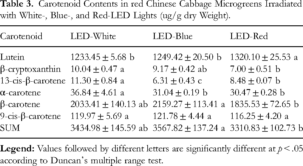
Using HPLC analysis, six carotenoids were identified and quantified in Chinese cabbage microgreens cultivated under white-, blue-, and red-LED illumination. Among these, β-carotene was the most abundant in all treatments. According to Duncan's multiple range test, the blue-LED treatment resulted in the highest total carotenoid content, which was significantly greater than that observed under red-LED light, whereas the white-LED group showed intermediate levels. Specifically, microgreens grown under red-LED exhibited the highest lutein content, significantly exceeding those grown under white- and blue-LEDs. In contrast, β-cryptoxanthin, 13-cis-β-carotene, and α-carotene were most abundant in microgreens under white-LED, with significantly lower levels detected in the red-LED group. For β-carotene, the highest concentration was observed in the blue-LED treatment, whereas 9-cis-β-carotene levels did not differ significantly among the three light conditions. These results demonstrate that the LED light quality has a pronounced effect on carotenoid accumulation in Chinese cabbage microgreens (Table 3).
Carotenoid Contents in red Chinese Cabbage Microgreens Irradiated with White-, Blue-, and Red-LED Lights (ug/g dry Weight).
Antibaterial Effect of Chinese Cabbage Microgreens Irradiated with White-, Blue-, and Red-LED Lights
The antibacterial efficacy of Chinese-cabbage microgreens cultivated under various LED light spectra was assessed using a disc diffusion assay (Figure 1 and Table 4). Extracts were prepared from microgreens grown for 10 d under white-, blue-, and red-LED illumination. For the tested pathogenic bacterial strains, extracts derived from microgreens exposed to red-LED light generally exhibited comparable or slightly superior antibacterial activity, as indicated by the inhibition zone diameters. In particular, Staphylococcus aureus (KCTC 3881) showed a significantly larger inhibition zone under red LED treatment compared with blue LED exposure (p < .05). In contrast, no statistically significant differences in inhibition zones were observed among LED treatments for Salmonella typhimurium, Micrococcus luteus, Bacillus cereus, or Vibrio parahaemolyticus, although minor variations in inhibition diameters were detected. Similarly, ten bacterial strains, including normal and methicillin-resistant pathogens, were evaluated for susceptibility (Table 4 and Figure 1). For methicillin-resistant strains, extracts obtained from microgreens exposed to red-LED light generally exhibited superior antibacterial effects, as evidenced by the largest inhibition zones. Specifically, P. aeruginosa strains 0225 and 0254 exhibited significantly greater inhibition when treated with red-LED-derived extracts than with blue-LED. Likewise, the inhibition zone for P. aeruginosa p01828 markedly increased under red LED irradiation relative to that under blue LED irradiation. Conversely, no statistically significant differences in inhibition were observed among the treatments with P. aeruginosa strains 1113 and 1731 (Table 4 and Figure 2). In summary, exposure to red-LED light appeared to enhance the antibacterial potential of Chinese cabbage microgreens, particularly against several P. aeruginosa strains. The diameter of the inhibition zones followed the order: Red > White > Blue, with several comparisons showing statistical significance (Figures 1 and 2).

Representative images showing antibacterial activities against pathogens of methanol extracts of red Chinese cabbage microgreens grown under different LED light illuminations.

Representative images showing antibacterial activities against multidrug-resistant pathogens of methanol extracts of red Chinese cabbage microgreens grown under different LED light illuminations.
Antibacterial Activity of Methanol Extracts of Chinese Cabbage Microgreens Irradiated with White-, Blue-, and Red-LED Lights.

The t-test showed a statistically significant difference (*** p < .001, ** p < .01, and * p < .05) compared with LED-Red.
Correlation Analysis
According to Pearson's correlation analysis, several metabolites exhibited significant positive correlations (r > 0, p ≤ .05) with bacterial strains (Figure 3). Specifically, sum of glucosinolates showed significant positive correlations with individual glucosinolates (4-methoxyglucobrassicin, progoitrin, glucoraphanin, glucoalyssin, and gluconapin, glucobrassicanapin) and with antibacterial responses against V. parahaemolyticus, P. aeruginosa strains 0225, 0254, 1378, and P01828. Furthermore, neoglucobrassicin was positively correlated with P. aeruginosa strain 1113, 4-hydroxyglucobrassicin showed a significant positive correlation with P. aeruginosa strain 1731, and progoitrin exhibited significant positive correlations with P. aeruginosa strains 0225, 0254, and 1378 (p ≤ .05). Carotenoids, including β-carotene, 9-cis-β-carotene, 13-cis-β-carotene, α-carotene, and β-cryptoxanthin, showed significant positive correlations with antibacterial effects against S. aureus, M. luteus, and P. aeruginosa strains 0826, 0225, and 0254. Specifically, β-carotene, 9-cis-β-carotene, and 13-cis-β-carotene were positively correlated with antibacterial effects against S. aureus and M. luteus. In addition, α-carotene showed a significant positive correlation with antibacterial activity against P. aeruginosa strains 0826. Furthermore, β-cryptoxanthin and 13-cis-β-carotene exhibited significant positive correlations with antibacterial activity against Pseudomonas aeruginosa strains 0225 and 0254 (p ≤ .05). These results indicate that carotenoids and glucosinolates are positively associated with antibacterial activity against various bacterial strains, supporting the role of these metabolite classes in enhancing antimicrobial potential.

Correlation matrix showing the relationships between metabolites and antibacterial effects in red Chinese cabbage microgreens grown under different LED light conditions. Each square represents the Pearson's correlation coefficient (r) for a given metabolite–antibacterial activity pair. The magnitude and direction of the correlation are indicated by the color intensity, ranging from deep blue (strong negative correlation) to deep red (strong positive correlation), as shown in the color scale.
Discussion
In the present study, nine glucosinolates (progoitrin, glucoraphanin, glucoalyssin, gluconapin, 4-hydroxyglucobrassicin, glucobrassicanapin, glucobrassicin, 4-methoxyglucobrassicin, and neoglucobrassicin) were identified in Chinese cabbage microgreens cultivated under red-, blue-, and white-LED lights. In addition, six carotenoids, namely lutein, β-cryptoxanthin, 13-cis-β-carotene, α-carotene, β-carotene, and 9-cis-β-carotene, were also detected. These findings are consistent with previous reports demonstrating the presence of phytochemicals in Brassica vegetables.4,25,26 A total of eight glucosinolates (progoitrin, glucoalyssin, gluconapin, 4-hydroxyglucobrassicin, glucobrassicanapin, glucobrassicin, 4-methoxyglucobrassicin, and neoglucobrassicin) and six carotenoids (lutein, β-cryptoxanthin, α-carotene, 13-cis-β-carotene, 9-cis-β-carotene, and β-carotene) were detected in oval- and rectangular-shaped Chinese cabbage. 25 Nine glucosinolates (progoitrin, glucoalyssin, gluconapin, 4-hydroxyglucobrassicin, glucobrassicanapin, glucobrassicin, 4-methoxyglucobrassicin, and neoglucobrassicin) and five carotenoids (lutein, β-carotene, 9Z-β-carotene, and 13Z-βcarotene) were detected in Chinese cabbage. 4 In addition, lutein, β-carotene, 9-cis-β-carotene were detected in red Chinese cabbage. 26 Therefore, red Chinese cabbage microgreens are a good source of carotenoids and glucosinolates.
According to carotenoid content analysis, blue-LED light irradiation showed the higher levels of carotenoids detected, such as lutein, β-cryptoxanthin, 13-cis-β-carotene, α-carotene, β-carotene, and 9-cis-β-carotene, compared with white- or red-LED light irradiation. In particular, the levels of β-carotene and 9-cis-β-carotene were higher in blue-LED-irradiated red Chinese cabbage microgreens. Blue light is primarily perceived by cryptochromes and phototropins, which activate downstream signaling pathways involving key transcription factors such as HY5 (LONG HYPOCOTYL 5). HY5 has been reported to directly regulate light-responsive genes and to interact with phytochrome-interacting factors (PIFs), modulating reactive oxygen species (ROS) signaling and antioxidant metabolism. 27 Moreover, HY5 plays a central role in maintaining photosystem stability under high-energy light conditions by activating ROS-scavenging systems. 28 In response to blue light, HY5 is activated and regulates the transcription of genes involved in carotenoid biosynthesis, including phytoene synthase (PSY) and phytoene desaturase (PDS), which are crucial for carotenoid production in tomato. 29 Compared with red light, blue light more effectively enhanced HY5 protein levels, carotenoid content (lycopene and β-carotene), and the expression of PSY1 and PDS. 29 Furthermore, long-term blue light activates transcription factors (BrHY5-2, BrMYB4, BrPIF4, BrMYB12) as well as induces carotenoid biosynthetic genes (BrPSY, BrLCYB, BrLCYE, BrVDE, and BrZEP), resulting in the accumulation of major carotenoids such as lutein, zeaxanthin, β-carotene, and lycopene in Chinese cabbage. 30 In addition, short-term blue light treatment enhanced β-carotene, violaxanthin, lutein, and glucosinolate accumulation in broccoli and cauliflower. 31 These observations align with the carotenoid analysis results obtained in the present study.
Glucosinolates are widely recognized for their roles in plant defense mechanisms and their health-promoting benefits in human nutrition. In the present study, red LED irradiation resulted in
The present study demonstrated that the methanol extracts of Chinese cabbage (B. rapa L. ssp. pekinensis) microgreens cultivated under red-, blue-, and white-LED lighting conditions exhibited notable antibacterial activity against multiple strains of Pseudomonas aeruginosa, including methicillin-resistant strains such as 0225, 0254, 0826, 1113, 1731, 1378, P01827, and P01828. Extracts derived from red LED-irradiated microgreens showed significantly enhanced inhibition zones against strains 0225, 0254, and P01828 compared to those treated with blue or white light. This is consistent with previous reports indicating that various cultivars of Brassica rapa, including Chinese cabbage, possess antimicrobial potential. For instance, the extracts of purple-colored and typical Chinese cabbage cultivars showed antimicrobial effects against methicillin-resistant P. aeruginosa (0826, 0225, 0254, 1113, 1731, 1827, and 1828), 25 as did the extracts of B. rapa ssp. narinosa (Tatsoi), B. rapa var. narinosa × chinensis (Dacheongchae), and B. rapa subsp. chinensis (Pakchoi). 35 Furthermore, the antimicrobial efficacy of kale microgreens against multidrug-resistant P. aeruginosa strains (1113, 1828, 1731, 0225, 0826, 1378, and p01827) has been reported. 7
In this study, red-LED treatment was the most effective for antibacterial effect, as the extract of red Chinese cabbage microgreens irradiated with red-LED showed higher levels of antimicrobial activity against methicillin-resistant P. aeruginosa. Interestingly, despite higher carotenoid levels under blue-LED light treatment, the microgreens grown under red-LED light showed significantly stronger antimicrobial activity. In contrast
Sulforaphane has been reported against antibiotic-resistant strains of Helicobacter pylori, 37 and isothiocyanates against methicillin-resistant Staphylococcus aureus. 38 Additionally, Wang et al (2021) reported that broccoli seedlings grown under red LED illumination accumulated higher levels of glucosinolates and sulforaphane than those grown under blue LED light, supporting an explanation for the stronger antibacterial activity observed under red-LED treatment in this study. 38 Carotenoids exert their antimicrobial properties primarily through interactions with proteins present in the outer membranes of bacterial cells, which can damage microbial cells. 39 Thus, microgreens grown under red-LED light may have stronger antimicrobial activity than those grown under blue-LED light. In future studies, CLSI-compliant MIC and MBC assays, together with standardized antibiotic positive controls, will be employed to more rigorously characterize the antibacterial activity of these microgreen extracts and to validate the mechanistic interpretation proposed in this study.
Traditional Chinese medicine and its extracts have been utilized for over 2000 years, serving as a foundational resource for modern pharmacology. Natural products and their active ingredients are increasingly recognized for their therapeutic potential in managing chronic and age-related conditions. For instance, recent studies have demonstrated that natural extracts can attenuate neuroinflammation by modulating gut microbiota in Alzheimer's disease. 40 Furthermore, natural components have shown promise in ameliorating aging-induced reproductive disorders 41 and treating cardiovascular aging by leveraging their potent bioactivity. 42 Consistent with these findings, the enhancement of glucosinolates and carotenoids in Chinese cabbage through optimized LED spectra further underscores the value of edible natural products as functional materials with significant biological efficacy.”
In further studies, gene expression analysis related to biosynthetic pathways, Minimum Inhibitory Concentration (MIC) and Minimum Bactericidal Concentration (MBC) assays, and qualitative and quantitative analysis of isothiocyanates are required to support the findings of the current study, which showed that LED lights significantly influenced the production of secondary metabolites (glucosinolates and carotenoids) and their antibacterial activities in red Chinese cabbage microgreens. Additionally, other phytochemicals, such as phenolics and anthocyanins, were not analyzed in this study. Thus, their potential synergistic effects should be investigated in the future to fully elucidate the functional properties of these microgreens.
Conclusion
In conclusion, this study demonstrates that red-LED irradiation significantly enhances glucosinolate content and antibacterial activity in red Chinese cabbage microgreens, whereas blue-LED light predominantly increases carotenoid accumulation. These findings suggest that manipulating LED light quality is an effective strategy for tailoring the production of health-promoting phytochemicals and optimizing the antimicrobial properties of Brassica microgreens, thus offering valuable insights into their functional food applications and commercial cultivation.
Footnotes
Ethical Approval
All experimental data in this study did not involve animals or humans; therefore, ethical approval was not required.
Consent for Publication
All of the authors are aware of and agree to the content of the article and their being listed as a co-author of the article.
Authors’ Contributions
Chang Ha Park: Conceptualization, Formal analysis, Project administration, Supervision, Writing-original draft, Writing-review & editing.
Jae Yoon Kim: Conceptualization, Formal analysis, Writing-original draft, Writing-review & editing.
Funding
The authors disclosed receipt of the following financial support for the research, authorship, and/or publication of this article: This work was supported by the Regional Innovation System & Education (RISE) program through the Chungnam RISE center, funded by the Ministry of Education (MOE) and the Chungcheongnam-do, Republic of Korea (2025-RISE-12-007).
Declaration of Conflicting Interests
The authors declared no potential conflicts of interest with respect to the research, authorship, and/or publication of this article.