
Abstract
Objectives
Oral mucositis is a common complication of chemotherapy and/or radiotherapy (CRT), often leading to severe pain, difficulty in eating, and weight loss, which can disrupt cancer treatment. Hangeshashinto (HST), a traditional Kampo medicine, has been reported to suppress CRT-induced oral mucositis. This study aimed to investigate the effects of HST on chemotherapy-induced oral mucositis and uncover its protective mechanisms.
Material and Method
The TR146 oral mucosa cell line was used. Cell growth with HST was evaluated using the MTT assay. The expression of cell proliferation-related genes was analyzed by quantitative real-time PCR. The effects of HST combined with cisplatin were assessed by cell cycle analysis.
Results
The MTT assay showed that HST reduced cell proliferation. PCR revealed that HST lowered the expression of CCND1 and CCND3. Cell cycle analysis indicated that HST promoted G0/G1 and G2/M phase arrest, reducing cell death and apoptosis after cisplatin treatment.
Conclusions
HST enhances resistance to cisplatin-induced cytotoxicity by regulating the cell cycle, suggesting its potential as a protective agent against chemotherapy-induced oral mucositis.
Introduction
Oral mucositis of cancer patients consists of erythematous and ulcerative lesions in the oral cavity following chemotherapy and/or radiotherapy (CRT). Oral mucositis is often very painful and undergo less nutrition intake and worse oral hygiene, as well as increasing the risk of local and systemic infection.1–4 Food consumption via oral intake is difficult for most patients receiving radiation therapy for head and neck cancer because of pain due to mucositis and a reduced appetite. As a result, they often receive nutrition through a gastrostomy tube or intravenously. Several studies have reported that patients with oral mucositis were more likely to have severe pain and weight loss.5,6 Previous studies have reported that some types of treatments, such as the maintenance of good oral hygiene, administration of narcotic analgesics, topical palliative mouth rinses, cryotherapy, and palifermin use, were effective for preventing CRT-induced oral mucositis; however, the efficacy of these treatments remains limited.7–10
Hangeshashinto (HST) is a Kampo medicine that is composed of a mixture of seven herbal extracts; it has already been approved by the health insurance system in Japan for the clinical treatment of oral mucositis. Previous clinical studies have reported that HST is effective for preventing CRT-induced oral mucositis. 11 The suggested mechanism is that HST prevents oral mucositis by the inhibition of the expression of PGE2, COX-2, and reactive oxygen species, and the induction of anti-microbial peptide production.12–15 HST has also been proposed to work via four modes of action, namely, anti-inflammatory, anti-oxidant, anti-bacterial, and wound healing activity.1,4
In some clinical facilities in Japan, the method of HST gargling has been used for preventing oral mucositis related CRT treatment without taking HST. However, the reason how HST prevent oral mucositis, the other word, the direct effect to oral epithelium is no unclear.
The purpose of this study was to explore the direct action of anti-mucositis activity of HST in terms of the cell cycle.
Materials and Methods
Hangeshashinto (HST)
The HST sample used was TSUMURA hangeshashinto extract granules (product no. 014, Lot. no. D06232) which is a pharmaceutical-grade preparation standardized by TSUMURA & CO., ensuring consistent quality and reproducibility. HST was kindly supplied by TSUMURA & CO. (Tokyo, Japan). 7.5 g HST contain 4.5 g of a dried extract mixed crude drugs; JP Pinellia Tuber (5.0 g), JP Scutellaria Root (2.5 g), Glycyrrhiza (2.5 g), JP Ginseng (2.5 g), JP jujube (2.5 g), JP Processed Ginger (2.5 g), JP Coptis rhizome (1.0 g) (JP: The Japanese Pharmacopoeia). The HST concentration in the present report refers to the concentration of the herbal mixture in the HST product. 16 HST was dissolved in sterile water up to 10 mg/mL as a stock solution, and then, this solution was filtered through a membrane filter with a pore size of 0.45 µm (#SLHVR25LS; Millipore, Billerica, MA, USA).
Cell Culture
TR146 is an oral epithelial cell line isolated from human buccal carcinoma cells. 17 The TR146 oral mucosa cell line was obtained from the European Collection of Authenticated Cell Cultures (ECACC), catalog number 1003230. TR146 cells were seeded at a density of 2.0 × 10⁵ cells/ml and cultured in Ham's F12 medium (#N4888; Sigma-Aldrich, St. Louis, MO) supplemented with 10% FBS, 100 U/mL penicillin (#15140122; Thermo Fisher Scientific, Waltham, MA, USA), and 100 µg/mL streptomycin (#P0781; Sigma-Aldrich). For the experiments, TR146 cells were cultured for 9 h in the medium and then cultured with HST (0-800 µg/mL) and/or 50 µM cisplatin (#033-20091; Wako Pure Chemical Industries, Ltd, renamed FUJIFILM Wako Pure Chemical Corporation, Osaka, Japan). Cisplatin was dissolved in dimethylformamide (DMF) (#045-29192; Wako Pure Chemical Industries, Ltd), and this solution was filtered through a membrane filter with a pore size of 0.45 µm (#SLHVR25LS; Millipore).
MTT Assay
The growth of TR146 cells was examined by the MTT assay. TR146 cells were suspended in the culture medium at a density of 2.0 × 10⁵ cells/ml and then seeded into 96-well plates (100 µL of the cell suspension in each well). TR146 cells were cultured for 9 h in the medium and then, HST (0-400 µg/mL) was added. After various incubation times, the cell proliferation was determined by colorimetric assay using the MTT reagent (#M-2128; Sigma-Aldrich). MTT solution (5 mg/mL) was prepared by dissolving MTT in PBS, and sterilized by filtering this solution using a 0.22-µm filter (#SLGV025LS; Millipore). The MTT solvent was prepared with 400 µl of 1 M HCl (#080-01066; Wako Pure Chemical Industries, Ltd), 10 mL tritonX-100 (#T8787; Sigma-Aldrich), and 90 mL of isopropanol (#32435-00; KANTO KAGAKU, Tokyo, Japan). The culture medium was removed from the wells, after which 90 µL of new culture medium was added to each well; then, 10 µL of the prepared MTT solution was added to each of the treated wells. After incubation at 37 °C and 5% CO2 for 2 h in the dark, 100 µL of the MTT solvent was added to each well, followed by thorough mixing. The plates were incubated at 37 °C and analyzed by the VARIOSCAN system (Thermo Electron Corporation, renamed Thermo Fisher Scientific). We determined the value of cell proliferation by subtracting the 560-nm absorbance reading with the 650-nm absorbance reading for the reference sample.
Quantitative Real-Time PCR
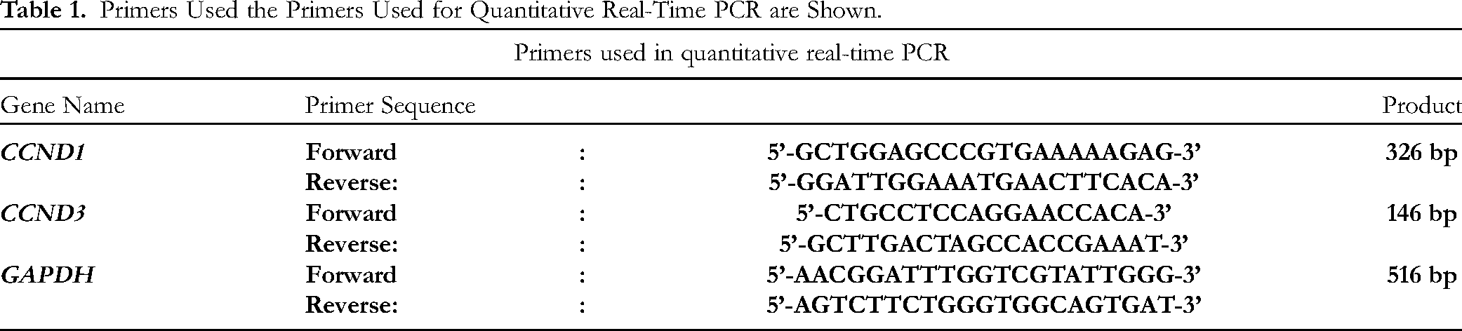
Total RNA was prepared by using the RNeasy Plus Mini Kit (#74136; Qiagen, Hilden, Germany) or RNeasy Plus Micro Kit (#74034; Qiagen) following the manufacturer's instructions, and cDNA was synthesized with Superscript III reverse transcriptase (#18080093; Invitrogen, Carlsbad, CA, USA). TB Green Advantage qPCR Premix (#639676; Takara Bio Inc., Shiga, Japan) and Applied Biosystems 7500 Fast Real-Time PCR System (Applied Biosystems, Foster City, CA) were used for performing the quantitative PCR. The primers used for the quantitative real-time PCR analysis are shown in Table 1. 18
Primers Used the Primers Used for Quantitative Real-Time PCR are Shown.
Cell Cycle Analysis
The cell cycle profile in control and 200 µg/mL HST-treated TR146 cells cultured in the presence or absence of 50 µM cisplatin for another 12 and 24 h, were analyzed by Click-iT Plus EdU Flow Cytometry Assay Alexa 488 Kits (#C10633; Invitrogen) for S-phase analysis and FxCycle violet stain (#F10347; Invitrogen) for DNA content analysis, following the manufacturer's instructions. The TR146 cells were suspended in the culture medium at a density of 2.0 × 10⁵ cells/ml and cultured for 9 h in the medium. Then, they were incubated in the presence or absence of 200 µg/mL HST and 50 µM cisplatin. Then, Click-iT EdU was added at a concentration of 20 µM at 3 h before the targeting time. After incubation for 3 h, the harvested cells were fixed with the Click-iT fixative for 15 min at room temperature. After washing with 1% BSA in PBS, the cells were treated with Click-iT permeabilization reagent, and EdU was labeled with Alexa Fluor 488 picolylazide for 30 min at room temperature, with protection from light. After washing with 1% BSA in PBS, the DNA was stained with FxCycle Violet Stain for 30 min at room temperature, with protection from light. The proportion of cells in the cell cycle was analyzed by flow cytometry on the FACS Aria II (Becton Dickson, Franklin. Lakes, NJ, USA). Data were analyzed using the FlowJo software (TreeStar, Ashland, OR, USA).
Statistical Analysis
The statistical significance of differences was determined by unpaired Student's t test. P values less than 0.05 were considered significant. Data are expressed as the mean value ± standard error (S.E).
Results
To elucidate the mechanisms of HST, cells from the TR146 cell line (oral mucosa cells) were used. After culturing the cells for 9 h, we added HST and performed the MTT assay at several time points. The MTT assay was chosen to evaluate the effect of HST on overall cell viability and metabolic activity, providing a baseline understanding of how HST influences cell proliferation. The cell proliferation was found to be stable at 3 and 9 h; however, it decreased significantly at 18 and 27 h in case of all HST concentrations compared with the control (Figure 1). These results suggest that exposure to HST suppressed the cell proliferation.

HST suppressed the cell proliferation/after culturing the TR146 cells for 9 h, HST was added to the medium, and MTT assay was conducted after 3, 9, 18, and 27 h. Means ± standard errors from three independent experiments are shown with error bars. Significance of the differences was estimated by using unpaired student's t-test. *p < 0.05.
Next, to elucidate the factors associated with the attenuation of cell proliferation by HST, we conducted quantitative real-time PCR on the representative cell cycle-related genes such as CCND1 and CCND3. Quantitative real-time PCR was used to examine changes in the expression of key cell cycle-related genes, allowing us to identify specific molecular targets affected by HST treatment. For quantitative PCR, the 3-h and 6-h time points were chosen based on the results of the MTT assay, which indicated that significant changes in cell viability occurred after 9 h of HST exposure. These findings suggested that differences in gene expression likely occur earlier, during the initial stages of HST treatment, before functional changes in cell viability are observed. Thus, we selected 3 h to capture early gene expression changes and 6 h to examine subsequent dynamics. As results, the mRNA expression of CCND1 and CCND3 was found to be reduced significantly after exposure to all HST concentrations for 3 h, but these reductions were not observed at the 6-h time-point (Figure 2A and B). These data suggest that HST affected the expression of cell cycle-related genes in a short time period, and regulated the cell cycle.

HST reduced the expression of cell cycle-related genes/(A and B): after culturing the TR146 cells for 9 h, HST was added to the medium, and quantitative real-time PCR analysis of CCND1 and CCND3 after 3 and 6 h was performed. Relative expression was normalized by using the GAPDH expression. Means ± standard errors from four independent experiments are shown with error bars. Significance of the differences was estimated by using paired student's t-test. *p < 0.05, **p < 0.01.
We hypothesized that one of the clinical advantages of HST use for CRT-induced oral mucositis was the regulation of the cell cycle in the oral epithelium. To confirm this hypothesis, we conducted cell cycle analysis following HST and/or cisplatin treatment. Cell cycle analysis was conducted to understand how HST modulates cell cycle progression, particularly in the context of its interaction with cisplatin. After culturing TR146 cells for 9 h, we added 200 µg/mL HST and/or 50 µM cisplatin. We conducted cell cycle analysis after exposing the cells to these compounds for 12 and 24 h. The 12 h time point was chosen to capture the initial effects of which forms DNA adducts and interstrand crosslinks during the S phase. These crosslinks prevent DNA replication, inducing an S-phase block and triggering apoptosis. The 24 h time point was selected to observe the cumulative impact of HST and/or cisplatin on cell cycle regulation and apoptosis. These intervals also align with the doubling time of TR146 cells (approximately 33 h), ensuring that critical phases of the cell cycle were assessed. This design enabled us to evaluate how HST enhances cell survival and reduces apoptosis under cytotoxic stress. As a result, the percentage of live cells did not change after HST and/or cisplatin treatment for 12 h. Furthermore, we investigated the cell cycle in live cells. HST reduced the attendance in S phase by half, while cisplatin thoroughly inhibited the attendance in S phase (Figure 3). At 24 h, the percentage of live cells did not change after HST treatment, and most of the cells were dead due to cisplatin. However, a considerable number of cells treated with both HST and cisplatin were still alive. Among the live cells, HST promoted G0/G1 and G2/M phase arrest and reduced the percentage of apoptotic cells, compared with the control. Cisplatin perfectly diminished the population of cells in the S phase and promoted apoptosis; however, the percentage of the cells in the G0/G1 and G2/M phase increased and that of apoptotic cells decreased after treatment with both HST and cisplatin. These results suggest that HST increased the resistance of the cells to cisplatin cytotoxicity, and the G0/G1 and G2/M phase arrest was related with its effect.

HST increased the resistance of cells to cisplatin cytotoxicity after culturing the TR146 cells for 9 h, we added 200 µg/mL HST and 50 µM cisplatin to the medium, and cell cycle analysis was conducted after 12 and 24 h. Cell cycle profiles were analyzed by flow cytometric analysis.
Discussion
This study investigated the direct effect of HST to oral mucosa cell. To elucidate the mechanism of HST, we conducted experiments such as the MTT assay, quantitative real-time PCR, and cell cycle analysis using the TR146 cell line. The combination of MTT assay, quantitative PCR, and cell cycle analysis allowed us to assess the effects of HST from multiple perspectives: cell viability, gene expression, and cell cycle progression. These complementary approaches provide a comprehensive understanding of HST's mechanisms of action. The results showed that HST suppressed the cell proliferation and also the expression of cell cycle-related genes, and finally promoted G0/G1 and G2/M phase arrest, resulting in the resistance of cells to cisplatin cytotoxicity.
Previous studies have reported how oral mucositis was induced by CRT, for instance, four phases have been proposed, namely, expression of free radicals, inflammation, infection, and wound healing. 1 It has already been known that HST regulates these four phases related to CRT-induced oral mucositis.1,3 Our in vitro findings showed that HST rendered the cells resistant to cisplatin cytotoxicity by regulating the cell viability and cell cycle (Figure 3), which confirmed the direct effect of HST on the oral region. To the best of our knowledge, this was the first detailed report suggesting that gargling HST may be useful for suppressing CRT-induced oral mucositis without taking HST, which may reduce side effects on liver and kidney function.
This study showed that HST played a key role to regulate cell cycle resulted in cisplatin-resistance. This result was biologically plausible because HST has originally been consumed for treating diarrhea, and like the direct action of HST on intestinal mucosa cells, its direct action on oral cells may be important for suppressing oral mucositis. Our finding that HST suppressed the cell cycle was enough to explain the resistance of cells to cisplatin cytotoxicity, because cisplatin forms crosslinks among DNA and prevents DNA cleavage in the S phase, thus activating signaling pathways culminating in apoptosis. 19 Because of cell cycle-regulated genes such as CCND1 and CCND3 are associated with cyclin-dependent kinases 4 and 6, which promote G1-S progression, 20 G0/G1 arrest may imply the downregulation of cell cycle-regulated genes such as CCND1 and CCND3, resulting in cisplatin resistance. 18 In addition, a study has reported that radioresistant Hela cells were maintained in the G0/G1 phase during the accumulation of irradiation, compared to standard Hela cells. 21 Our findings align with recent studies suggesting that HST modulates pathways involved in oxidative stress and inflammation. Additionally, emerging research on the cell cycle-regulating properties of herbal medicines supports the observed effects of HST in this study. For example, recent publications highlight the interplay between cyclin D regulation and chemoresistance, underscoring the importance of targeting such pathways.22,23
Interestingly, our data show that HST promoted G0/G1 and G2/M phase arrest. DNA synthesis events in the S phase and mitosis events in the M phase need relatively long preparation periods, pausing at interval phases until the arrangement of suitable cell environments. If certain factors interrupt these preparatory phases before their completion, the cell would restart the preparatory phase from the beginning. In this study, we demonstrated that HST significantly reduced the expression of CCND1 and CCND3 after exposure to HST for 3 h, but these reductions were not observed at the 6-h time-point (Figure 2A and B). At 3 h without HST, the TR146cells, which have a doubling time of approximately 33 h, 24 were metabolically active, showing higher expression levels of cell cycle-related genes. By 6 h, however, cellular activity naturally declines, likely due to changes in cell proliferation dynamics. Notably, in the presence of HST, this decline in activity is mitigated, and cellular activity appears to be maintained or even enhanced. These findings suggest that HST may regulate the cell cycle by slowing its progression, which is consistent with the observed promotion of G0/G1 and G2/M phase arrest. By moderating the speed of the cell cycle, HST could provide a protective mechanism, enabling cells to better withstand environmental stress and resist the cytotoxic effects of agents like cisplatin.
This effect is consistent with the observed promotion of G0/G1 and G2/M phase arrest in our study, indicating that HST may contribute to a more controlled and gradual cell cycle progression. Such modulation may be a protective mechanism, allowing cells to better cope with environmental stress or cytotoxic agents like cisplatin. Furthermore, in most oral mucosa cells, the preparation for entering the S phase needs about 8 h; thus, gargling HST thrice a day is reasonable in terms of cell cycle regulation for preventing CRT-induced oral mucositis. It may be more effective to use an adherent ointment containing HST if the direct contact between oral mucosa cells and HST is important for preventing oral mucositis. On the contrary, HST has the potential to abrogate the promising effects of CRT for head and neck cancer because of the resistance to cisplatin. Thus, it is reasonable for patients to use local application by gargling HST for the purpose of protecting oral mucosa cells, not tumor cells located in oral region, against CRT. Future studies will be conducted to elucidate the expression change of signal pathways associated with apoptosis following HST treatment. HST's effects on CCND1 and CCND3 suggest its involvement in pathways regulating G1/S transition. Potential pathways include the PI3 K/AKT and MAPK signaling cascades, which are known to influence cyclin D expression and cell cycle arrest. Further studies are needed to confirm the involvement of these pathways in HST-mediated chemoprotection. As a limitation, this study was conducted in vitro using the TR146 oral mucosa cell line, which may not fully replicate in vivo conditions. Further in vivo studies are needed to confirm the protective effects of HST against cisplatin-induced cytotoxicity. Additionally, future research should investigate the roles of individual bioactive compounds to better understand their contributions to these effects.
Conclusion
This study demonstrated that HST induced the resistance of oral mucosa cells to cisplatin cytotoxicity by regulating the cell cycle. It has already been shown that there are four modes of action by which HST prevents chemotherapy-induced oral mucositis; here, we found a new fifth mode: direct cell cycle-regulatory activity. These data suggest that these five elements work in cooperation to prevent chemotherapy-induced oral mucositis.
Footnotes
Acknowledgments
We are very grateful to the Biomedical Research Core of Tohoku University Graduate School of Medicine for the use of biochemical measuring instruments and to TSUMURA & CO. for providing hangeshashinto for research use.
Author Contribution
Toru Tamahara, the first author, conducted the cell experiments and drafted the manuscript. Ryutaro Arita, as a specialist in Kampo medicine, contributed to the experimental design and manuscript preparation by providing expertise on the medical validity of the study.
Declaration of Conflicting Interests
The authors declared no potential conflicts of interest with respect to the research, authorship, and/or publication of this article.
Funding
The authors disclosed receipt of the following financial support for the research, authorship, and/or publication of this article: This work was supported by the JSPS KAKENHI Research Grant, Grant-in-Aid for Scientific Research (C) (24K13105).
Ethical Approval
Ethical Approval is not applicable for this article.
Statement of Human and Animal Rights
This article does not contain any studies with human or animal subjects.
Statement of Informed Consent
There are no human subjects in this article and informed consent is not applicable