
Abstract
Objective
Literature reveals a significant deficiency in scientific investigations regarding the use of Bambusa vulgaris extract (BVE) for the treatment of neuropsychiatric disorders, particularly those characterized by anxiety and depression. Thus, this study was designed to examine the anxiolytic and antidepressant-like properties of BVE using mouse models, while also elucidating the potential underlying mechanisms responsible for the observed antidepressant effects of the extract.
Methods
Validated behavioral assessments were employed to assess the anxiolytic effects (including the hole-board test (HBT), open field test (OFT), elevated plus maze test (EPMT), and light/dark exploration test (LDET)) as well as the antidepressant effects (comprising the forced swim test (FST) and tail suspension test (TST)) of the extract. Chronic mild stress paradigms were employed to induce depressive-like behaviors in the mouse subjects. The mice were administered treatments consisting of distilled water (10 mL/kg; serving as a negative control), fluoxetine (20 mg/kg; standard drug), and BVE (at varying doses of 100, 200, and 400 mg/kg). The concentrations of biogenic amine neurotransmitters (noradrenaline and serotonin) and cyclooxygenase-2 (COX-2) within the brain were measured using ELISA kits. Antioxidant biomarkers were assessed through the application of standardized commercial assay kits.
Results
BVE significantly diminished anxiety levels in mouse subjects, as demonstrated by an enhancement in exploratory activities in the light/open enclosures and an increased aversion to darker compartments. Furthermore, BVE significantly alleviated depression-like behaviors in mice, which was reflected by an increased latency to immobility and a reduction in the overall duration of immobility. BVE enhanced noradrenaline and serotonin neurotransmission, suppressed oxidative species, and inhibited COX-2 activity.
Conclusion
The results obtained from this study suggest that BVE demonstrates anxiolytic and antidepressant properties, the latter being mediated through the enhancement of biogenic amine neurotransmitters (noradrenaline and serotonin), inhibition of COX-2 activity, and suppression of oxidative radicals within the brain.
Introduction
Depression involves intricate interactions among neurochemical systems, stress hormones, and neuroplasticity.1,2 The manifestations of this condition indicate disturbances in affective state, cognitive processes, and physiological health. Depression is associated with numerous neurochemical dysregulations within the brain, although the precise mechanisms underlying these disorders are complex and diverse. 3 Serotonin, a neurotransmitter, is fundamentally associated with regulating mood, sleep, and appetite.4,5 Decreased concentrations of serotonin are frequently linked to depressive manifestations, which elucidates the rationale behind the therapeutic approach of many antidepressants, such as SSRIs (Selective Serotonin Reuptake Inhibitors) that seek to elevate serotonin levels within the central nervous system.6,7 Targeting noradrenaline is one of several pharmacological strategies to address depression, especially when serotonin-focused treatments alone are insufficient. Norepinephrine is involved in the physiological response to stress and maintaining alertness. A decrease in norepinephrine activity has been associated with symptoms of fatigue, decreased motivation, and a reduced capacity to experience pleasure.8,9 Dopamine is essential for reward, motivation, and pleasure. 10 The dysregulation of dopamine pathways, especially in regions such as the mesolimbic pathway, has been implicated in the deficits of pleasure, motivation, and interest that are frequently observed in individuals experiencing depression.11,12
Dysfunctions in the Glutamate and GABA neurotransmitter systems, notably an excess of glutamate or a deficiency of GABA, have been associated with mood dysregulation, cognitive impairments, and anxiety, all of which may play a role in the manifestation of depressive disorders. 13 The involvement of the enzyme COX-2 in the pathophysiology of depression has been documented, particularly regarding its involvement in neuroinflammation, and its role in exacerbating oxidative stress. 14 The role of chronic inflammation is increasingly acknowledged as a contributing factor in the etiology of depression. 15 Elevated concentrations of pro-inflammatory prostaglandins within the central nervous system, such as IL-6, TNF-α, and COX-2, are frequently detected in patients diagnosed with depression. 16 It has also been documented that COX-2 and its metabolic byproducts may inhibit the bioavailability of serotonin, an essential neurotransmitter implicated in the modulation of mood, and such inhibition may exacerbate the manifestation of depressive symptoms. 17 The strategic targeting of COX-2 and its related inflammatory pathways represents a promising avenue for therapeutic intervention, particularly in cases of depression characterized by inflammatory mechanisms.
Furthermore, oxidative stress has been implicated in the onset and exacerbation of depressive disorders through its effects on cerebral inflammation, mitochondrial integrity, and the equilibrium of neurotransmitters. 18 Over-expression of reactive oxygen species (ROS) can result in cellular, protein, lipid, and DNA damage, thereby contributing to a spectrum of health complications, including psychiatric conditions such as depression. 19 Modulation of oxidative stress may serve as a viable therapeutic approach for depression.
Anxiety disorders represent the most widespread mental health conditions on a global scale, impacting individuals of all age demographics, and substantially hindering daily functioning and overall quality of life. 20 Notwithstanding the substantial prevalence of these disorders, a significant number of cases remain unaddressed, particularly among children and the elderly, thereby emphasizing the necessity for increased awareness, and more effective anxiolytic remedies. Therefore, there exists a burgeoning interest among the public regarding the use of natural remedies for various health concerns, notably including mental health disorders. Extensive ethnobotanical investigations on Bambusa vulgaris species have elucidated its application in the management of neuropsychiatric conditions, including anxiety and depression, and this could potentially pave the way for the discovery of cheaper and more effective anxiolytic and antidepressant therapies.
Bambusa vulgaris, commonly called “Golden Bamboo” or ordinary Bamboo,” represents one of the most extensively cultivated bamboo species globally. 21 This species is classified within the Poaceae family of grasses and is indigenous to tropical and subtropical climates. Characterized by its rapid growth and clumping growth form, this species features striking golden-yellow culms that frequently exhibit green striations. It can reach heights of 10–20 meters (33–66 feet), with culms exhibiting diameters ranging from 6–10 centimetres (2.4–4 inches). The leaf is lanceolate and displays a vibrant green colouration. 21 In various traditional medicinal practices, infusions derived from the leaves and shoots of Bambusa vulgaris extract are employed for their anti-inflammatory and antipyretic effects. 21 Additionally, the sap obtained from the stems and leaves is occasionally used in topical applications for its wound-healing properties. 22 The plant is also used as a gentle laxative and for the regulation of menstrual flow in females. 23 A comprehensive review of the ethnobotanical significance of B. vulgaris has elucidated its efficacy in the treatment and management of malaria, diabetes mellitus, inflammatory conditions, constipation, measles, hepatitis, as well as renal and neurological ailments. 24 Adebayo et al 25 highlighted the neuroprotective attributes of Bambusa vulgaris against neuropsychiatric disorders, including convulsions, memory impairment, and anxiety. Liu et al 26 demonstrated in their findings the cognitive enhancement potential of Bambusa vulgaris. Murtala et al 27 elucidated that the leaf extract of Bambusa vulgaris ameliorates the motor impairments and non-motor co-morbidities induced by haloperidol by mitigating the pathophysiological anomalies linked to the advancement of the disorder. Nevertheless, there exists a lack of scientific literature concerning the application of Bambusa vulgaris in the management of neuropsychiatric conditions, particularly anxiety and depressive disorders. Consequently, this investigation aimed to explore the anxiolytic- and antidepressant-like effects of the hydroethanol leaf extract of B. vulgaris in mouse models, along with elucidating the potential mechanisms underlying the antidepressant effects of the plant.
Materials and Methods
Drugs and Chemicals
Fluoxetine (Grams Pharmaceuticals Ltd, Lagos, Nigeria), Ethanol (Nosak Distilleries Limited, Lagos, Nigeria), Noradrenaline & Serotonin ELIZA kits (Elabscience, Wuhan, China) and COX-2 ELIZA kit (Elabscience, Wuhan, China), Antioxidant biomarkers commercial ELIZA kits (Elabscience, Wuhan, China).
Plant Material
Fresh leaf specimens of Bambusa vulgaris were procured from the Sagamu area of Ogun State, Nigeria. Mr Adeoti O. Adekunle, a taxonomist affiliated with the Department of Pharmacognosy, Faculty of Pharmacy, Olabisi Onabanjo University, Sagamu, Ogun State, Nigeria, conducted the botanical classification and verification. A voucher specimen was subsequently archived in the institutional herbarium.
Extraction
The standardized procedures for extraction, as described by Murtala et al 27 were meticulously adhered to. Fresh leaves of B. vulgaris were subjected to air-drying until stable weight measurements were achieved. The leaves were pulverized, weighed (550 g), and immersed in 1.5 L hydroethanol (1:1) for 72 h. The extract underwent decantation and filtration using muslin cloth, followed by filtration through Whatman filter paper. The extraction, decantation, and filtration processes were repeated twice with the resultant residues. The cumulative extract filtrates were subjected to evaporation until dryness at 40 °C under reduced pressure, yielding dark brown solids with an approximate yield of 12.72%. The desiccated extract was then weighed and reconstituted in distilled water to achieve the requisite working concentrations before administration to the experimental subjects. Murtala et al 27 reported an absence of abnormal alterations in the general behavior of the animals and mortality during the acute oral LD50 estimation of over 2 g/kg of B. vulgaris extract.
Animals
Albino mice, weighing between 20 and 25 grams, of both genders were procured from the Faculty of Pharmacy Animal Center at Olabisi Onabanjo University located in Sagamu, Ogun State. The animals were housed in adequately ventilated and sanitary cages, maintained under standardized environmental conditions, and provided unrestricted access to a conventional rodent pellet diet supplied by Jafel Agro Services in Sagamu, Ogun State, and water. A period of acclimatization lasting 14 days was implemented before the initiation of the experimental procedures. Ethical clearance was secured from the Animal Care and Use Research Ethics Committee (ACUREC) of CMUL, designated as CMUL/ACUREC/10/24/1607. The rights and well-being of the animals were safeguarded in accordance with the Guidelines for the Care and Use of Laboratory Animals. To mitigate the variability associated with sex differences in the mice used for this study, an equal ratio of Male to Female mice (3:3) was maintained across the various experimental groups.
Gas Chromatography-Mass Spectrometry (GC–MS) ANALYSIS
Gas chromatography-mass spectrometry (GC-MS) analysis was performed to obtain a chemical fingerprint. The GC-MS analysis was conducted as described by Vidhya et al 28 using Agilent Technologies (Santa Clara, CA, USA) gas chromatography systems (specifically, the GC-7890A/MS-5975C model; MSD - 5975C, injector - 7683B series). The parameters employed included an initial temperature (maintained for 2 min) of 100 °C and a final temperature (increased at 10 °C/min) of 270 °C. The volume of the injected extract amounted to 1 μL of a concentration of 0.2 g/mL. The heater's temperature was maintained at 250 °C, while the pressure was recorded at 3.2652 psi using a split less injection mode. The analytical system was outfitted with an HP-5MS column (30 m × 320 μm × 0.25 μm) and employed helium as the carrier gas, which exhibited a purity of 99.9%. The established flow rate was 1.4963 mL/min, correlating to an average velocity of 45.618 cm/s. The compounds present in the extract were identified by comparing retention times and mass spectra of authentic samples obtained via gas chromatography with the mass spectra from the National Institute of Standards and Technology (NIST) database in the United States.
Neuropharmacological Evaluations
A distinct cohort of mice was used for the various behavioral assessments conducted in this investigation.
Anxiolytic Activity
Hole-Board Test
The hole-board apparatus consists of a wooden panel measuring 40 cm × 40 cm, featuring four uniformly spaced apertures, each with a diameter of 1 cm and a depth of 2 cm. One hour after the oral administration of the extract to five distinct cohorts, each comprising six mice (totalling 30 mice), which included a control group receiving distilled water at a dose of 10 mL/kg, a standard group treated with fluoxetine at 20 mg/kg, and test groups administered B. vulgaris at doses of 100, 200, and 400 mg/kg, each mouse was strategically placed in one corner of the hole-board and meticulously monitored for five minutes. The frequency and duration of head dips and the number of sectional crossings within the designated observation timeframe were systematically documented for each mouse.29,30
Open-Field Test
One hour following the oral administration of the control group with distilled water at 10 mL/kg, the standard group with fluoxetine at 20 mg/kg, and the experimental groups with B. vulgaris extract at doses of 100, 200, and 400 mg/kg (including a total of 30 mice), each mouse was carefully placed at the center of the apparatus.The experimental apparatus was constructed from wood, measuring 50 cm in length, 50 cm in width, and 25 cm in height. The flat base of the enclosure was partitioned into 8 cm × 8 cm segments, resulting in a total of 16 squares. Among these, sixteen squares were designated as the centre, while the remaining squares, located adjacent to the walls, were classified as the periphery. The frequency of squares traversed, instances of rearing, and occurrences of assisted rearing (where the fore paws make contact with a wall of the enclosure) during the designated observation period of 5 min were meticulously documented. A mouse was considered to have transitioned from one square to another when all four paws successfully crossed the boundary.31,32
Elevated Plus Maze Test
The elevated plus maze consists of two open arms and two enclosed arms, each measuring 50 × 10 × 40 cm, raised to a height of 50 cm. Distilled water (10 mL/kg, p.o.), B. vulgaris (100, 200, and 400 mg/kg, p.o.), and fluoxetine (20 mg/kg, p.o.) were administered to five groups comprising six mice each (a total of 30 mice). One hour after the administration, each mouse was placed at the maze's central locus and observed for a duration of 5 min. The total time spent by each mouse in both the open and closed arms of the maze, along with the frequency of entries, was meticulously recorded throughout the observation period.33,34
Light/Dark Exploration Test
The light/dark box constitutes a rectangular apparatus measuring 50 × 25 × 25 cm, partitioned into two distinct compartments (light and dark). In the course of this experimental protocol, a total of thirty mice were systematically allocated into five groups, each comprising six mice. The vehicle (distilled water at a dose of 10 mL/kg), the standard (fluoxetine at a dose of 20 mg/kg), and the extract (at a dose of 100, 200, and 400 mg/kg) were administered via the oral route. Subsequently, one hour after administration, each mouse was placed within the illuminated section of the light/dark box. The parameters measured included latency (the duration required for the animal to first transition into the dark compartment), the frequency of entries into both the light and dark compartments, the cumulative time spent in the light compartment, as well as the observable frequency of rearing and assisted rearing, all documented within a five-minute observation window. This investigation was conducted based on the inherent aversion exhibited by rodents towards brightly illuminated environments.35,36
Antidepressant Activity
Forced Swim Test
The antidepressant protocol was executed with minimal modifications. All animals were administered treatment once daily for 14 days, and 30 min following the treatment, the animals were subjected to immobilization for one hour to induce restraint stress.The mice were allocated randomly into five groups, each consisting of six animals (totalling 30 mice), and were subjected to distinct treatments as follows: Group 1 received distilled water (10 mL/kg, administered orally); Group 2 was treated with fluoxetine (20 mg/kg, administered orally); and Groups 3 to 6 were administered extracts at dosages of 100, 200, and 400 mg/kg (also via oral administration). One hour after treatment on the fourteenth day, each mouse was compelled to swim sequentially in an open cylindrical vessel (with a diameter of 10 cm and a height of 25 cm) containing 19 cm of water for 5 min. The latency and total duration of immobility were meticulously observed and documented. An individual mouse was deemed immobile when it ceased vigorous movements and remained afloat without motion in the water, engaging only in minimal movements necessary to maintain its head above the water's surface. A reduction in the duration of immobility serves as an indicator of an antidepressant-like effect.37,38
Tail Suspension Test
The methodology used in the present investigation resembles those elucidated by Yan et al 39 and Son et al 40 The treatment was administered following the protocol above for the Forced Swim Test (FST). Sixty minutes after treatment on the 14th day, each mouse was sequentially suspended on a retort stand positioned 50 cm above the ground, facilitated by adhesive tape affixed approximately 1 cm from the distal end of the tail. Each mouse's latency and cumulative immobility duration were meticulously documented during the 5-min observation interval. A mouse was deemed immobile if it refrained from any attempts to elevate its head and maintained a downward head posture for a duration exceeding 5 s.
Brain Tissue Preparation for Biochemical Evaluations
Following the behavioral assessment on day 14, the animals were euthanized, and the brain was expeditiously excised and preserved at −20 °C for subsequent quantification of amine neurotransmitters, COX-2 enzyme activity, and levels of antioxidant biomarkers. The brain tissues were subjected to homogenization in a phosphate buffer solution (PBS) with a ratio of tissue weight (g) to PBS volume (mL) of 1:9, using a glass homogenizer on ice. The resultant brain homogenates were centrifuged at 5000 × g for 5 min at 4 °C to isolate the supernatant. The supernatant was subsequently stored at −80 °C. The concentrations of noradrenaline and serotonin were quantified using an ELISA kit (Elabscience, Wuhan, China), while COX-2 levels were assessed via a COX-2 ELISA kit (Elabscience, Wuhan, China).
Determination of Amine Neurotransmitters Levels and Cyclooxygenase-2 Enzyme (COX-2) Activities
The quantification of serotonin and noradrenaline levels, along with the assessment of COX-2 activity in brain homogenates, was carried out using the ELISA technique provided by Elabscience (Wuhan, China), in accordance with the manufacturer's protocol. In summary, the concentrations of Noradrenaline, Serotonin, and COX-2 within brain tissue were quantified by adding 100 μL of the brain tissue sample to each well containing rat-derived captured antibodies for Noradrenaline, Serotonin, and COX-2, which had been allowed to incubate overnight (18 h, 4◦C). The resulting mixture was incubated on a shaker at room temperature (25 ◦C) for 2 h at approximately 500 rpm within a 96-well plate. Subsequently, 100 μL of biotinylated goat polyclonal anti-rat Noradrenaline, Serotonin, and COX-2 detection antibody, as well as avidin-horseradish peroxidase (avidin-HRP) solutions, were introduced to each well. The plates designated for Noradrenaline, Serotonin, and COX-2 were sealed and incubated for 63 min at 25 ◦C. Following this incubation, 100 μL of the chromogenic substrate [3, 3′, 5, 5′-tetramethylbenzidine (TMB)] was added to each well for the assay of Noradrenaline, Serotonin, and COX- 2, and the mixture was incubated in darkness for 15 min at 25 ◦C prior to the addition of 100 μL of the stop solution. The absorbance measurements for Noradrenaline, Serotonin, and COX-2 were conducted at 450 nm within 15 min using the Spectramax M-5 (Molecular Devices, Sunnyvale, CA) multifunctional microplate reader, equipped with Softmax Pro v 5.4 (SMP 5.4). The concentrations of Noradrenaline, Serotonin, and COX-2 extracted from the brain tissue were extrapolated from established standard curves and expressed in picograms per milligram of protein (pg/mg protein). 41
Brain Antioxidant Assay
Evaluation of Glutathione (GSH) Content
The methodology described by Kazak and Coskun 42 was adopted to quantify the concentration of glutathione (GSH) within brain tissues. Briefly, a volume of 0.4 mL of brain homogenate was mixed with 0.4 mL of 20% trichloroacetic acid (TCA) and gently agitated by swirling. Subsequently, the resultant suspension underwent centrifugation at 4 °C for a period of 20 min at 5400 g. Following this procedure, 0.25 mL of the obtained supernatant was combined with 2 mL of 0.6 mmol/L 5,5′-dithio-bis (2-nitrobenzoic acid) (DTNB), with the total volume of the solution being adjusted to 3 mL using a phosphate buffer (0.2 mol/L, pH 8.0). The absorbance of this solution was recorded at 412 nm, using a blank reagent consisting of 2 mL of 0.6 mmol/L DTNB mixed with 1 mL of phosphate buffer (0.2 mol/L, pH 8.0) as a reference, conducted via spectrophotometric analysis. The concentration of reduced GSH present in the brain tissues was quantified and expressed as nanomoles per milligram of protein (nmol/mg protein).
Evaluation of Malondialdehyde (MDA) Levels
The quantification of malondialdehyde (MDA) concentration within cerebral tissues was carried out in accordance with the standardized methodology described by Ohkawa et al 43 In summary, an aliquot of 0.4 mL of brain tissue was combined with 1.6 mL of Tris-potassium chloride (Tris-KCl) buffer, subsequent to which 0.5 mL of a 30% trichloroacetic acid (TCA) solution was incorporated. Following this, 0.5 mL of 0.75% thiobarbituric acid (TBA) was introduced into the mixture, which was then incubated in a water bath for duration of 45 min at a temperature of 80 °C. Upon the conclusion of the incubation period, the mixture was subjected to cooling on ice and then centrifuged at 1200 g for 15 min. The resulting clear supernatant was subsequently collected, and its absorbance was measured against a reference blank of distilled water at a wavelength of 532 nm. The concentration of MDA was computed using the molar extinction coefficient of 1.56 × 10^5 mol/L/cm, with the resulting value articulated as nanomoles of MDA per milligram of protein (nmol MDA mg−1 protein).
Determination of Catalase Levels
The enzymatic activity of catalase (CAT) was assessed by using the technique explicated by Sinha 44 The absorbance at 240 nm was determined through the addition of 3 mL of phosphate-buffered saline with 10.5 mL of brain tissue homogenate supernatant. The kinetics of hydrogen peroxide (H2O2) decomposition at 240 nm serves as the theoretical underpinning of the assay. The resultant findings are expressed in terms of kilograms of protein.
Determination of Superoxide Dismutase Levels (SOD)
The methodology articulated in Gao et al 45 was employed to evaluate the activity of superoxide dismutase (SOD) within brain tissue homogenates. The SOD reaction mixture was constituted using 0.1 mM ethylenediaminetetraacetic acid (EDTA), 0.3 mM xanthine, 0.6 mM nitro blue tetrazolium (NBT), and 50 μL of xanthine oxidase. This reaction mixture was subsequently dissolved in a 10 mM sodium phosphate buffer at pH 7.4, with the solution maintained in an ice bath. Thereafter, 20 μL of brain supernatant (sample) was added to the wells containing the reaction mixture. The reaction mixture was then incubated at ambient temperature or at 37 °C for duration of 15 min. The absorbance was quantified at 560 nm with the aid of spectrophotometer.
Statistical Analysis
The results from this investigation were articulated as mean ± SEM The data underwent statistical analysis using One-way ANOVA, followed by Dunnett's post-hoc tests, employing GraphPad Prism 6 Software (GraphPad Software Inc., CA, USA). Statistical significance was established at P < 0.05.
Results
Gas Chromatography-Mass Spectrometry (GC–MS) ANALYSIS
The Gas Chromatography-Mass Spectrometry (GC-MS) analysis of B. vulgaris demonstrated the identification of hexadecanoic acid, ethyl ester, phytol, ethyl oleate, 3,5-octadecene, cyclopropane, cyclododecane, 35,9-eicosene, pentadecanoic acid, and methyl ester (Figure 1).

The GC-MS spectrum of Bambusa vulgaris showing 32 peaks. hexadecanoic acid and ethyl ester (peak #13.627), phytol (peak #14.583), ethyl oleate (peak #14.978), 3,5-octadecene and cyclohexadecane (peak #10.074), cyclopropane and cyclododecane (peak #7.985), 35,9-eicosene (peak #11.933), pentadecanoic acid, hexadecanoic acid and methyl ester (peak #13.084).
Bambusa vulgaris Increased the Frequency of Head Dips and Crossed Sections (HBT)
The application of One-way ANOVA revealed a statistically significant augmentation in the frequency of head dips (P < 0.05, 0.01; 100–400 mg/kg) and sectional crossings (P < 0.05; 200–400 mg/kg) induced by the extract when juxtaposed with the distilled water-treated mice subjects (Table 1). Furthermore, B. vulgaris administered at a dose of 400 mg/kg resulted in a non-significant elevation (P > 0.05) in the duration of head dips in comparison to the control group (Table 1). The reference pharmacological agent, fluoxetine, elicited a statistically significant increase in the frequency of head dips (P < 0.001; 20 mg/kg) and sectional crossings (P < 0.05) when compared to the negative control (Table 1).
Effect of Hydroethanol Leaf Extract of B. vulgaris in the Hole-Board Test in Mice.
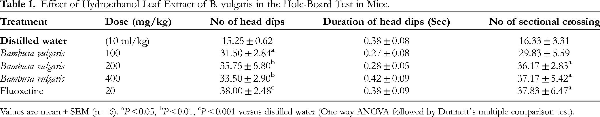
Values are mean ± SEM (n = 6). aP < 0.05, bP < 0.01, cP < 0.001 versus distilled water (One way ANOVA followed by Dunnett's multiple comparison test).
Bambusa vulgaris Increased the General Square Crossings and Rearing (OFT)
B. vulgaris significantly enhanced the frequency of general square crossings (P < 0.05; 400 mg/kg) as well as the incidence of rearing (P < 0.01, 0.001; 200–400 mg/kg) in comparison to the control group (Table 2). The administration of fluoxetine (20 mg/kg) resulted in a significant increase in the frequency of general square crossings (P < 0.05) and assisted rearing (P < 0.0001); additionally, it produced a non-significant increase (P > 0.05) in the occurrence of rearing relative to the control group (Table 2).
Effect of Hydroethanol Leaf Extract of B. vulgaris in the Open Field Test in Mice.

Values are mean ± SEM (n = 6). aP < 0.05, bP < 0.01, cP < 0.001, dP < 0.0001 versus distilled water (One way ANOVA followed by Dunnett's multiple comparison test).
Bambusa vulgaris Increased the Time Spent and Number of Entry into Open Arms and Decreased the Time Spent and Number of Entries into Closed Arms (EPMT)
B. vulgaris significantly increased the duration spent in open arms (P < 0.01; 400 mg/kg) and the number of entries into open arms (P < 0.05; 400 mg/kg) compared to the negative control (Table 3). In terms of closed arms, B. vulgaris significantly reduced the frequency of entries into closed arms (P < 0.05; 100 & 400 mg/kg) as well as the duration spent in closed arms (P < 0.0001, 0.05; 200–400 mg/kg) when compared to the distilled water-treated cohort (Table 3). Fluoxetine (20 mg/kg) significantly increased the frequency of entries into open arms (P < 0.05); concurrently, it decreased both the number of entries into closed arms (P < 0.05) and the duration spent in closed arms (P < 0.0001) relative to the distilled water-treated group (Table 3).
Effect of Hydroethanol Leaf Extract of B. vulgaris in the Elevated Plus Maze Test in Mice.

Values are mean ± SEM (n = 6). aP < 0.05, bP < 0.01, dP < 0.0001 versus distilled water (One way ANOVA followed by Dunnett's multiple comparison test).
Bambusa vulgaris Increased the Latency of Entry into the Dark box, Time Spent in the Light box, and the Number of Entries into the Light box; Decreased the Number of Entries into the Dark box (LDET)
Bambusa vulgaris significantly prolonged the latency of entry into the dark box (P < 0.05; 100 mg/kg), the duration spent in the light box (P < 0.05; 200–400 mg/kg), and the frequency of assisted rearing (P < 0.001; 200 mg/kg) in comparison to the control (Table 4). Concerning the frequency of explorations into both compartments, the extract significantly decreased the frequency of explorations into the dark box (P < 0.01; 100 mg/kg) while concurrently increasing the frequency of explorations into the light box (P < 0.05, 0.001; 200–400 mg/kg) when compared to the negative control (Table 4). The extract, across all administered doses, elicited a non-significant decrease (P > 0.05) in the duration spent in the dark compartment relative to the negative control. Fluoxetine (20 mg/kg) significantly augmented the time spent in the light box (P < 0.01), the frequency of rearing (P < 0.05), assisted rearing (P < 0.001), and the frequency of explorations into the light box (P < 0.01) in comparison to the distilled water-treated group (Table 4).
Effect of Hydroethanol Leaf Extract of B. vulgaris in Light/Dark Exploration Test in Mice.

Values are mean ± SEM (n = 6). aP < 0.05, bP < 0.01, cP < 0.001 versus distilled water (One way ANOVA followed by Dunnett's multiple comparison test).
Bambusa vulgaris Increased the Latency of Immobility and Decreased the Duration of Immobility in FST
One-way ANOVA revealed a statistically significant increase in the onset/latency of immobility (P < 0.05; 400 mg/kg) as well as a notable reduction in the duration of immobility (P < 0.001–0.05; 100–400 mg/kg) attributable to the extract when compared to the distilled water control group (Table 5). Fluoxetine (20 mg/kg) induced a significant prolongation (P < 0.05) in the duration of immobility in contrast to the distilled water treatment (Table 5).
Effects of Hydroethanolic Leaf Extract of B. vulgaris in Forced Swim Test in Mice.

Values are mean ± SEM (n = 6). aP < 0.05, bP < 0.01, cP < 0.001 versus distilled water (One way ANOVA followed by Dunnett's multiple comparison test).
Bambusa vulgaris Delayed the Onset of Immobility and Reduced Total Duration of Immobility in TST
Bambusa vulgaris extract resulted in a significant delay in the onset of immobility (P < 0.05; 200 mg/kg) along with a significant decrease in the duration of immobility (P < 0.05; 100–200 mg/kg) relative to the negative control group (Table 6). Fluoxetine (20 mg/kg) also demonstrated a significant delay in the latency of immobility (P < 0.05) and a marked reduction in the duration of immobility (P < 0.001) when compared to the negative control group (Table 6).
Effects of Hydroethanolic Leaf Extract of B. vulgaris in Tail Suspension Test in Mice.

Values are mean ± SEM (n = 6). aP < 0.05, bP < 0.01, cP < 0.001 versus distilled water (One way ANOVA followed by Dunnett's multiple comparison test).
Bambusa vulgaris Ameliorates Depression-Like Symptoms in Mice by Up-Regulating Noradrenaline and Serotonin Functions and Down-Regulating COX-2 Enzyme Activity
Regarding the influence of the extract on noradrenaline function within the brain, Bambusa vulgaris significantly elevated (P < 0.001–0.01; 200–400 mg/kg) the concentration of noradrenaline in the brain relative to the distilled water control group (Figure 2). The maximal effect was noted at a dose of 200 mg/kg of the extract. Concerning serotonin function within the brain, B. vulgaris elicited a significant enhancement (P < 0.01; 400 mg/kg) in serotonin concentration in comparison to the negative control group (Figure 3). Simultaneously, fluoxetine (20 mg/kg) provoked a significant increase (P < 0.05) in serotonin concentration in the brain when assessed against the distilled water control group (Figure 3). With respect to COX-2 enzyme activity, B. vulgaris produced a significant reduction (P < 0.01; 400 mg/kg) in COX-2 enzyme activity in the brain as opposed to the distilled water control group (Figure 4). Concurrently, fluoxetine resulted in a significant decrease (P < 0.0001; 20 mg/kg) in COX-2 activity in the brain when compared to the vehicle control (Figure 4).

Assessment of noradrenaline function in the brain on the elucidation of possible mechanism(s) of antidepressant-like effect of B. vulgaris in the FST. Values are Mean ± SEM (n = 6). bP ˂ 0.01, cP < 0.001 versus distilled water (one-way ANOVA followed by Dunnett's multiple comparison tests).

Assessment of serotonin function in the brain on the elucidation of possible mechanism(s) of antidepressant-like effect of B. vulgaris in the FST. Values are Mean ± SEM (n = 6). bP ˂ 0.01 versus distilled water (one-way ANOVA followed by Dunnett's multiple comparison tests).

Assessment of COX-2 enzyme activity in the brain on the elucidation of possible mechanism(s) of antidepressant-like effect of B. vulgaris in the FST. Values are Mean ± SEM (n = 6). bP ˂ 0.01 versus distilled water (one-way ANOVA followed by Dunnett's multiple comparison tests).
Bambusa vulgaris Improves Depression-Like Symptoms Through Inhibition of Oxidative Stress in Mice
Bambusa vulgaris induced a significant reduction in malondialdehyde (MDA) levels (P < 0.01, 0.001; 100–400 mg/kg); and an elevation in glutathione (GSH) (P < 0.05; 100–200 mg/kg), catalase (P < 0.01; 200 mg/kg), and superoxide dismutase (SOD) levels (P < 0.01; 200–400 mg/kg) in the brain relative to the distilled water control group (Figure 5–8).

Assessment of MDA level in the brain on the elucidation of possible mechanism(s) of antidepressant-like effect of B. vulgaris in the FST. Values are Mean ± SEM (n = 6). bP ˂ 0.01, cP < 0.001 versus distilled water (one-way ANOVA followed by Dunnett's multiple comparison tests).

Assessment of GSH level in the brain on the elucidation of possible mechanism(s) of antidepressant-like effect of B. vulgaris in the FST. Values are Mean ± SEM (n = 6). aP ˂ 0.05 versus distilled water (one-way ANOVA followed by Dunnett's multiple comparison tests).

Assessment of catalase level in the brain on the elucidation of possible mechanism(s) of antidepressant-like effect of B. vulgaris in the FST. Values are Mean ± SEM (n = 6). bP ˂ 0.01 versus distilled water (one-way ANOVA followed by Dunnett's multiple comparison tests).

Assessment of SOD level in the brain on the elucidation of possible mechanism(s) of antidepressant-like effect of B. vulgaris in the FST. Values are Mean ± SEM (n = 6). bP ˂ 0.01 versus distilled water (one-way ANOVA followed by Dunnett's multiple comparison tests).
Discussion
Depression constitutes a prevalent psychiatric condition that adversely affects individuals’ overall quality of life and interpersonal relationships. The currently accessible therapeutic agents, comprising both conventional antidepressants for depression, isolated anxiety, or co-occurring depression and anxiety, have achieved notable successes; however, these advancements are not without a series of challenges. The use of these conventional pharmacological agents has been significantly characterized by life-threatening adverse effects, instances of remission, treatment resistance, withdrawal symptoms, financial implications, patient preferences, and interactions with concomitant medications.46,47 In light of these concerns, promoting and standardizing natural product remedies, particularly those derived from plant sources, which exhibit enhanced pharmacological properties while devoid of these harmful effects, is imperative.
In this study, the ethanol extract obtained from the leaves of Bambusa vulgaris, administered over a continuous period of 14 days, demonstrated notable antidepressant-like effects in mice subjected to stress. Furthermore, B. vulgaris also displayed significant anxiolytic-like effects in the animal subjects.
Research has indicated that pharmacological substances (eg anxiolytics) mitigate anxiety through the enhancement of exploratory head-dipping behaviour and the frequency of sectional crossings. 36 Takeda et al 48 and Kashdan et al 49 posited that exploration typically indicates diminished anxiety levels. In the present investigation, B. vulgaris significantly augmented the frequency of head dips (100–400 mg/kg) and sectional crossings (200–400 mg/kg) while exhibiting a non-significant increase in the duration of head dips. The results observed from the hole-board assessment imply that B. vulgaris possesses anxiolytic properties.
Ajao and Akindele 50 and Akindele et al 51 described the anxiolytic effects of medicinal plants based on the augmentation of exploratory behaviours (including general square crossings, centre square crossings, rearing, and assisted rearing) observed in the open field test. In our analysis, B. vulgaris elicited a significant enhancement in general square crossing (400 mg/kg) and the incidence of rearing (200–400 mg/kg), akin to the effects observed with fluoxetine. The increased exploratory behaviors of the mouse subjects in the open field test signify potential anxiolytic activity of the extract.
The Elevated Plus Maze (EPM) assessment is a metric for gauging anxiety in rodent populations. It is predicated upon the dichotomy between an animal's inherent exploratory inclination and its intrinsic apprehension of open, exposed environments.52,53 Existing literature suggests that rodents experiencing anxiety and fear exhibit a natural repulsion towards open compartments, resulting in a diminished duration of exposure.54,55 In this investigation, B. vulgaris significantly prolonged the time spent in open arms (400 mg/kg) and the number of entries into open arms (400 mg/kg), paralleling the effects seen with fluoxetine. Furthermore, B. vulgaris significantly decreased the frequency of entries into closed arms (100 & 400 mg/kg) and the duration spent within closed arms (200–400 mg/kg), mirroring the effects of fluoxetine. These findings substantiate the anxiolytic properties of the extract. This research endeavour, in alignment with the findings of Adebayo et al, 56 underscores the potential of Bambusa vulgaris as a novel therapeutic strategy for anxiety, thereby sparking intrigue and hope among the scientific community.
The rodent's intrinsic aversion to brightly illuminated, open spaces and innate exploratory tendencies constitute the foundational psychological principle underlying the light/dark exploration test of anxiety in rodent models.57,58 An increased duration spent in the light compartment or a higher frequency of entries denotes reduced anxiety levels.57–59 In this study, Bambusa vulgaris significantly extended the latency of entry into the dark box (100 mg/kg), the duration spent in the light box (200–400 mg/kg), and the frequency of assisted rearing (200 mg/kg). Additionally, the extract significantly diminished the frequency of explorations into the dark box (100 mg/kg) while concurrently amplifying the frequency of explorations into the light box (200–400 mg/kg), consistent with the observations made with fluoxetine. These findings, which align with the conclusions drawn by Murtala and Akindele, 36 provide reassurance and confidence in the anxiolytic potential of the extract.
The Forced Swim Test (FST) and the Tail Suspension Test (TST) represent two extensively employed behavioral assays in rodent models for the assessment of depression-like behavior.60,61 The behavioral metric evaluated in these experimental frameworks is immobility, which is construed as an indicator of despair or depression-like states.62,63 In the FST, B. vulgaris (100–400 mg/kg) and fluoxetine notably delayed immobility onset and diminished immobility duration. The extract's effect was comparable to the standard pharmacological agent, fluoxetine. In the case of the TST, which exhibits increased pharmacological sensitivity relative to the FST,64,65 Bambusa vulgaris demonstrated a significant delay in the onset of immobility (200 mg/kg) alongside a considerable reduction in the duration of immobility (100–200 mg/kg), akin to the effects observed with fluoxetine.
Pharmacological interventions aimed at modulating noradrenaline transmission exhibit considerable promise as a viable therapeutic strategy for the treatment of depression. In our investigation, Bambusa vulgaris significantly elevated (200–400 mg/kg) noradrenaline concentrations in the brain. The peak effect was observed at a dose of 200 mg/kg of the extract. This observation implies a potential role of noradrenaline in the underlying mechanism of antidepressant activity associated with B. vulgaris, shedding light on its mechanism of action and potential as a therapeutic agent.
Antidepressant agents that enhance serotonin transmission predominantly function by elevating serotonin concentrations within the brain, thereby contributing to the alleviation of depressive symptoms through the enhancement of mood, emotional regulation, sleep, and cognitive performance. The up-regulation of serotonin via medicinal botanicals presents an intriguing pathway for the formulation of natural antidepressants. Investigations on Costus afer, Murtala et al 34 and Hypericum perforatum, Gupta et al 66 revealed substantial increases in serotonin levels within the central nervous system, which underpin their antidepressant-like effects by modulating serotonin and dopamine levels. In our study, B. vulgaris elicited a significant increase (400 mg/kg) in serotonin concentration, mirroring the effects observed with fluoxetine. This result not only suggests a possible involvement of serotonin up-regulation within the mechanical framework of the antidepressant activity associated with B. vulgaris, but also instills optimism about its potential as a natural antidepressant.
The inhibition of COX-2 (cyclooxygenase-2) carries significant implications for understanding the mechanisms underlying antidepressant agents, as contemporary research indicates a plausible correlation between inflammation and depression. Elevated levels of COX-2 and pro-inflammatory cytokines are frequently detected in individuals diagnosed with depression.67,68 In this investigation, B. vulgaris induced a significant reduction (400 mg/kg) in COX-2 enzyme activity within the brain, an effect that parallels that of fluoxetine. Wang et al 69 highlighted the role of BDNF and COX-2 in the antidepressant mechanisms of catalpol in rats subjected to chronic unpredictable mild stress. Adzic et al 70 and Su et al 71 established that COX-2 inhibition may effectively diminish neuroinflammation, accelerate the therapeutic response to antidepressant treatments, and present a promising adjunct to conventional depression therapies. This finding underscores the involvement of the COX-2 enzyme in the mechanistic pathways underlying the antidepressant activity exhibited by B. vulgaris.
Studies have shown that the inhibition of oxidative biomarkers may correlate with the treatment of depressive disorders.72,73 Bhatt et al 74 elucidated a relationship between increased levels of reactive oxygen species (ROS) and the manifestation of depressive disorders. In addition, research has shown elevated levels of oxidative stress markers (eg, malondialdehyde) and reduced levels of antioxidant enzymes (eg, superoxide dismutase) in individuals with depression. 75 This suggests that oxidative damage plays a significant role in the pathophysiology of depression.In the present investigation, Bambusa vulgaris was found to induce a significant decrease in malondialdehyde (MDA) levels (100–400 mg/kg), alongside an increase in glutathione (GSH) levels (100–200 mg/kg), catalase levels (200 mg/kg), and superoxide dismutase (SOD) levels (200–400 mg/kg) within the brain. The results of this study imply that the reduction in oxidative biomarkers may be intricately connected to the underlying mechanism of the extract's antidepressant activity.
The Gas Chromatography-Mass Spectrometry (GC-MS) analysis of B. vulgaris revealed the identification of hexadecanoic acid, ethyl ester, phytol, ethyl oleate, 3,5-octadecene, cyclopropane, cyclododecane, 35,9-eicosene, pentadecanoic acid, and methyl ester. Ganesan et al 76 demonstrated the antioxidant properties of hexadecanoic acid, which are linked to its ability to neutralize free radicals and mitigate oxidative damage associated with various disease processes. Furthermore, the anti-inflammatory properties of hexadecanoic acid through the modulation of inflammatory pathways, have also been documented. 77 Phytol is noted for its diverse biological activities, including antioxidant, anti-inflammatory, neuroprotective, anxiolytic, sedative, and immunomodulatory effects.78,79 The neuroprotective effects of ethyl oleate through the reduction of inflammation and oxidative stress within the brain has been documented. 80 Additionally, existing literature has reported the neuroprotective roles of pentadecanoic acid and methyl ester, primarily via the inhibition of oxidative stress and the suppression of pro-inflammatory cytokines, including cyclooxygenase-2, interleukins, and tumor necrosis factor-alpha (TNF-α). 81 Consistent with these findings, the anxiolytic and antidepressant activities observed in Bambusa vulgaris extract in this study may be attributed to the presence of hexadecanoic acid, phytol, ethyl oleate, pentadecanoic acid, and methyl ester in the leaf extract of B. vulgaris.
With regards to the limitations of the study, bioactivity-guided fractionation would have elucidated the precise phytochemicals responsible for the observed pharmacological activities. Furthermore, elucidating the molecular pathways or receptor-level mechanisms that underpin the anxiolytic and antidepressant properties of BVE could have enhanced the understanding of the underlying mechanisms.
Conclusion
The current study's findings indicate that the Bambusa vulgaris leaf extract exhibits anxiolytic and antidepressant activities. The antidepressant activity observed is ascribed to the extract's capacity to modulate the neurotransmitter profile by enhancing noradrenaline and serotonin transmission in conjunction with its anti-inflammatory and antioxidant properties through the inhibition of COX-2 activity and the reduction of oxidative stress. The identification of the anxiolytic and antidepressant properties of Bambusa vulgaris emphasizes the significance of investigating traditional medicinal plants in the pursuit of novel therapeutic strategies for mental health disorders. Subsequent research should prioritize isolating and characterizing the bioactive compounds responsible for these observed effects.
Footnotes
Acknowledgements
The authors express their profound gratitude to Dr Abayomi Ajayi of the University of Ibadan, Nigeria, for his invaluable scientific guidance. The authors also thank Mr Osipitan Luqman from the Department of Pharmacology, Nigeria, for his diligent efforts.
ARRIVE Guidelines
The authors can confirm that ARRIVE guidelines were adhered to in this study.
Author Contribution
Abdullahi A. Murtala, Oyinloye E. Oladapo, Farouk A. Oladoja, Olashore H. Adedeji, Luqman O. Ogunjimi, Wasiu E. Olooto, and Oluwatosin O. Soyinka were instrumental in the conceptualization, methodological design, experimental execution, data analysis, and manuscript preparation; Farouk A. Oladoja, Abdullahi A. Murtala, Fageyinbo M. Samuel, Abayomi S. Fapomle, and Oluwatoyin O. Shonde were tasked with the plagiarism assessment, editorial oversight, and project management; Luqmon E. Osipitan, Abdullahi A. Murtala, Olusola O. Joseph, Oderinde I. Afolashade, and Onaolapo R. Onifade contributed to the methodological approach, experimental implementation, and data evaluation.
Declaration of Conflicting Interests
The authors declared no potential conflicts of interest with respect to the research, authorship, and/or publication of this article.
Ethical Approval
Ethical authorization was obtained from the Animal Care and Use Research Ethics Committee (ACUREC) of CMUL, and the handling of animals was conducted in accordance with the stipulations outlined in the United States National Academy of Sciences Guide for the Care and Use of Laboratory Animals.
Informed Consent
This is not directly relevant to animal studies. The authors conformed with the guidelines and regulations that promote the welfare and humane treatment of animals.
Funding
The authors received no financial support for the research, authorship, and/or publication of this article.