
Abstract
Piceatannol is a stilbenoid, which has shown bioactivities in various cell culture models. However, its stability in cell culture medium is not clear. Here, UPLC-MS/MS was applied in situ to analyze the degradation products of piceatannol in Dulbecco's Modified Eagle's Medium (DMEM) and cell culture to investigate the compound's stability in DMEM. During the incubation with cell culture medium (at 4 and 37 °C), several piceatannol derivatives, such as an oxidation product (m/z 243.06), a reduction product (m/z 247.09), dimers (m/z 485.12 and 487.14) and trimers (m/z 727.18) were detected, which demonstrated the instability of piceatannol in cell culture conditions. To confirm if the new products during the incubation were generated due to the instability of piceatannol, ascorbic acid was added. The presence of ascorbic acid could significantly slow the degradation rate of piceatannol and the generation of piceatannol derivatives, which proved that the new products were generated by the degradation of piceatannol and indicated that the instability of piceatannol might be related to its antioxidant activity.
Introduction
Piceatannol (trans-34,3′,5′-tetrahydroxystilbene) is a natural stilbenoid found in wine and grapes. Stilbenoids are a class of polyphenols that has attracted much attention. Polyphenols have been shown to possess anticancer, antimicrobial, anti-inflammatory, and antioxidant activities.1-11 Some benefits of polyphenols have been reported in cell models. Usually, the compounds are directly added to the culture medium with cells, which are then studied by various approaches. However, the fate of polyphenols in cells was ignored. The culture medium in the cell culture experiment could affect the stability of the compounds. Our previous study demonstrated that polyphenols with a catechol structure were unstable in Dulbecco's Modified Eagle's Medium (DMEM) at 37 °C.12,13 However, the stability of piceatannol in cell culture medium has still not been investigated. In this study, we studied the stability of piceatannol in cell culture medium by an in situ UPLC-MS/MS analysis. During the incubation under different conditions, the degradation characteristics of piceatannol were investigated. Several degradation products were detected and initially identified.
Results
Stability of Piceatannol in Dimethyl Sulfoxide and Methanol
Piceatannol was found to be stable in dimethyl sulfoxide (DMSO) at 37, 4 and −20°C for 2 weeks. As expected, the higher the storage temperature, the more unstable was the piceatannol. Piceatannol was more unstable when kept in methanol than in DMSO. Derivatives of piceatannol with a m/z of 243.06 had already been formed within the 2 weeks of storage in methanol, even at −20 °C (Figure 1).

TIC chromatogram of piceatannol in DMSO and methanol at different temperatures. (A) Mass range 80.00 to 1500.00 (Max relative abundance: 15 000 000). (B) Mass range 245.00 to 245.10 (Max relative abundance: 5000 000). Abbreviations: TIC, total ion current; DMSO, dimethyl sulfoxide.
the Stability of Piceatannol in DMEM
An in situ analysis was performed to determine the stability of piceatannol in DMEM at 37 °C. The samples were automatically injected into the UPLC-MS-MS system every 35 min. The changes are shown in Figure 2. The peak at m/z 245.08 (tR 12.95 min) disappeared after 350-min incubation at 37 °C (Figure 3A). At the same time, piceatannol was converted to several new products with m/z of 243.06 (tR 12.95, 13.59), 247.09 (tR 12.92), 485.12 (tR 10.71, 12.96, 13.66), 487.14 (tR 10.69, 12.84, 13.52, 16.51, 18.07, 20.81), and 727.18 (tR 7.93, 9.86, 10.79, 13.49, 14.63). The time points of the peaks that appeared and disappeared are shown in Table 1.
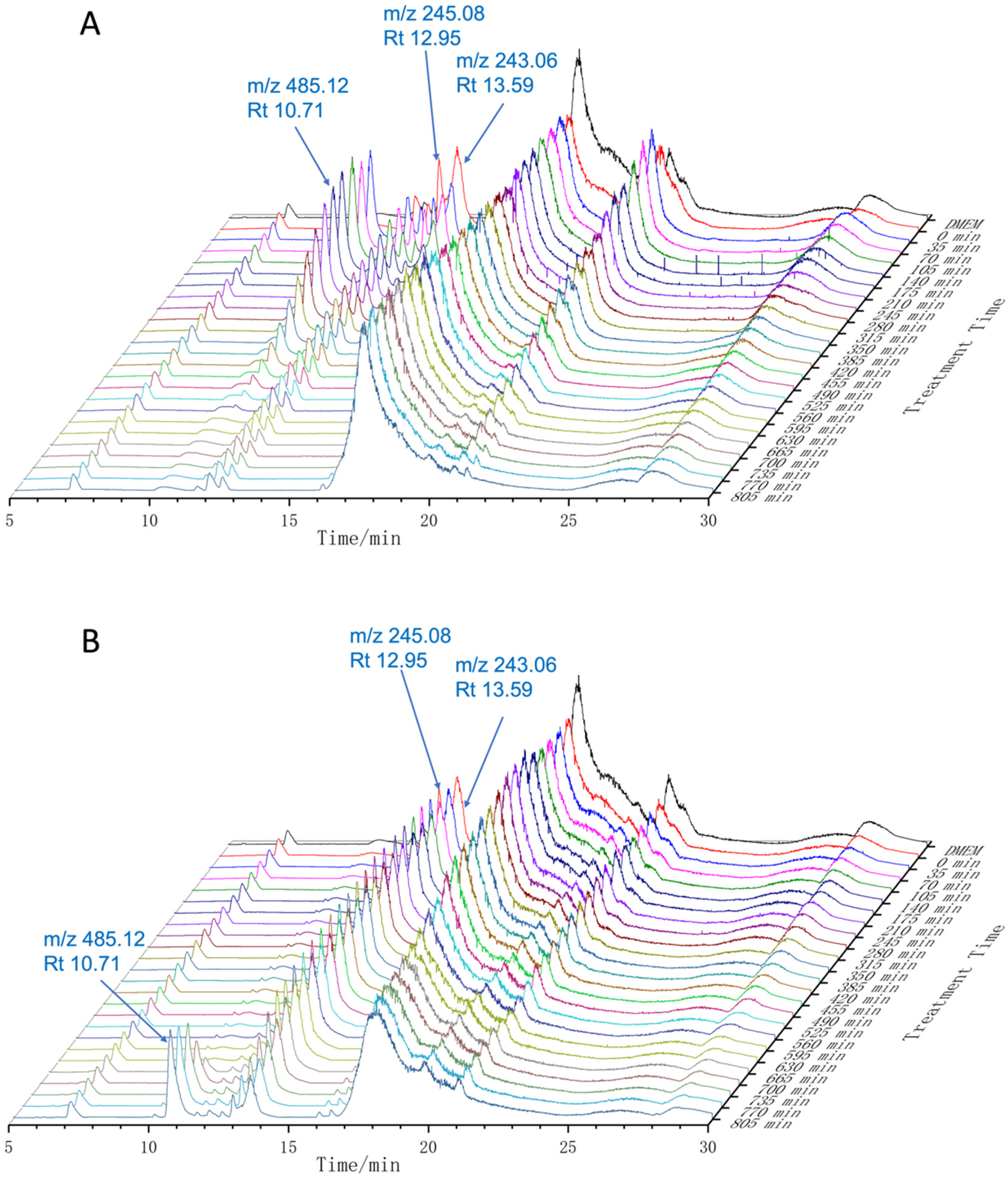
TIC chromatogram (mass range 80.00-1500.00) of piceatannol in DMEM at 37 °C in the absence (A) and presence (B) of ascorbic acid. Max relative abundance 30 000 000. Abbreviations: TIC, total ion current; DMEM, Dulbecco’s Modified Eagle’s Medium.

TIC chromatogram of piceatannol in DMEM and blank DMEM at 37 °C in the absence (A, C, E, G, I, K) and presence (B, D, F, H, J, L) of ascorbic acid. Abbreviations: TIC, total ion current; DMEM, Dulbecco’s Modified Eagle’s Medium.
The Appearance and Disappearance Time Points of Piceatannol Derivatives in Dulbecco’s Modified Eagle’s Medium (DMEM) Medium.

Effect of Ascorbic Acid on the Stability of Piceatannol in DMEM
In the presence of ascorbic acid, the peak of piceatannol (m/z 245.08, tR 12.95) stayed longer than without ascorbic acid (Figure 3B). Without ascorbic acid, the peak of piceatannol started to decay by the time it was added to DMEM at 37 °C. However, 0.5 mM ascorbic acid delayed the degradation of piceatannol until 630 min after the launch of the experiment. In addition to piceatannol, the peaks at m/z 243.06 (tR 12.95 and 13.59), 247.09 (tR 12.92) and 487.14 (tR 12.84) showed a similar trend as piceatannol. Ascorbic acid delayed the generation of the peak at m/z 487.14 (tR 13.52) from the beginning to 665 min after the experiment had started. However, it did not slow the rate of decline of this compound, and its peaks still started to drop within 70 min after its appearance. Without ascorbic acid, the peaks at m/z 485.12 (tR 10.71) and 487.14 (tR 10.69) appeared within 70 min and began to decline from 175 min. Ascorbic acid slowed the generation and degradation of these compounds, as their peaks appeared at 665 min and did not show any sign of decay until 805 min.
The peak at m/z 727.18 (tR 7.93, 9.86, 10.79 and 14.63 min) appeared at the third injection (70 min), then reached its highest intensity at the fourth injection (105 min) and dropped to zero before 385 min without ascorbic acid. However, the presence of ascorbic acid delayed its appearance time points to 665 min and these peaks still existed before the experiment was ended (805 min).
The intensity of peaks at m/z 485.12 (tR 12.96 and 13.66), 487.14 (tR 20.81, 13.52) and 727.18 (tR 13.49) were stable within 630 min in the presence of ascorbic acid, then started to increase after this time point. However, without ascorbic acid, the increment of these peaks happened at 70 min and then disappeared at 385 min. The time points of peaks that appear and disappear in the presence of ascorbic acid are shown in Table 1.
Characterization of New Products of Piceatannol in DMEM
Several new peaks appeared due to the degradation of piceatannol in DMEM at 37 °C. These derivatives were grouped by their molecular ion peaks [M + H] at m/z 243.06 (A), 247.09 (B), 485.12 (C), 487.14 (D) and m/z 727.18 (E). The characterization of the new products was based on the precise mass of the quasimolecular ion and characteristic fragment ions. However, there were still structures that could not be identified only from mass spectra. For the undefined compounds, the possible structures, and the comparison of the actual and predicted MS2 are listed in Table 2.
Comparison of Actual MS2 and Predicted MS2 of m/z 485.12.

Compound A (m/z 243.06)
Compound A (m/z 243.06) was predicted as the main oxidation product of piceatannol. In DMEM at 37 °C, the catechol was oxidized to a quinone then formed piceatannol-O-quinone. Piceatannol-O-quinone might be the possible structure of the compound with a m/z of 243.06. The double bond of piceatannol might also be oxidized to a triple bond then become dehydro-piceatannol. Major daughter ions were detected, such as those at m/z 224.94, 214.95, m/z 196.94 and m/z 172.91. Therefore compound A should be piceatannol-O-quinone. The predicted fragmentation pathway and MS2 are shown in Figure 4A.

The MS2 and predicted structure of (A) compound A (m/z 243.06), (B) compound B (m/z 247.09).
Compound B (m/z 247.09)
The peaks of compound B (m/z 247.09) referred to the reduction product of piceatannol. Two hydrogen atoms were donated by the liquid phase to piceatannol, generating dihydro-piceatannol. According to the total ion current (TIC) chromatography of the ion at m/z 247.09, there was only 1 newly generated compound. The predicted fragmentation pathway and MS2 are shown in Figure 4B.
Compound C (m/z 485.12)
The peaks of compound C (m/z 485.12) are assumed to be due to the dimers of piceatannol. Because of the instability of piceatannol, it would form dimers easily. The TIC chromatography in the mass range from 485.10 to 485.20 suggested 3 isomers had been generated because of the same m/z, but different retention times. These 3 compounds were defined as the dimers of piceatannol based on the daughter ion signals. The main daughter ions of the peak at m/z 485.12 were at m/z 467.10, 449.10, 375.09, and 243.06. The daughter ions at m/z 467.10 and 449.10 were correlated, referring to a dimer losing 1 and 2 hydrogen oxide groups. When the dimer lost a dihydroxybenzene group, it would produce daughter ions with m/z values of 375.09. The daughter ions at m/z 243.06 might belong to 1 piceatannol. The possible structures of C compounds were found from previously reported literature and are shown in Figure 5 and the comparison with MS2 is listed in Table 2.

The structure of piceatannol and the predicted structures of piceatannol derivatives.
Dimers of Piceatannol (m/z 487.14)
The peaks at m/z 487.14 are assumed to be dimers of piceatannol referred to its secondary spectrum. Piceatannol forms dimers in a liquid solution quickly due to its instability. According to the TIC chromatography of mass range 487.15 to 487.20, there were 5 isomers generated, which shared the same m/z, but with different retention times. The main daughter ions of the m/z 487.12 ion were at m/z 469.13, 377.10, 365.10, 255.05, and m/z 243.06, representing the signature fragments of piceatannol dimers (Table 3). The daughter ion at m/z 377.10 might belong to a dimer after losing a dihydroxybenzene group when this fragment lost 1 more such group (m/z 255.05). The ions at m/z 365.10 might be the dimer fragment after losing a dihydroxytoluene group. The daughter ion at m/z 243.06 referred to the fragment of piceatannol.
Comparison of Actual MS2 and Predicted MS2 of m/z 487.14.

There were several ways for piceatannol to form a dimer by losing 2 hydrogen atoms. Because the oxidized piceatannol is dehydrogenated and rearranged to different radicals, the coupling of different piceatannol radicals would form dimers. An 8-O-4′ coupling of piceatannol can form a quinone methide, which is then rearomatized into a new 6-membered ring (compound 7).
14
The 8 to 10′ coupling of piceatannol could result though a similar way of compound 7 to form a new 5-membered ring (compound 8).
14
7-O-5′ coupling of piceatannol generated compound 9
Trimers of Piceatannol (m/z 727.18)
The peaks at m/z 727.18 were assumed to be trimers of piceatannol according to its daughter ions. All compounds producing an m/z 727.18 ion had daughter ions at m/z 709.17, 617.14, 599.13, 485.12, 375.09, and 243.06 (Table 4). The possible structures of group E compounds were found from previously reported literature and are shown in Figure 5.
Comparison of Actual MS2 and Predicted MS2 of m/z 727.18.

Previous studies have shown that piceatannol derivatives could have multiple biological activities. Eight piceatannol dimers synthesized through biotransformation of piceatannol showed their potential for α-glucosidase inhibition activity. 15 Scirpusin B, a dimer of piceatannol, was reported to show a better vasorelaxant effect in rat thoracic aorta than piceatannol via NO derived from the endothelium. 18 Cyperusphenol D, a trimer of piceatannol, was found to have an effect in suppressing the growth of human T-cell leukemia Jurkat cells. 19
Conclusions
As there must be a relatively long time between sample collection and UPLC-MS/MS analysis, the stability of piceatannol was determined when it was kept in different solutions at different temperatures for 2 weeks. The result proved that the storage temperature and solution could affect the stability of piceatannol through long-term storage, and the importance of sample storage for the biological study is beyond doubt.
Since most cell culture experiments are performed at 37 °C, the stability of piceatannol was studied in DMEM at 37 °C; piceatannol was unstable under these conditions. Previous studies showed that other polyphenols, such as quercetin and EGCG, were unstable in the cell culture medium.20,21 It is interesting to note that in most of the cell culture studies, the activity might not be due to the compound added to the cell culture system, but to its degradation products. As piceatannol is a potent antioxidant, it can be oxidized easily, especially at higher temperatures. The result from the current study showed that piceatannol might be oxidized to a quinone form or multiple piceatannol molecules might form different types of dimers or trimers (Table 5). Earlier studies have shown that dimers and trimers of piceatannol could have multiple biological activities.
The Source of Predicated Structure of Piceatannol Derivatives.

Methods and Materials
Chemicals
Piceatannol (98%) was obtained from Macklin Bio Chem Technology Co. Ltd. HPLC grade methanol, acetonitrile, acetic acid (>99%, AR) and DMSO were purchased from Merck, hydrochloric acid (34%-37%, AR) from Fisher Scientific, DMEM high glucose liquid medium from Life Technologies (Gibco BRL), and fetal bovine serum, trypsin-EDTA, penicillin-streptomycin from Professional Health Trading Co. Ltd. Deionized water was prepared using a Milli Q Integral water purification system (Millipore). Ascorbic acid was provided by Aladdin Industrial Co. Ltd.
Stability of Piceatannol in DMSO and Methanol
Piceatannol was dissolved in either DMSO or methanol to obtain a solution of 5 mM in autosampler vials. The solutions were placed at −20, 4 and 37 °C for 2 weeks, respectively. Before analysis, the solutions were diluted to a concentration of 0.5 mM with methanol. An aliquot (5 μL) was automatically injected into a Thermo Scientific LTQ Orbitrap XL hybrid FT Mass Spectrometer.
Stability of Piceatannol in DMEM
Piceatannol was dissolved in DMSO to obtain a standard solution (5 mM), which was kept at −20 °C in the dark. An in situ analysis based on UPLC-MS/MS was used to analyze the stability of piceatannol in DMEM at 37 °C. DMEM (900 μL) and 100 μL of piceatannol standard solution (5 mM) were mixed in an autosampler vial and immediately placed in the UPLC autosampler, which was kept at 37 °C. Every 35 min, a 5 μL aliquot was automatically injected into a Thermo Scientific LTQ Orbitrap XL hybrid FT Mass Spectrometer.
Effect of Ascorbic Acid on the Stability of Piceatannol in DMEM
Ascorbic acid was dissolved in MilliQ water to obtain a solution of 10 mM before being used. In an autosampler vial, 850 μL of DMEM and 50 μL of ascorbic acid solution (10 mM) were mixed, then 100 μL of piceatannol standard solution (5 mM) was added and mixed well. Immediately, an aliquot (5 μL) was automatically injected into a Thermo Scientific LTQ Orbitrap XL hybrid FT Mass Spectrometer.
UPLC-LTQ Orbitrap XL Hybrid FT Mass Analysis
For qualitative analysis, the assay was performed using an ultimate 3000 hyperbaric liquid chromatography system coupled to a Thermo Scientific LTQ Orbitrap XL hybrid FT Mass Spectrometer via an ESI interface. The chromatography system consisted of an autosampler, a diode array detector, a column compartment and 2 pumps. Xcalibur and Mass Frontier 7.0 software packages were used for data collection and analysis. Liquid chromatographic separations were performed using a Waters ACQUITY UPLC HSS T3 column (2.1 × 150 mm, 1.8 µm). The mobile phase consisted of 0.1% formic acid in water (solvent A) and acetonitrile (solvent B). The samples were eluted with the following linear gradient: 15% B at 0 to 5 min, 15% to 25% B at 5–10 min, 25% to 40% B at 10 to 20 min, 40% to 45% B at 20 to 25 min, 45% to 15% B at 25 to 30 min. After that, the mobile phase was returned to the initial condition and held for 5 min so that system could equilibrate for the next injection. The flow rate was 0.3 mL/min. The temperature-controlled column oven was set at 25 °C and the autosampler was kept at different temperatures. Positive ionization (ESI) modes were used in the analysis. The capillary temperature was 350 °C, sheath gas (N2) flow rate 40 psi, aux gas flow rate 10 psi, and the ion spray voltage was set at 3.5 kV. In the FT cell, full MS scans were acquired in the range of m/z 50 to 1500 with a mass resolution of 30,000. The MS/MS experiments were set as a data-dependent scan. 28
Footnotes
Acknowledgements
The authors would like to thank Professor Lunzhao Yi from Kunming University of Science and Technology, China for chemical structure analysis.
Authors’ Contributions
Meng Sam Cheong contributed toward the stability assay and the preparation of the manuscripts. Hui Cao carried out data analysis. Wai San Cheang and Jianbo Xiao supervised the experiments and checked the descriptions in the manuscript. Lutfun Nahar and Satyajit D Sarker revised the manuscript. All authors have read and approved the final manuscript.
Declaration of Conflicting Interests
The authors declared no potential conflicts of interest with respect to the research, authorship, and/or publication of this article.
Funding
The authors disclosed receipt of the following financial support for the research, authorship, and/or publication of this article: This work is supported by Ramón y Cajal grant (RYC2020-030365-I). Lutfun Nahar gratefully acknowledges the financial support of the European Regional Development Fund-Project ENOCH (No. CZ.02.1.01/0.0/0.0/16_019/0000868).