
Abstract
Plagiochasma appendiculatum, a thalloid liverwort, contains high levels of bisbibenzyls, aromatic compounds with potent antitumor as well as antifungal activities. In the present study, rapid growth callus was induced from the thallus of P. appendiculatum, and optimal culture conditions, including medium, temperature, pH, and plant growth regulators for callus production were evaluated. Under optimal culture conditions, the biomass of the callus doubled with a sigmoidal growth curve after 15 days. Differentiation and plant regeneration were studied on a medium supplemented with different plant hormones (α-naphthaleneacetic acid [NAA], 6-benzyladenine [6-BA], and 2,4-dichlorophenoxyacetic acid [2,4-D]). NAA and 6-BA stimulated rhizoid and thallus differentiation, respectively, whereas 2,4-D inhibited the differentiation of thallus and rhizoid. Different metabolic profiles of callus, differentiated thallus, and thallus in the soil were studied by high-performance liquid chromatography. The results showed that both the callus and thallus could synthesize bisbibenzyls. In addition, the kinds and content of bisbibenzyl differed significantly between the callus and thallus. In conclusion, P. appendiculatum thallus cultured in vitro possesses the ability to biosynthesize bisbibenzyl, and it may be utilized for the mass production of specific bisbibenzyls in an appropriate growth environment.
Introduction
Bryophytes can be detected within most ecosystems, except the sea. They can be divided into 3 coordinate phyla: Anthocerotophyta (hornworts), Bryophyta (mosses), and Marchantiophyta (liverworts).
1
Liverworts produce numerous products, including terpenoids,
2
lignin,
3
flavonoids,
4
and bibenzyls, such as lunularic acid (

Structures of bibenzyl (
Plagiochasma appendiculatum Lehm., family Aytoniaceae, is a thalloid liverwort, widely distributed worldwide. It is used by traditional healers to treat burns, boils, eczema, cuts, and wounds, as well as cutaneous diseases.16–20 Furthermore, it is suggested to be an antioxidant and sterilizing agent,
21
and has been used by the Chinese, Indian, and Native Americans since olden times. This liverwort is a rich source of bisbibenzyls, including marchantin A-C, riccardin C (
Although products isolated from liverworts exhibit excellent bioactivities, the source of materials is limited because thalloid liverworts have a tiny morphology, and there is a difficulty in collecting pure samples in large quantities. In addition, to prevent the deterioration of the environment where it grows and to ensure that ecological balance is maintained, harvesting large quantity of wild plants is difficult. Chemical synthesis of bisbibenzyls has been employed, but it does not have economic attractiveness due to its complex nature and low production rate.32–34 This problem could be mitigated by developing plant tissue culture of the species. Bisbibenzyls and terpenoids have been successfully isolated from cultured liverworts.35,36 Sesquiterpenoids were found in Porella vernicosa cultured in vitro, and the volatile constituents of the suspension culture of P. vernicosa and those of the field-collected plants were compared. 37 This biotechnological technique provides an alternative extraction procedure using cells, tissues, or organs in an aseptic, controllable, and automatized manner, resulting in large-scale cultivation. To establish a callus that can produce the best contents of high-value secondary metabolites, determining optimal tissue culture conditions is of great importance. 38 Furthermore, it is essential to be able to differentiate and regenerate the cultures. Metabolic differences in tissues might be correlated with in vitro morphogenesis; but they are usually visually identified, leading to subjectivity. However, it is verified after long-term culture. Therefore, it is valuable to study the content of bisbibenzyls within P. appendiculatum tissues at diverse morphogenetic stages.
In this study, a rapid growth callus was obtained from P. appendiculatum thallus, and the effect of different tissue culture conditions on callus production was evaluated. In addition, the high efficiency of plant regeneration systems from the callus was calculated. The distribution pattern of bisbibenzyls in different cultures was analyzed. The present study established an approach to provide sufficient biomass of taxonomically clean entities and high-value secondary metabolites.
Results and Discussion
Callus Initiation
Callogenesis of P. appendiculatum demonstrated that callus initiation, growth, aspect, and consistency differed according to the type and concentration of plant growth regulators (PGRs) employed. The thallus produced a more developed callus when cultured in a medium that contained 6-benzyladenine (BA; 0.5 mg/L).
The explants cultured on BA produced excellent callus with green and hard aspects, whereas those cultured within the medium that contained 2,4-dichlorophenoxyacetic acid (2,4-D) exhibited callus that was dark green and hard in the center and green and friable at the ends. In contrast, explants cultured in medium with α-naphthaleneacetic acid (NAA) resulted in less callus; however, a more enlarged thallus and a large number of rhizoids were present on the back instead (Figure 2).
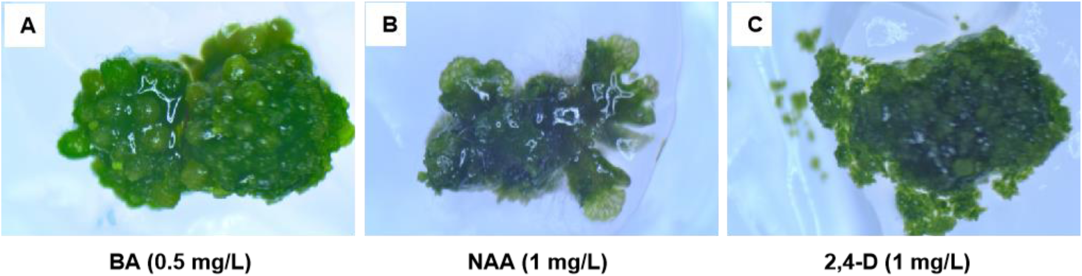
Morphological aspects of callus of Plagiochasma appendiculatum grown on MS medium supplemented with (A) BA, (B) NAA, and (C) 2,4-D.
Growth Measurement
This work adopted growth curves for determining the optimal transplantation period. To be specific, P. appendiculatum callus growth exhibited a frequently seen sigmoidal pattern that was divided into 3 stages (Figure 3A). Of them, the lag phase was observed till day 3 after inoculation, while the exponential phase took place within days 3 to 15 following inoculation, when maximum cell division was being seen. Growth deceleration was observed within days 15 to 18 following inoculation. In contrast, the total bisbibenzyls (TB) content continuously increased within days 3 to 15 following inoculation, with highest levels being obtained at the 15th day after inoculation (Figure 3C). Based on the above findings, transplantation must be conducted within days 12 to 15 during the culture period.

Callus growth curve of Plagiochasma appendiculatum grown on MS medium supplemented with 0.5 mg/L BA.
Optimization of Tissue Culture Conditions
The results of callus growth and TB content are shown in Figure 4. The callus cultured under 25 °C and with a 12-h/12-h light–dark cycle generated through cool white fluorescence (25 μM·m−2 s−1) on solidified Murashige and Skoog culture medium (MS) medium supplemented with BA as PGRs at pH 6 resulted in the optimal growth of P. appendiculatum callus and formation of bisbibenzyls.

Effects of the medium, temperature, pH, and PGRs on the growth of P. appendiculatum callus and TB content. The values are means ± standard deviation of 3 replicates, TB content was based on DW.
Three kinds of media, MS, Gamborg culture medium (B5), and Gui and Ma culture medium (N6), were selected for trials to select a suitable callus growth medium. The results are shown in Figure 4A. In each medium, fresh weight (FW) and dry weight (DW) and TB content were the highest in the MS group. According to the biomass and TB content, the MS medium was confirmed with the highest effectiveness for callus growth.
Callus showed sensitivity to temperature; at temperatures either lower or higher than 25 °C, both callus biomass and TB content decreased significantly (Figure 4B). This could be attributed to the activity of a key enzyme involved in bisbibenzyl synthesis, phenylalanine ammonia lyase, being the highest at 25 °C, as has been suggested for the accumulation of secondary metabolites in other species. 39
The optimal pH for culturing the callus was found to be pH 6.0. When the pH was higher than 6.0, the callus biomass decreased significantly (Figure 4C). This result indicated that the callus growth was inhibited when the pH was above 6.0. However, the TB content was insensitive to pH; when the pH was higher than 6.0, the TB content was not significantly different.
The effects with PGRs showed statistical significance: NAA, BA, and 2,4-D on biomass and TB on the content of the callus (Figure 4D). The greatest callus production could be acquired within the medium that contained 2,4-D at 0.5 mg/L in each accession. With BA at either 0.25 or 1.0 mg/L, high callus generation, second only to 0.5 mg/L of 2, 4-D was seen; however, at 2,4-D <0.5 or >0.5 mg/L, callus biomass decreased significantly.
The highest TB content was obtained in the medium that contained BA at 0.25 mg/L, followed by 1.0 and 0.5 mg/L BA. By contrast, the callus that grew in medium that contained 2, 4-D had the lowest TB content, and when the concentration of 2,4-D grew higher, the TB content gradually decreased. Another notable difference observed between NAA, BA, and 2,4-D was associated with the bisbibenzyls found within the callus. With BA, we detected 6 different bisbibenzyls in the callus, whereas 4 types were detected with NAA and 2,4-D; neither riccardin D nor marchantin C was present.
Differentiation and Regeneration
Observations were performed every 15 days after the culture started, and the following variables were considered: callus induction, rhizoid formation, and thallus formation (Figure 5). The callus cultured under 25 °C and 12-h/12-h light–dark cycle conditions generated through cool white fluorescence (25 μM·m−2 s−1) on solidified MS medium that contained BA (0.5 mg/L) as a PGR at pH 6 resulted in the optimal thallus formation of P. appendiculatum.

Morphogenetic developments in tissue cultures of P. appendiculatum on MS medium supplemented with different PGRs.
At day 15 following inoculation, callus cultured on NAA and 2,4-D retained the callus status, and no thallus primordia were observed (Figure 5A and C). After 45 days, the callus cultured on NAA turned brown and produced significant rhizoids around the callus (Figure 5D, Table 1), whereas the callus cultured on 2,4-D was enlarged and green, and had less (0.25) or no (0.5 mg/L) surrounding rhizoids (Figure 5F, Table 1).
Re-Differentiation of Plagiochasma appendiculatum Callus on MS medium Supplemented With Different PGRs (45 Days).

Differentiation of callus cultures to thallus primordia was observed after 15 days (Figure 5B). Completely developed thallus formed after 45 days of the initial inoculation in a regeneration medium that contained BA at 0.5 mg/L (Figure 5E). This completely developed thallus had dichotomous branching with significant rhizoids on the reverse side. In a previous study, the spores of P. appendiculatum were used to induce aseptic cultured thallus. 40 In this study, surface sterilized thallus was used as explants to induce callus, and then differentiated into aseptic cultured thallus. The use of surface sterilized thallus is not limited by the period of plant development. The induction period of thallus from callus was also shorter than that from spores.
Large variations in bisbibenzyl types and contents were observed during differentiation and regeneration (callus, differentiated culture, and thallus). The callus had higher isomarchantin C (
The biosynthesis of secondary metabolites in a plant is closely related to cell division and differentiation. 41 The difference in the types and contents of bisbibenzyls in P. appendiculatum callus could be attributed to the stimulation of biosynthesis of bisbibenzyls by PGRs. Cytokinin and auxin contained within the medium can regulate callus differentiation and division, while cytokinins are responsible for stimulating protein production efficiency and suppressing cell expansion, thus impacting the cell expansion–cell division balance, as well as secondary metabolite formation.
Based on the above results, the growth phase within callus cultures has a vital effect on regulating biosynthetic pathways. Secondary metabolite accumulation and production are suggested to be different in different plant organs, culture stages of morphogenetic tissues, and morphological stages. Similarly, abietane diterpene antioxidizing agents (carnisol as well as carnosic acid) existed only within Salvia officinalis shoot 42 and Rosmarinus officinalis 43 in vitro cultures, rather than within callus, hairy roots or suspension.
Materials and Methods
Plant Materials
P. appendiculatum was procured from E’mei Mountain, Sichuan province, China, and identified by Dr Xuesen Wen. Plant materials were planted in an agricultural greenhouse in an experimental area of the Department of Natural Product Chemistry, Shandong University, China. The thallus maintenance and propagation were conducted through gemmae growth, as well as sexual hybridization within the greenhouse performed under 25 °C and with a 12-h/12-h light–dark cycle.
Callus Initiation
The young thallus was utilized to be the explant source to establish P. appendiculatum tissue culture in vitro. The thallus was surface sterilized for a 30 s period in 0.5% mercury chloride by agitation, followed by subsequently rinsing 3 to 4 times in sterile deionized water.
After the extra water was removed, the thallus was cut into segments of approximately 0.5 cm2. These were placed in 50 mL conical flasks that contained MS culture medium (15 mL) 44 containing NAA (1.0 mg/L), BA (0.5 mg/L), and 2,4-D (1.0 mg/L), as well as 3% sucrose (w/v), followed by solidification using 0.8% agar. The medium was adjusted to pH 6.0 with 1 M NaOH before solidification. Culture incubation was conducted at 25 ± 1 °C and with 12-h/12-h light–dark conditions generated through cool white fluorescence (25 μM·m−2 s−1) within a plant growth chamber.
Growth Curve
The callus was cultured at 25 °C and with 12-h/12-h light–dark conditions generated through cool white fluorescence (25 μM·m−2 s−1) on solidified MS medium. This contained BA (0.5 mg/L) as PGR at pH 6. Sampling was performed every 3 days after the culture started. The evaluated variables were FW and DW and the contents of TB. All values were obtained in triplicate.
Optimization of Tissue Culture Conditions
Different media, temperatures, pH, and PGRs were used in this experiment. The experiment was designed as follows:
The screening of culture medium was designed using 3 kinds of media, MS, 44 B5, 45 and N6, 46 that contained BA at 0.5 mg/L.
Culture temperature and pH were screened using 5 diverse temperatures (10, 17, 25, 32, 38 °C), along with 3 pH values (pH 6, 7, and 8), in a medium that contained BA at 0.5 mg/L.
PGRs were screened using 3 NAA, BA, and 2,4-D concentrations (all 0.25, 0.5, 1.0 mg/L, respectively).
To each culture medium 3% sucrose (w/v) was added, followed by solidification using 0.8% agar. Each culture was subject to incubation at 25 ± 1 °C and under 12-h/12-h light–dark conditions generated through cool white fluorescence (25 μM·m−2 s−1) within a plant growth chamber at a subculture interval of 15 days. The evaluated variables were FW and DW and the contents of TB. All values were obtained in triplicate.
Differentiation and Regeneration
The regeneration media were designed with different NAA, 6-BA, and 2,4-D contents (all 0.25 and 0.5 mg/L). To each medium 3% sucrose (w/v) was added, followed by solidification using 0.8% agar. Subsequently, callus was transferred onto different regeneration media, followed by incubation at 25 ± 1 °C and with 12-h/12-h light–dark conditions generated through cool white fluorescence (25 μM·m−2·s−1) within a plant growth chamber.
The culture period was 60 days. Evaluations were performed every 15 days after the culture was started, and the following variables were considered: callus induction, rhizoid formation, and thallus formation.
Biomass Determination
To measure FW, all calli in flasks were removed, followed by excessive moisture removal through gentle pressing onto filter paper, and then the weight was measured. To determine DW, the calli within flasks were removed, air dried for 24 h at ambient temperature, and the DW determined.
Secondary Metabolite Extraction
Dried samples were ground to a fine powder (about 0.1 g), followed by extraction using 1 mL methanol with a 40 min ultrasonic process. The sample was later centrifuged for 15 min at 10,000 rpm, the supernatant collected and passed through a 0.22 µm filter. The filtrate was subject to high-performance liquid chromatography (HPLC) analysis using a C18 column.
High Performance Liquid Chromatographic Analysis of Bisbibenzyls
Bisbibenzyls were analyzed using HPLC. An Agilent 1260 system (Agilent Technologies) was adopted for HPLC using a Phenomenex LUNA 5µ C18 column (4.6 × 250 mm) and multi-wavelength diode array detector. Two solvent systems were used: (A) water that contained 0.05% formic acid, and (B) 100% acetonitrile that contained 0.05% formic acid. The separation program was: 0 to 45 min: 40% to 67% B; 45 to 50 min: 90% B; 50 to 55 min: 40% B. This work set the injection volume, flow rate, and detection wavelength at 20 µL, 1.0 mL/min, and 280 nm, separately. Each peak was detected based on UV spectra and retention time; these were compared with appropriate standards. Bisbibenzyl amounts were determined based on standard calibration curves.
The mixed standards containing lunularic acid (
Statistical Analysis
Each experiment was conducted 2 times with identical conditions in triplicate. In the graph, error bars indicated ± SD. SPSS25.0 (IBM) was employed for statistical analysis. Means were examined through one-way analysis of variance (GML procedure). P ≤ .05 stood for statistical significance.
Conclusions
This work suggests that in vitro cultures of P. appendiculatum thallus may biosynthesize bisbibenzyl, and may be used to synthesize specific bisbibenzyls on a large scale in an appropriate growth environment. Tissue cultures provide an appropriate biological system under controllable environmental conditions, and morphogenetic events within this system can be modulated through adjusting the contents of PGRs within the nutrient medium, and thus pharmaceutically important plant metabolites are produced rapidly.
Footnotes
Authors' Contributions
H-X.L. designed the experiments. Y.Z. and X-S.W. performed the experiments. Y.Z., R.N., and A-X.C. analyzed the data and wrote the paper. All the authors analyzed the results and edited and approved the final version of the paper.
Declaration of Conflicting Interests
The authors declared no potential conflicts of interest with respect to the research, authorship, and/or publication of this article.
Funding
The authors disclosed receipt of the following financial support for the research, authorship, and/or publication of this article: This research was funded by Shandong Administration of Traditional Chinese Medicine (2019-0989), Shandong Provincial Natural Science Foundation, China (No. ZR2012CQ032), Independent Innovations Foundation of Shandong University (No. 2012TS104).
Ethical Approval
Not applicable, because this article does not contain any studies with human or animal subjects.
Statement of Human and Animal Rights
This article does not contain any studies with human or animal subjects.
Statement of Informed Consent
Not applicable, because this article does not contain any studies with human or animal subjects.