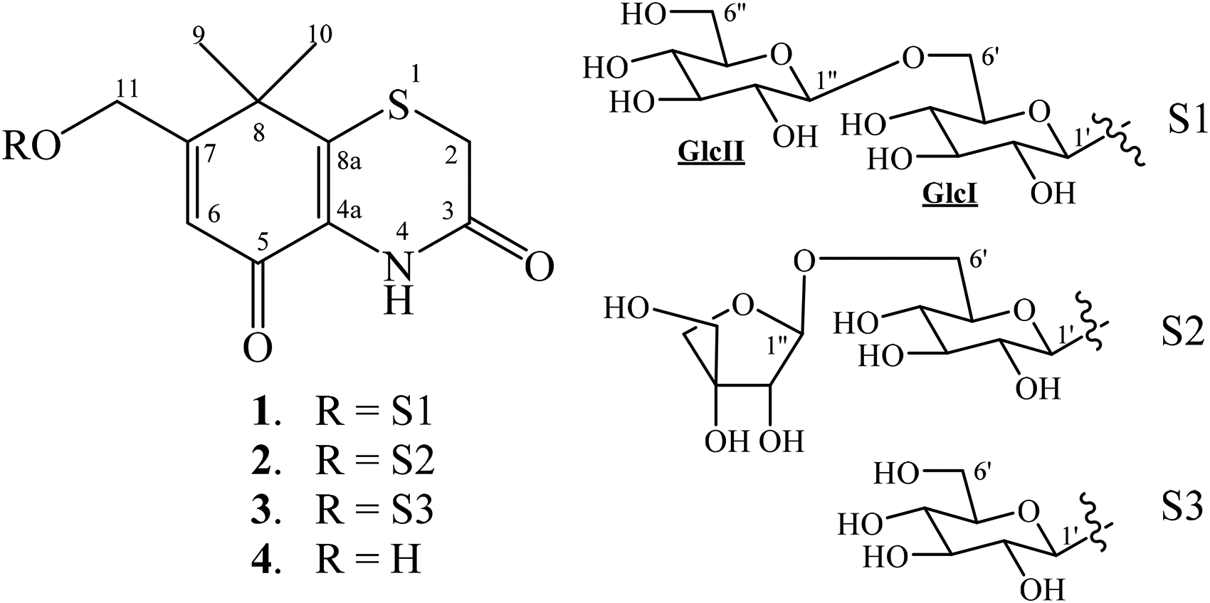
A total of 4 thiazinedione derivatives including 1 new thiazinedione glycoside (1) and 3 known compounds (2-4) were isolated from the fruits of Xanthium strumarium L. Their chemical structures were determined as 7-hydroxymethyl-8,8-dimethyl-4,8-dihydrobenzo[1,4]-thiazine-3,5-dione-11-O-[β-D-glucopyranosyl-(1→6)-O-β-D-glucopyranoside] (1), 7-hydroxymethyl-8,8-dimethyl-4,8-dihydrobenzo[1,4]-thiazine-3,5-dione-11-O-[β-D-apiofuranosyl-(1→6)-O-β-D-glucopyranoside] (2), xanthiside (3), and xanthiazone (4) by extensive nuclear magnetic resonance spectroscopic and high-resolution electron spray ionization mass spectrum analysis and by comparison of the spectral data with those reported in the literature. Compounds 3 and 4 exhibited cytotoxic activity against lung carcinoma (SK-LU-1), human breast carcinoma (MCF-7), hepatocellular carcinoma (HepG2), and skin melanoma (SK-Mel-2) cell lines with half-maximal inhibitory concentration (IC50) values ranging from 27.0 ± 1.1 to 43.2 ± 1.8 µM.
Xanthium strumarium L. (Asteraceae) is a common and well-known traditional herbal medicine in China and Vietnam.1,2 The fruits of X strumarium, named as “Cang-Er-Zi” or “Chang-Er-Zi” in China and “Ke Dau Ngua” in Vietnam, were traditionally used to treat many diseases such as arthritis,3 urticaria,4 headache,5 rheumatism, nasal sinusitis, gastric ulcer, and bacterial and fungal infections.1,2 The main chemical constituents of X strumarium are sesquiterpenoids, coumarins, thiazides, phenylpropenoids, lignanoids, steroids, glycosides, flavonoids, anthraquinones, and naphthoquinones.1,2,6-12 Some of these compounds exhibited useful medicinal properties such as diuretic, anthelmintic, antifungal, anti-inflammatory, antidiabetic, anticancer, and other activities.2,6-9,11 Continuing research on the bioactive compounds from the fruits of X strumarium12 has led to the isolation and structural elucidation of 1 new and 3 known thiazinedione compounds, and their cytotoxic activity on some cancer cell lines; these results are reported in this paper.
Results and Discussion
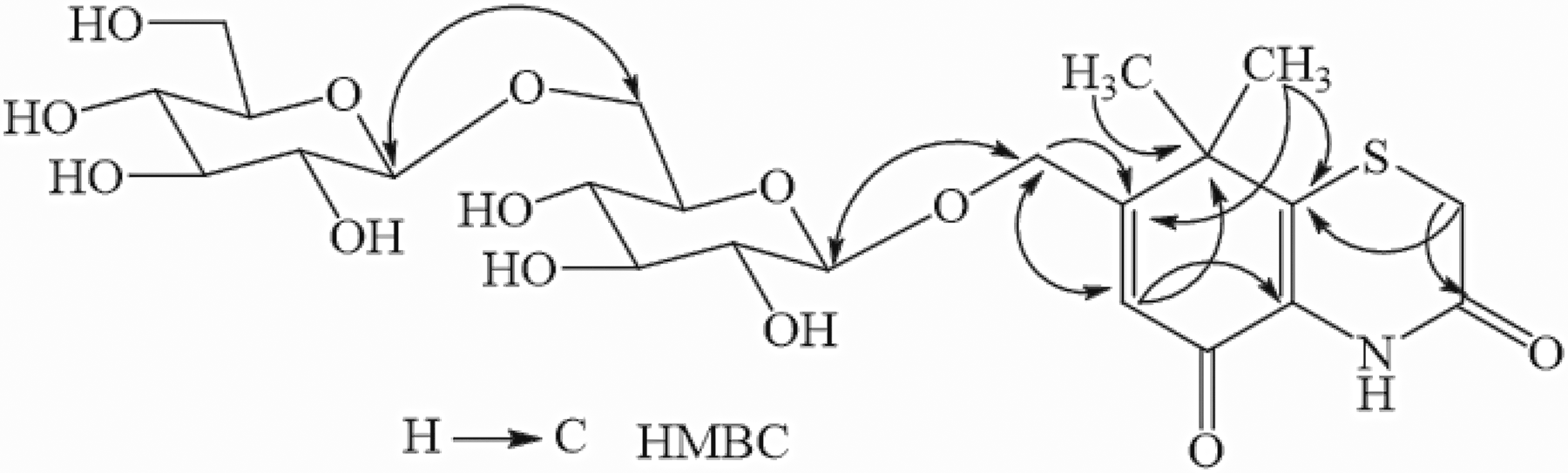
Compound 1 was obtained as a white amorphous powder. The molecular formula of 1 was determined to be C23H33NO13S based on a quasimolecular ion peak at m/z 562.15880 [M–H]− (Calcd. for C23H32NO13S, 562.15998, Δ = −2.1 ppm) in the high-resolution electron spray ionization mass spectrum (HR-ESI-MS) (Supplemental Figure S1). The nuclear magnetic resonance (1H NMR), 13C NMR, and heteronuclear single quantum coherence/correlation (HSQC) spectra of 1 (Supplemental Figures S2 to S6) showed the presence of 2 methyl singlets [δH 1.50 (6H, s)/δC 27.6 and 27.4], 1 olefinic proton [δH 6.69 (s)/δC 123.2], an oxygenated methylene group [δH 4.78 and 4.53 (each, 1H, dd, J = 16.0, 1.5 Hz), 1 methylene singlet at δH 3.51 (2H)/δC 29.8], 2 carbonyl groups at δC 177.1 and 164.6, and 4 quaternary carbons at 43.5, 130.9, 143.5, and 167.4. In addition, 2 glucopyranosyl sugars were identified by anomeric signals at δH 4.40 (1H, d, J = 7.5 Hz)/δC 104.0) and δH 4.40 (1H, d, J = 7.5 Hz)/δC 105.1), and 2 oxygenated methylene groups at δH 4.18 (1H, dd, J = 11.5, 2.0 Hz) and 3.80 (1H, dd, J = 11.5, 6.5 Hz)/δC 70.1) and δH 3.88 (1H, dd, J = 11.5, 2.0 Hz) and 3.67 (1H, dd, J = 11.5, 5.5 Hz)/δC 62.8), as well as 8 oxygenated methine carbons at δC 71.6, 71.7, 75.1, 75.2, 77.3, 78.0 × 2, and 78.1. The above evidence suggested that the aglycon of compound 1 was xanthiazone, with 2 glucopyranosyl sugars. Comparing the NMR data of 1 with that of xanthiside found that compound 1 has one more glucose moiety, attaching to C-6′ of the first glucose unit (Table 1).13 This suggestion perfectly matched the molecular formula of C23H33NO13S indicated by the HR-ESI-MS, and was further confirmed by heteronuclear multiple bond correlation (HMBC) spectra (Supplemental Figures S7 to S10). The HMBC from H-2 (δH 3.51) to C-3 (δC 164.6)/C-8a (δC 143.5), from 2 methyl groups (δH 1.50) to C-7 (δC 167.4)/C-8 (δC 43.5)/C-8a, and from H-6 (δH 6.69) to C-4a (δC 130.9)/C-8 (δC 43.5)/C-11 (δC 68.1) indicated that the aglycone chemical structure of 1 was xanthiazone. In addition, the HMBC from H-11 (δH 4.78 and 4.53) to C-1′ (δC 104.0), from H-1′ (δH 4.40) to C-11 (δC 68.1), from H-6′ (δH 4.18 and 3.80) to C-1′′ (δC 105.1), as well as from H-1′′ (δH 4.40) to C-6′ (δC 70.1) confirmed the sugar moiety as glucopyranosyl-(1→6)-O-glucopyranoside, which was attached to C-11 by an ether linkage (Figure 1). The β-configurations of the glycosidic bonds were assigned based on the coupling constants of the anomeric protons H-1′ and H-1′′ (J = 7.5 Hz). All the NMR assignments of 1 were made by analyzing the HSQC and HMBC spectra, 1H–1H coupling constants, as well as by comparison of its NMR data with the corresponding data of xanthiside, as shown in Table 1.13 Acid hydrolysis of 1 obtained D-glucose, which was identified by thin-layer chromatography (TLC) comparison with an authentic sample and from its positive optical rotation sign.14 Consequently, the chemical structure of compound 1 was established as 7-hydroxymethyl-8,8-dimethyl-4,8-dihydrobenzo[1,4]-thiazine-3,5-dione-11-O-[β-D-glucopyranosyl-(1→6)-O-β-D-glucopyranoside], a new compound named xanthistrumoside A (Figure 2).
Important heteronuclear multiple bond correlation (HMBC) of compound 1.
Chemical structures of compounds 1 to 4.
1H NMR and 13C NMR Spectroscopic Data for Compounds 1 and 3 in Deuterated Methanol.
Pos.
1
3
δC
δH (mult., J in Hz)
δC
δH (mult., J in Hz)
2
29.8
3.51 (s)
29.8
3.51 (s)
3
164.6
–
164.6
–
4a
130.9
–
130.1
–
5
177.1
–
177.1
–
6
123.2
6.69 (dd, 1.5, 1.5)
123.2
6.69 (dd, 1.5, 1.5)
7
167.4
–
167.3
–
8
43.5
–
43.4
–
8a
143.5
143.4
9
27.6
1.50 (s)
27.5
1.50 (s)
10
27.4
1.50 (s)
27.2
1.48 (s)
11
68.1
4.78 (dd, 16.0, 1.5)
67.7
4.80 (dd, 16.0, 1.5)
4.53 (dd, 16.0, 1.5)
4.55 (dd, 16.0, 1.5)
GlcI
1′
104.0
4.40 (d, 7.5)
103.8
4.39 (d, 7.5)
2′
75.2
3.23 (dd, 9.0, 7.5)
75.1
3.24 (dd, 9.0, 7.5)
3′
78.0
3.33*
78.2
3.33*
4′
71.7
3.31*
71.7
3.30*
5′
77.3
3.53 (m)
78.1
3.38 (ddd, 9.0, 6.0, 1.5)
6′
70.1
4.18 (dd, 12.0, 2.0)
62.9
3.92 (dd, 12.0, 1.5)
3.80 (dd, 12.0, 6.5)
3.69 (dd, 12.0, 6.0)
GlcII
1′′
105.1
4.40 (d, 7.5)
2′′
75.1
3.21 (dd, 9.0, 7.5)
3′′
78.1
3.30*
4′′
71.6
3.31*
5′′
78.0
3.40 (m)
6′′
62.8
3.88 (dd, 11.5, 2.0)
3.67 (dd, 11.5, 5.5)
Abbreviation: NMR, nuclear magnetic resonance.
* indicates overlapped signals.
The known compounds were identified as 7-hydroxymethyl-8,8-dimethyl-4,8-dihydrobenzo[1,4]-thiazine-3,5-dione-11-O-[β-D-apiofuranosyl-(1→6)-O-β-D-glucopyranoside] (2),15 xanthiside (3), and xanthiazone (4).13 The NMR spectral data of compounds 2 to 4 were consistent with those previously reported in the literature (Supplemental Figures S11 to S19).
Compounds 1 to 4 were evaluated for their cytotoxic effects on lung carcinoma (SK-LU-1), human breast carcinoma (MCF-7), hepatocellular carcinoma (HepG2), and skin melanoma (SK-Mel-2) cell lines at a concentration of 100 µM. As shown in Supplemental Table S1, compounds 3 and 4 (100 µM) displayed highly cytotoxic effects against SK-LU-1, MCF-7, HepG2, and SK-Mel-2 cell lines with cell death percentages ranging from 65.1 ± 2.1 to 86.3 ± 2.1%. Further dose-dependent experiments found that compounds 3 and 4 induced cytotoxic activity against SK-LU-1, MCF-7, HepG2, and SK-Mel-2 cell lines with half-maximal inhibitory concentration (IC50) values of 35.2 ± 2.1, 41.2 ± 1.3, 37.6 ± 1.3, and 41.5 ± 2.3 µM (for compound 3), and 28.1 ± 2.3, 35.9 ± 1.6, 43.2 ± 1.8, and 27.0 ± 1.1 µM (for compound 4), respectively, compared to the IC50 values of the positive control compound, ellipticine: 1.41 ± 0.10, 1.54 ± 0.12, 1.65 ± 0.12, 1.71 ± 0.19 µM, respectively (Supplemental Table S2). These results suggested that compounds 3 and 4 exhibited weak cytotoxic activity, while compounds 1 and 2 were inactive on the tested cell lines.
Material and Methods
General Experimental Procedures
The NMR spectra were obtained on a Bruker Avance III 500 MHz spectrometer, and HR-ESI-MS on an Agilent 6530 Accurate Mass Q-TOF system. Optical rotation was recorded on a Jasco P-2000 polarimeter. Column chromatography was performed using either silica gel or reversed-phase C-18 resins as adsorbents. Thin-layer chromatography was carried out on precoated silica gel 60 F254 and/or RP-18 F254S plates. Compounds were visualized under UV irradiation (254 and 365 nm) and by spraying with H2SO4 solution (5%) followed by heating with a heat gun.
Plant Material
The fruits of X strumarium L. were collected in Yen My District, Hung Yen Province, Vietnam in December 2019 and identified by Dr Nguyen The Cuong, Institute of Ecology and Biological Resources. A voucher specimen (NCCT-P91) was deposited at the Institute of Marine Biochemistry, VAST.
Extraction and Isolation
The dried fruits of X strumarium (10.0 kg) were ground and ultrasonically extracted 3 times with methanol (MeOH) at room temperature (each, 20 L MeOH, 30 min). After filtration, the solvent was evaporated in vacuo to give the MeOH extract (220 g). This crude was suspended in water (2 L) and successively partitioned with hexane, dichloromethane, and ethyl acetate to give hexane (155 g), dichloromethane (12 g), ethyl acetate extracts (15 g), and the water layer. The water layer was chromatographed on a Diaion HP-20 column eluting with water to remove sugar components, then increasing concentrations of methanol in water (25%, 50%, 75%, and 100%, v/v) to obtain 4 fractions, KN1–KN4. The KN2 fraction (12.0 g) was separated on a silica gel column, eluting with dichloromethane/methanol (15/1, 10/1, 5/1, and 1/1, v/v) to give 5 subfractions KN2A–KN2E. KN2C (4.3 g) was chromatographed on an RP-18 column eluting with acetone/water (2/5, v/v) to give 4 subfractions, KN2B1–KN2B4. KN2B2 (158 mg) was purified on a high-performance liquid chromatography (HPLC) column (J'sphere ODS H-80, 250 mm × 20 mm column) eluting with 25% acetonitrile to yield 1 (17.0 mg) and 2 (14.0 mg). KN2B3 (235 mg) was purified on an HPLC column (J'sphere ODS H-80, 250 mm × 20 mm column) eluting with 35% acetonitrile to yield 3 (39.0 mg). KN2B4 (310 mg) was purified on an HPLC column (J'sphere ODS H-80, 250 mm × 20 mm column) eluting with 45% acetonitrile to yield 4 (85.0 mg).
Xanthistrumoside A (1)
White amorphous powder,
: −25.3 (c 0.1, MeOH); HR-ESI-MS: m/z 562.15880 [M–H]− (Calcd. for C23H32NO13S, 562.15998); 1H NMR (CD3OD, 500 MHz) and 13C NMR (CD3OD, 125 MHz) data are given in Table 1.
Acid Hydrolysis of Compound 1
Compound 1 (5.0 mg) was dissolved in 5.0 mL of 6 N HCl and heated at 60°C for 1.5 h. After cooling, the mixture was extracted with EtOAc. The aqueous layer was concentrated in vacuo followed by TLC examination and optical rotation measurement. The optical rotation of the glucose of 1 was
: + 44.3 (c 0.1, H2O). By comparing this value with that of D-glucose:
: + 45.5 (c 0.1, H2O), the glucose in compound 1 was determined to have a D-configuration.
Cytotoxic Assay
Refer to Supplemental material.
Conclusions
A total of 1 new thiazinedione glycoside (1) and 3 known thiazinedione compounds (2-4) were isolated from the seeds of X strumarium. Their chemical structures were determined by extensive analysis of HR-ESI-MS and NMR spectra and by comparison of these data with those reported in the literature. Compounds 3 and 4 exhibited cytotoxic activity against SK-LU-1, MCF-7, HepG2, and SK-Mel-2 cell lines with IC50 values of 35.2 ± 2.1, 41.2 ± 1.3, 37.6 ± 1.3, and 41.5 ± 2.3 µM (for compound 3), and 28.1 ± 2.3, 35.9 ± 1.6, 43.2 ± 1.8, and 27.0 ± 1.1 µM (for compound 4).
Footnotes
Declaration of Conflicting Interests
The authors declared no potential conflicts of interest with respect to the research, authorship, and/or publication of this article.
Funding
The authors disclosed receipt of the following financial support for the research, authorship, and/or publication of this article: This work was supported by the Vietnam Academy of Science and Technology (grant no. NVCC38.02/21-21).
ORCID iD
Phan V. Kiem
Supplemental Material
Supplemental material for this article is available online.
Trial Registration
Not applicable, because this article does not contain any clinical trials.
References
1.
ChiVV. Dictionary of Vietnamese Medicinal Plants. Medicine Publishing House; 2012:1185-1186.
2.
FanWFanLPengC., et al. Traditional uses, botany, phytochemistry, pharmacology, pharmacokinetics and toxicology of Xanthium strumarium L.: A review. Molecules. 2019;24:359. doi:10.3390/molecules24020359
3.
QinLHanTLiHZhangQZhengH. A new thiazinedione from Xanthium strumarium. Fitoterapia. 2006;77(3):245-246. doi:10.1016/j.fitote.2006.02.001
4.
YinRHBaiXFengT, et al. Two new compounds from Xanthium strumarium. J Asian Nat Prod Res. 2016;18(4):354-359. doi:10.1080/10286020.2015.1099525
5.
HanTLiHZhangQ, et al. New thiazinediones and other components from Xanthium strumarium. Chem Nat Compd. 2006;42(5):567-570. doi:10.1007/s10600-006-0215-2
6.
TongCChemRHLiuDCZengDSLiuH. Chemical constituents from the fruits of Xanthium strumarium and their antitumor effects. Nat Prod Commun. 2020;15(8):1-5. doi:10.1177/1934578X20945541
7.
WenYLLiMJYeZJ, et al. Compounds isolated from the fruits of Xanthium strumarium, including a new neo-lignan, and their anticancer effects. Nat Prod Commun. 2020;15(12):1-4. doi:10.1177/1934578X20982782
8.
FerrerJPZampiniICCuelloAS, et al. Cytotoxic compounds from aerial organs of Xanthium strumarium. Nat Prod Commun. 2016;11(3):371-374. doi:10.1177/1934578X1601100313
9.
KimYSKimJSParkSH, et al. Two cytotoxic sesquiterpene lactones from the leaves of Xanthium strumarium and their in vitro inhibitory activity on farnesyltransferase. Planta Med. 2003;69(4):375-377. doi:10.1055/s-2003-38879
10.
MangelSMNareshKSKuldipSD. Xanthanolides from Xanthium strumarium. Phytochemistry. 1992;32(1):206-207. doi:org/10.1016/S0031-9422(00)90678-2
11.
LinBZhaoYHanP, et al. Anti-arthritic activity of Xanthium strumarium L. extract on complete Freund’s adjuvant induced arthritis in rats. J Ethnopharmacol. 2014;155(1):248-255. doi:10.1016/j.jep.2014.05.0238
12.
KiemPVHoangNHThuVKTaiBHNhiemNX. Diterpene glycosides and phenolic compounds from the fruits of Xanthium strumarium. Vietnam J Chem. 2020;58(5):649-654. doi:10.1002/vjch.2020000061
13.
DaiYHCuiZLiJLWangD. A new thiaziedione from the fruits of Xanthium sibiricum. J Asian Nat Prod Res. 2008;10(4):303-305. doi:org/10.1080/10286020701833495
14.
LaurenceVNRenetaGNicolasB, et al. Triterpenoid saponins from the roots of Gypsophila trichonoma wender. Phytochemistry. 2013;90:114-127. doi:10.1016/j.phytochem.2013.03.001
15.
JiangHYangLLiuC, et al. Four new glycosides from the fruits of Xanthium sibiricum patr. Molecules. 2013;18(10):12464-12473. doi:10.3390/molecules181012464
Supplementary Material
Please find the following supplemental material available below.
For Open Access articles published under a Creative Commons License, all supplemental material carries the same license as the article it is associated with.
For non-Open Access articles published, all supplemental material carries a non-exclusive license, and permission requests for re-use of supplemental material or any part of supplemental material shall be sent directly to the copyright owner as specified in the copyright notice associated with the article.