This review will summarize the authors’ studies on the reassignments of structures of 9 natural polyoxygenated sterols (24S)-24-ethylcholest-8-ene-3β,5α,6β,7α-tetrol (1), (24S)-24-ethylcholest-8(14)-ene-3β,5α,6β,7α-tetrol (2), (22E)-24-methylcholesta-8(14),22-diene-3β,5α,6β,7α-tetrol (3), 5β,6β-epoxy-(22E)-ergosta-8,22-diene-3β,7β-diol (4), (22E)-ergosta-7,22-diene-3β,5α,6β,9α,14α-pentol (5), 3β,5α,6β,8β,14α-pentahydroxy-(22E)-ergost-22-en-7-one (6), 5β,6β-epoxy-24-methylenecholesta-8,24(28)-diene-3β,7α,11α-triol (7), 6β-acetoxy-(22E)-10α-ergosta-7,22-diene-3β,5α-diol (8), and 8α,9α-epoxy-(22E)-ergosta-6,22-diene-3β,5α,14α-triol (9). The structures of 1 to 9 have been reassigned as (24S)-5α,6α-epoxy-24-ethylcholest-8-ene-3β,7α-diol (16), (24S)-5α,6α-epoxy-24-ethylcholest-8(14)-ene-3β,7α-diol (17), 5α,6α-epoxy-(22E)-ergosta-8(14),22-diene-3β,7α-diol (13), 5α,6α-epoxy-(22E)-ergosta-8,22-diene-3β,7α-diol (12), (22E)-ergosta-7,22-diene-3β,5α,6β,9α,14β-pentol (25), 5α,6α;8α,14α-diepoxy-3β-hydroxy-(22E)-ergost-22-en-7-one (18), 5α,6α-epoxyergosta-8,24(28)-diene-3β,7α,11α-triol (21), 6β-acetoxy-(22E)-ergosta-7,22-diene-3β,5α-diol (26), and 8α,14α-epoxy-(22E)-ergosta-6,22-diene-3β,5α,9α-triol (28), respectively, from the results of careful reexamination of the published 1H and 13C NMR spectral data.
Sterols are among the most studied groups of natural products with interest commencing in the 19th century and continuing through to the present.1-3 Sterols occur in all major groups of organisms, from fungi to humans, as secondary metabolites.4 In a continuation of our studies on the chemical constituents from Japanese mushrooms, 28 new polyoxygenated sterols have been obtained and characterized.5 On the other hand, structural misassignments of natural products are prevalent in the literature.6-8 Determination of structure is a fundamental pillar of the discipline of chemistry.9 However, misinterpretations of spectroscopic and/or physical data have often been known to result in structural misassignments.10 Recently, we have reported the reassignments of structures of 5 natural polyoxygenated sterols: (24S)-24-ethylcholest-8-ene-3β,5α,6β,7α-tetrol (1),11 (24S)-24-ethylcholest-8(14)-ene-3β,5α,6β,7α-tetrol (2),11 (22E)-24-methylcholesta-8(14),22-diene-3β,5α,6β,7α-tetrol (3),12 5β,6β-epoxy-(22E)-ergosta-8,22-diene-3β,7β-diol (4),13 and (22E)-ergosta-7,22-diene-3β,5α,6β,9α,14α-pentol (5)14 (Figure 1). Thereafter, continued literature survey on the polyoxygenated sterols from natural sources was conducted, and it was revealed that the structures of 4 sterols, 3β,5α,6β,8β,14α-pentahydroxy-(22E)-ergost-22-en-7-one (6),15 5β,6β-epoxyergosta-8,24(28)-diene-3β,7α,11α-triol (7),16 6β-acetoxy-(22E)-10α-ergosta-7,22-diene-3β,5α-diol (8),17 and 8α,9α-epoxy-(22E)-ergosta-6,22-diene-3β,5α,14α-triol (9),18 would need to be reassigned (Figure 1). In this review, we summarize our studies on the reassignments of structures of 1 to 9.
Structures of compounds 1 to 9.
Reassignments of Structures of (24S)-24-Ethylcholest-8-Ene-3β,5α,6β,7α-Tetrol, (24S)-24-Ethylcholest-8(14)-Ene-3β,5α,6β,7α-Tetrol, (22E)-24-Methylcholestra-8(14),22-Diene-3β,5α,6β,7α-Tetrol, and 3β,5α,6β,8β,14α-Pentahydroxy-(22E)-Ergost-22-En-7-One
First of all, we describe the structural reassignment of 4 sterols having 3β,5α,6β,7α-tetrahydroxylated and 3β,5α,6β,8β,14α-pentahydroxylated structures: (24S)-24-ethylcholest-8-ene-3β,5α,6β,7α-tetrol (1), (24S)-24-ethylcholest-8(14)-ene-3β,5α,6β,7α-tetrol (2), (22E)-24-methylcholesta-8(14),22-diene-3β,5α,6β,7α-tetrol (3), and 3β,5α,6β,8β,14α-pentahydroxy-(22E)-ergost-22-en-7-one (6).
In 1995, Costantino et al19 reported 2 new polyoxygenated sterols 1 and 2 from the marine sponge Neofibularia nolitangere (Figure 2). In 2006, Sun et al20 reported a new one 3 from the marine-derived fungus Penicillium sp. (Figure 2). Although there is no report on the biological activities of 1 and 2, it is reported that 3 has potent cytotoxicity against human liver cancer cell (Hep G) with IC50 value of 10.4 µg/mL.20 The 3β,5α,6β,7α-tetrahydroxylated structures of 1 to 3 were proposed on the basis of 1-dimensional (1D) and 2-dimensional (2D) nuclear magnetic resonance (NMR) data.19,20 During our studies on the sterol constituents from Japanese mushrooms,5 we reported 2 polyoxygenated sterols having the same A/B ring system as those of 1 to 3, (22E)-ergosta-8,22-diene-3β,5α,6β,7α-tetrol (10) and (22E)-ergosta-8(14),22-diene-3β,5α,6β,7α-tetrol (11), from the edible mushroom Grifola frondosa (Figure 2).21 Comparison of the NMR data of 1 and 2 for the A/B ring system with those of 10 and 11, respectively, indicated that the reported data do not fit with the proposed structures 1 and 2. Similarly, comparison of the NMR data of 3 for the A/B ring system with that of 11 indicated that the reported data also do not fit with the proposed structure 3. Consequently the structures for 1 to 3 are doubtful and reexamination of structures of 1 to 3 was necessary.
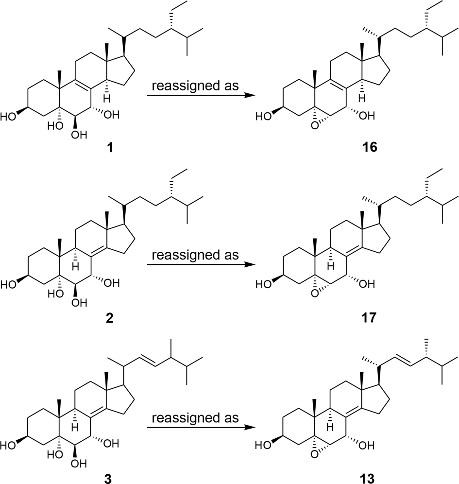
Structures of compounds 1 to 3 and 10 to 17.
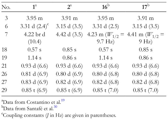
Although compounds 1 and 2, and 10 and 11 differ in the structures of the side chain at C-17, we have already confirmed that the differences in the side chain at C-17 have little effect on the chemical shift values in the NMR spectrum derived from the A/B ring system of polyoxygenated sterols.5 In the 1H and 13C NMR data of 1 to 3, and 10 and 11 (supplemental Tables S1 and S2 for 10 and 11), there were large differences in the chemical shift values for skeletal positions C-3, C-5, C-6, C-7, and C-19. In particular, the 13C NMR chemical shift values δC 65.6 (C-5) and 62.6 (C-6) in 1, δC 67.8 (C-5) and 61.3 (C-6) in 2, and δC 67.8 (C-5) and 61.3 (C-6) in 3 were attributed to those of an epoxide ring,22-27 suggesting that C-5 and C-6 of 1 to 3 were not connected to hydroxy groups. The 13C NMR chemical shift values for skeletal positions C-5 and C-6 of 1 to 3 were calculated using Kobayashi’s parameters for polyhydroxy steroids.28 Calculated values of C-5 and C-6 of 1 were δC 73.8 and 76.9, respectively. Those of 2 and 3 were δC 77.3 (C-5) and 80.4 (C-6). Kobayashi28 concluded that deviations of 2.5 ppm between the predicted and calculated δC values would be sufficient for adoption, or rejection, of a particular structure. Deviations of C-5 and C-6 of 1 were 8.2 and 14.3 ppm, respectively, and those of 2 and 3 were 9.5 and 19.1 ppm. The calculated values confirmed that C-5 and C-6 of 1 to 3 were not connected to hydroxy groups. On the other hand, we reported 2 polyoxygenated sterols having an epoxide ring at C-5 and C-6, 5α,6α-epoxy-(22E)-ergosta-8,22-diene-3β,7α-diol (12) and 5α,6α-epoxy-(22E)-ergosta-8(14),22-diene-3β,7α-diol (13), from G. frondosa (Figure 2).21 The functionality of the A/B ring system such as 12 and 13 was confirmed by the synthesis of 5α,6α-epoxycholest-8-ene-3β,7α-diol (14) from 5α,8α-epidioxy sterol utilizing microwave irradiation-induced isomerization as the key step29 and an x-ray diffraction experiment performed on 5α,6α-epoxycholest-8(14)-ene-3β,7α-diol-3,7-diacetate (15)24 (Figure 2). The 1H and 13C NMR data of 12 and 13 for skeletal positions C-3, C-5, C-6, C-7, and C-19 were identical with those of 1 and 2, respectively. Also, the NMR data of 3 for skeletal positions C-3, C-5, C-6, C-7, and C-19 were identical with that of 13. In 2002, Santafé et al30 reported 2 new sterols having the same A/B ring system as those of 12 and 13, (24S)-5α,6α-epoxy-24-ethylcholest-8-ene-3β,7α-diol (16) and (24S)-5α,6α-epoxy-24-ethylcholest-8(14)-ene-3β,7α-diol (17), from the marine sponge Polymastia tenax (Figure 2). The published 1H (Table 1) and 13C NMR (Table 2) data for 16 and 17 were in agreement with those of 1 and 2, respectively. Furthermore, the 1H (Table 3) and 13C NMR (Table 4) data of 13 were in agreement with those of 3. Thus, the structures of 1, 2, and 3 have been reassigned as 16, 17, and 13, respectively (Figure 3).11,12
1H NMR Chemical Shifts of Compounds 1, 2, 16, and 17 in CDCl3.
The molecular formula of 13, 16, and 17 differs from that of 1 to 3 by the loss of 1 molecule of H2O. Mass spectrometry (MS) experiments frequently provide ion peaks following the loss of H2O, especially in electron impact (EI) mass spectrum. Misreading these ion peaks has the potential to cause structure misassignment when the molecules have more than 2 hydroxy groups.10 Sterols having a 3β,5α,6β,7α-tetrahydroxylated structure such as 10 and 11 did not show a molecular ion in the EI mass spectrum and showed the dehydration ion due to the loss of 1 molecule of H2O (supplemental material for 10 and 11). However, the dehydration ion of sterols having an above structure correspond to the molecular ion for the sterols having a 3β,7α-dihydroxy-5α,6α-epoxy structure such as 12 and 1321 (supplemental material for 12 and 13). The above-mentioned misreading of the MS data was observed in structure determination of 1 to 3.19,20 Careful interpretation of the MS data is an important point to be noticed in structure determination of polyoxygenated sterols in order to avoid deriving a wrong structure.11,12
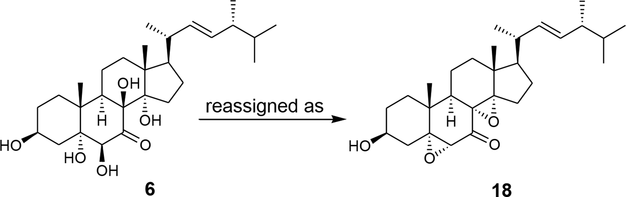
Furthermore, as a result of searching for literature related to the above polyoxygenated sterols, we found that the structure of 3β,5α,6β,8β,14α-pentahydroxy-(22E)-ergost-22-en-7-one (6) from the liquid culture of the basidiomycete Ganoderma applanatum would need to be reassigned (Figure 4).15
Structures of compounds 6 and 18.
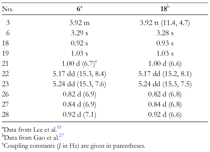
The molecular formula of 6 was determined to be C28H46O6 by high-resolution (HR) fast atom bombardment mass spectrum (m/z 443.3149, calcd 443.3161 for [(M+H)-2H2O]+) and NMR data. Regarding compound 6, Lee et al15 concluded that “The presence of five hydroxyls in the molecule was estimated from the ion peaks appearing at m/z 442 [M - 2H2O]+, 399 [(M - 2H2O) - C3H7]+, 317 [(M - 2H2O) - side chain (C9H17)]+, 299 [(M - 2H2O) - (side chain + H2O)]+, 281 [(M - 2H2O) - (side chain + 2H2O)]+ and 263 [(M - 2H2O) - (side chain + 3H2O)]+ in the EI mass spectrum.” However, as mentioned above, it is difficult to determine the number of hydroxy groups from the fragment ion peaks of the molecule such as polyhydroxy sterols in case no molecular ion was observed. On the other hand, Gao et al27 reported a new sterol, 5α,6α;8α,14α-diepoxy-3β-hydroxy-(22E)-ergost-22-en-7-one (18), from the mangrove fungus Aspergillus awamori (Figure 4). The published 1H (Table 5) and 13C NMR (Table 6) data for 18 were in agreement with those of 6. Compound 6 did not show the molecular ion in the EI mass spectrum and showed the dehydration ion due to the loss of 2 molecules of H2O at m/z 442.15 However, the dehydration ion at m/z 442 in the EI mass spectrum of 6 corresponds to the molecular ion for 18.25 Therefore, the proposed structure of 6 has been reassigned as 18 (Figure 5).
1H NMR Chemical Shifts of Compounds 6 and 18 in CDCl3.
Reassignments of Structures of 5β,6β-Epoxy-(22E)-Ergosta-8,22-Diene-3β,7β-Diol and 5β,6β-Epoxyergosta-8,24(28)-Diene-3β,7α,11α-Triol
In this section, we describe the structural reassignment of 2 polyoxygenated sterols having 3β,7β-dihydroxy-5β,6β-epoxy and 3β,7α,11α-trihydroxy-5β,6β-epoxy structures, 5β,6β-epoxy-(22E)-ergosta-8,22-diene-3β,7β-diol (4) and 5β,6β-epoxyergosta-8,24(28)-diene-3β,7α,11α-triol (7).
In 2016, Su et al31 reported a new sterol 4 from the gorgonian Pinnigorgia sp. (Figure 6). Compound 4 was shown to significantly inhibit the accumulation of the proinflammatory inducible nitric oxide synthase and cyclooxygenase protein in lipopolysaccharide (LPS)-stimulated RAW264.7 macrophage cells.31 The 3β,7β-dihydroxy-5β,6β-epoxy structure of 4 was proposed on the basis of 1D and 2D NMR data. As mentioned earlier, we already described the structure of 12 (Figure 6).21 The structural similarity between 4 and 12 is striking; however, the 5α,6α-epoxy-7α-hydroxy structure of 12 was substituted with a 5β,6β-epoxy-7β-hydroxy structure in 4. In contrast, a close investigation of the spectroscopic data for 4 revealed a level of equivalency with 12 that was inconsistent with this structural divergence. Reassignment of the structure of 4 was therefore pursued.
Structures of compounds 4 and 12.
Regarding compound 4, Su et al31 concluded that “comparison of these data with spectral data for a known sterol 12 revealed that these two compounds are isomers except the configurations of 5,6-epoxy-7-hydroxy groups.” “Key nuclear Overhauser effect spectroscopy (NOESY) correlations for 4 showed interactions between H-4β/H3-19 and H-4α/H-6. Also, H-7 showed a correlation with H-6. Thus, H-6 and H-7 should be located on the α-face and the oxygen of the 5,6-epoxide must be positioned on the β-face (Figure 7).” Although Su et al31 recorded nuclear Overhauser effect (NOE) correlation between H3-18 and H3-19, it is impossible for the structure 10 (Figure 7). Furthermore, the NOESY correlations showed in 4 were also found to be the case for 12 (Figure 7). The B ring of 12 adopts a boat-type conformation, as a result of the incorporation of 5α,6α-epoxide ring and double bond between C-8 and C-9 (Figure 7).22 When H-6 and H-7 were in pseudo-equatorial position and pseudo-axial position, respectively, of the boat-type conformation, the distances between H-4α and H-6, and H-6 and H-7 were close enough to produce the NOE correlation (Figure 7). The NOE was also observed between H3-19 and H-7. Further, the 1H (Table 7) and 13C NMR (Table 8) data of 4 were in agreement with those of 12. From the above data, the structure of 4 has been reassigned as 12 (Figure 8).13
Conformations and NOEs (full-line arrows) for compounds 4 and 12.
1H NMR Chemical Shifts of Compounds 4 and 12 in CDCl3.
cSignals reassigned by the authors are indicated by underlining.
Reassignment of compound 4.
Furthermore, as a result of searching for literature related to the above compound, we found that the structure of 5β,6β-epoxyergosta-8,24(28)-diene-3β,7α,11α-triol (7) from the marine sponge Dysidea herbacea would need to be reassigned (Figure 9).16
Structures of compounds 7 and 19.
Compound 7 was purified as the triacetate derivative 19 (Figure 9).16 Compound 19 was used to investigate its structure. In the difference NOE experiment, irradiation of the signal of H3-19 caused NOE enhancement in the signals of H-4β, H-7, H-11, and H3-18 (Figure 10). Furthermore, irradiation of the signal of H-6 caused NOE enhancement in the signals of H-4α and H-7 (Figure 10). Among these, the NOEs observed between H3-19 and H-7, H3-19 and H-11, and H3-19 and H3-18 are impossible for the structure 19. However, when the 5β,6β-epoxide ring of 19 was changed to the 5α,6α-epoxide ring (20), the distances between H3-19 and H-7, H3-19 and H-11, and H3-19 and H3-18 were close enough to produce the NOEs (Figure 10). Especially, as mentioned earlier, the NOE observed between H3-19 and H-7 implies that the B ring of 19 adopts a boat-type conformation, as a result of the incorporation of 5α,6α-epoxide ring and double bond between C-8 and C-9.22 Accordingly, compound 19 was revised to 20, and therefore, the structure of 7 has been reassigned as the new structure 5α,6α-epoxyergosta-8,24(28)-diene-3β,7α,11α-triol (21) (Figure 11). The 3β,7α,11α-trihydroxy-5α,6α-epoxy moiety is unprecedented in the natural sterols previously known.
Conformations and NOEs (full-line arrows) for compounds 19 and 20.
Reassignment of compound 7.
Reassignments of Structures of (22E)-Ergosta-7,22-Diene-3β,5α,6β,9α,14α-Pentol and 6β-Acetoxy-(22E)-10α-Ergosta-7,22-Diene-3β,5α-Diol
In this section, we describe the stereochemical reassignment of the hydroxy group at C-14 of (22E)-ergosta-7,22-diene-3β,5α,6β,9α,14α-pentol (5) and the methyl group at C-10 of 6β-acetoxy-(22E)-10α-ergosta-7,22-diene-3β,5α-diol (8).
In 2008, Zhang et al32 reported a new sterol 5 from the spores of the medicinal mushroom Ganoderma lucidum (Figure 12). The 3β,5α,6β,9α,14α-pentahydroxylated structure of 5 was proposed on the basis of NOESY spectrum.32 However, the stereochemistry of the hydroxy group at C-14 was tentatively assigned as α configuration based on the fact that the C/D ring system of ergostane-type sterols are generally trans-fused.32 During our studies on the sterol constituents from Japanese mushrooms,5 we reported a sterol having the same C/D ring system as that of 5, 3β,5α,9α,14α-tetrahydroxy-(22E)-ergosta-7,22-dien-6-one (22), from the edible mushroom Pleurotus ostreatus (Figure 12).33 Comparison of the 1H and 13C NMR data of 5 for the C/D ring system with those of 22 indicated that the reported NMR data do not fit well with the proposed structure 5. Although compounds 5 and 22 differ in the substituents at C-6, we have already confirmed that the differences in the substituent at C-6 have little effect on the chemical shift values in the NMR spectrum derived from the C/D ring system of polyoxygenated sterols.5 Therefore, we undertook a reexamination of the structure of 5.
Structures of compounds 5 and 22 to 24.
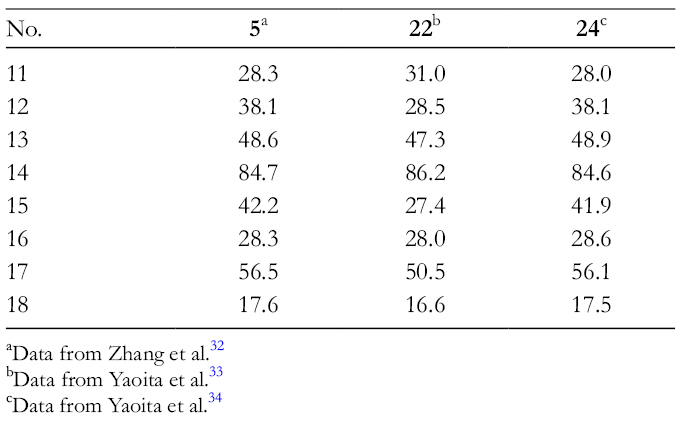
In the 13C NMR data of 5 and 22 (supplemental Table S3 for 22), there were large differences in the chemical shift values for skeletal positions C-12, C-15, and C-17 (Table 9). In particular, the 13C NMR chemical shift values at C-12 and C-17 in 22 were attributed to the γ-gauche effect of the 14α-hydroxy group, as compared with 3β,5α,9α-trihydroxy-(22E)-ergosta-7,22-dien-6-one (23)33 (supplemental Table S3 for 23), suggesting that the difference in the C/D ring system between 5 and 22 was traced to differences in the stereochemistry of the hydroxy group at C-14. On the other hand, we reported a sterol, 3β,5α,9α,14β-tetrahydroxy-(22E)-ergosta-7,22-dien-6-one (24), the epimer of 22 at C-14, from the edible mushroom Tricholoma matsutake (Figure 12).34 The 13C NMR data of 24 (supplemental Table S3 for 24) for skeletal positions C-11 to C-18 were coincident with that of 5 (Table 9). Therefore, the orientation of the C-14 hydroxy group of 5 should be revised as β. As additional proof, in comparison with the reported 1H NMR data of the H3-18 of 5 recorded in CD3OD with that of C5D5N,32 a pyridine-induced deshielding effect35-37 was observed, as was the case with 24,34 supporting that the C-14 hydroxy group of 5 has a β configuration (Figure 13). Furthermore, in the 1H NMR data of 5,30 the chemical shifts of H-1α, H-3, H-4β, and the H3-19 in C5D5N were shifted downfield by the pyridine-induced deshielding effect,35-37 confirming the presence of a 3β,5α,6β,9α-tetrahydroxy grouping in the A/B ring system of 5 (Figure 13). Accordingly, the structure of 5 has been reassigned as the new structure (22E)-ergosta-7,22-diene-3β,5α,6β,9α,14β-pentol (25) (Figure 14).14 The 3β,5α,6β,9α,14β-pentahydroxy moiety is unprecedented in the natural sterols previously known.
13C NMR Chemical Shifts of Skeletal Positions C-11 to C-18 Within Compounds 5, 22, and 24 in C5D5N.
Pyridine-induced deshieldings (full-line arrows) in revised structure 25.
Reassignment of compound 5.
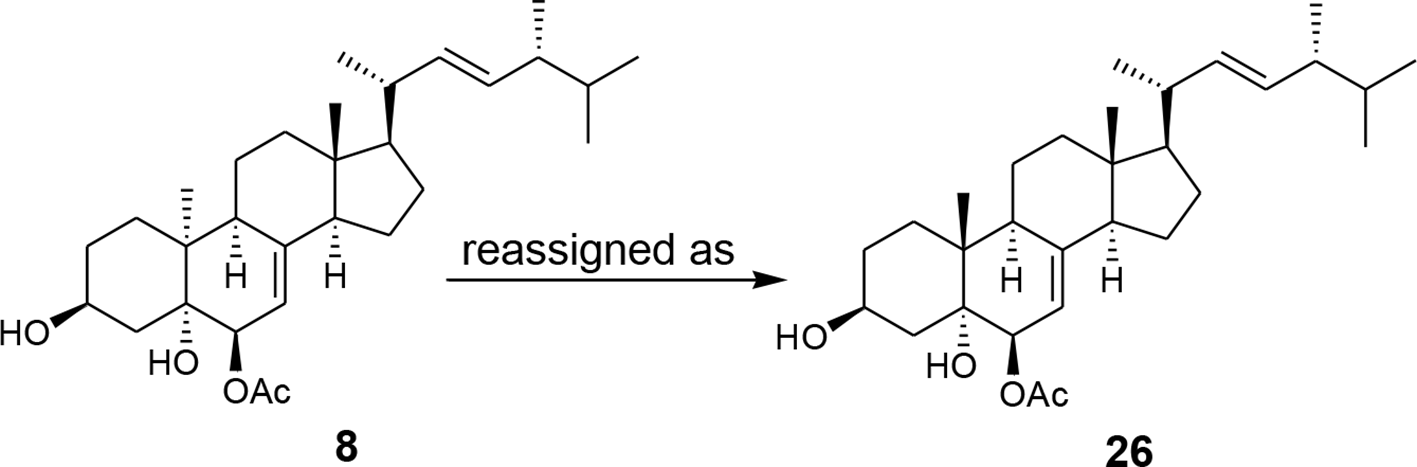
Next, in 2017, Qiao et al17 reported a new sterol 8 from the cultures of Eurotium rubrum, an endophytic fungus isolated from the marine green alga Codium fragile (Figure 15). The unusual 10α-ergostane skeleton of 8 was proposed on the basis of 2D NMR data.15 Regarding the stereochemistry of 8, Qiao et al17 concluded that “In the NOESY spectrum (Figure 16), the observed correlations between H3-18/H-3, H-6 and between H3-18/H-20 indicated H-3, H-6, C-18 and C-20 to be located on the same face of the molecule.” “However, H3-19 was axial and opposite to H-3 and H-6, while H-9 was oriented the same as H3-19 and H3-21 by the NOESY correlations between H-9/H3-19 and between H3-19/H3-21.” It is well known that the NOE can be very useful in stereochemical studies because it provides information about the spatial proximity of nuclei of hydrogen. However, some of the above NOEs cannot be explained for the stereochemistry of 8. Especially, although Qiao et al17 recorded NOE correlations between H3-18 and H-3, and H3-18 and H-6, it is impossible for the structure 8, because the distances between these hydrogens are far and not enough to produce the NOE correlation (Figure 16). On the other hand, Zhang et al38 reported a sterol, 6β-acetoxy-(22E)-ergosta-7,22-diene-3β,5α-diol (26), the epimer of 8 at C-10, from Colletotrichum sp., an endophytic fungus isolated from Ilex canariensis, an evergreen shrub from the island of Gomera (Figure 15). The published 1H (Table 10) and 13C NMR (Table 11) data for 26 were in agreement with those of 8. Therefore, the proposed structure of 8 has been reassigned as 26 (Figure 17).
Structures of compounds 8 and 26.
NOEs (full-line arrows) for compound 8.
Reassignment of compound 8.
1H NMR Chemical Shifts of Compounds 8 and 26 in CDCl3.
cSignals reassigned by the authors are indicated by underlining.
Reassignment of Structure of 8α,9α-Epoxy-(22E)-Ergosta-6,22-Diene-3β,5α,14α-Triol
In this final section, we describe the structural reassignment of 8α,9α-epoxy-(22E)-ergosta-6,22-diene-3β,5α,14α-triol (9) to its regioisomer.
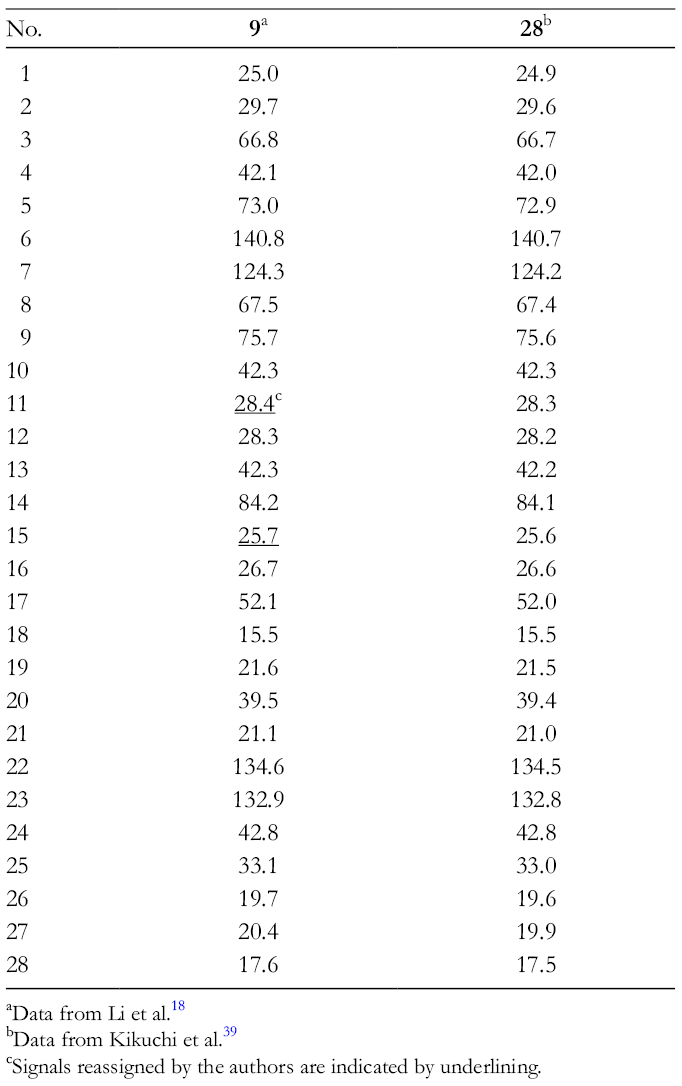
In 2015, Li et al18 reported a new polyoxygenated sterol 9 from the fruiting bodies of edible mushroom Hericium erinaceum (Figure 18). Compound 9 exhibited inhibitory activity against tumor necrosis factor α secretion in LPS-stimulated murine RAW264.7 macrophage cells.16 The 8α,9α-epoxy-3β,5α,14α-trihydroxylated structure of 9 was proposed on the basis of 1D and 2D NMR data.16 Regarding compound 9, Li et al18 concluded that “Both 1H and 13C NMR spectroscopic data of 9 had some similarity to compound 27 (Figure 18), except for substituent groups at C-8, 9 and 14.” “A double bond between C-8 (δC 123.9) and C-14 (δC 149.0) was not observed, however, as it was oxidized to form the 8,9-epoxy (δC 67.5 and 75.5) and 14-OH (δC 84.2) moiety.” On the other hand, Kikuchi et al39 reported 8α,14α-epoxy-(22E)-ergosta-6,22-diene-3β,5α,9α-triol (28), the regioisomer of 9, from the fruiting bodies of edible mushroom Pleurotus eryngii (Figure 18). The published 1H (Table 12) and 13C NMR (Table 13) data for 28 were in agreement with those of 9. Furthermore, Kikuchi et al39 also reported that an x-ray diffraction analysis of 28 was conducted to confirm the position of the hydroxy and epoxy groups, and it established the relative stereostructure of 28. From the above, the structure of 9 has been reassigned as 28 (Figure 19).
Structures of compounds 9, 27, and 28.
1H NMR Chemical Shifts of Compounds 9 and 28 in CDCl3.
cSignals reassigned by the authors are indicated by underlining.
Reassignment of compound 9.
Conclusion
In this review, we summarized our studies on the reassignments of structures of 9 misassigned polyoxygenated sterols 1 to 9. The structural assignment of natural products supports research in multitude of disciplines that may lead to new therapeutic agents or new understanding of disease biology.40 However, structural misassignments of natural products are prevalent in the literature.6-8 Total synthesis can eventually and strongly support the real chemical structure of natural products.41 Only NMR assignment sometimes causes misassignment of the structures. Therefore, we want to point out the importance of careful interpretation of spectroscopic and/or physical data and eliminating preconceptions in interpretation of the data, especially when determining the structure of new compounds isolated from natural sources.
Supplemental Material
Supplementary material - Supplemental material for Misassigned Polyoxygenated Sterols and Reassignments of Their Structures
Supplemental material, Supplementary material, for Misassigned Polyoxygenated Sterols and Reassignments of Their Structures by Yasunori Yaoita and Koichi Machida in Natural Product Communications
Footnotes
Author’s Note
This paper is dedicated to Professor Chiaki Kuroda on the occasion of his 65th birthday.
Declaration of Conflicting Interests
The author(s) declared no potential conflicts of interest with respect to the research, authorship, and/or publication of this article.
Funding
The author(s) received no financial support for the research, authorship, and/or publication of this article.
ORCID iD
Yasunori Yaoita
References
1.
FieserLF.FieserM. Steroids. New York: Reinhold; 1959:1-945.
2.
GoadJL.AkihisaT. Analysis of Sterols. London: Blackie Academic & Professional; 1997:1-437.
3.
LednicerD. Steroid Chemistry at a Glance. West Sussex: Wiley; 2011:1-144.
4.
DewickPM. Medicinal Natural Products. West Sussex: Wiley; 2002:232-285.
5.
YaoitaY.KikuchiM.MachidaK. Terpenoids and sterols from some Japanese mushrooms. Nat Prod Commun. 2014;9(3):419-426.doi:10.1177/1934578X1400900332
6.
KutateladzeAG.KuznetsovDM.BeloglazkinaAA.HoltT. Addressing the challenges of structure elucidation in natural products possessing the oxirane moiety. J Org Chem. 2018;83(15):8341-8352.doi:10.1021/acs.joc.8b01027
7.
KutateladzeAG.HoltT.ReddyDS. Natural products containing the oxetane and related moieties present additional challenges for structure elucidation: A DU8+ computational case study. J Org Chem. 2019;84(12):7575-7586.doi:10.1021/acs.joc.9b01005
8.
KutateladzeAG.KrenskeEH.WilliamsCM. Reassignments and corroboration of oxo-bridged natural products directed by OSE and DU8+ NMR computation. Angew. Chem. Int. Ed.. 2019;58(21):7107-7112.doi:10.1002/anie.201902777
9.
JensenWP.PalenikGJ.SuhIH. The history of molecular structure determination viewed through the Nobel Prizes. J Chem Educ. 2003;80(7):753-761.
10.
AmagataT. Misassigned structures: Case examples from the past decade. In: ManderL.LiuH-W., eds.Comprehensive Natural Products II. Volume 2. London, United Kingdom: Elsevier; 2010:581-621.
11.
YaoitaY.KikuchiM.MachidaK. Structure revision of two polyoxygenated sterols from the marine sponge Neofibularia nolitangere. Nat Prod Commun. 2015;10(6):881-883.doi:10.1177/1934578X1501000622
12.
YaoitaY.MachidaK. Structure Revision of (22E)-24-methylcholesta-8(14),22-diene-3β,5α,6β,7α-tetraol from the marine-derived fungus Penicillium sp. Nat Prod Commun. 2016;11(7):947-948.
13.
YaoitaY.MachidaK. Structure revision of 5β,6β-epoxy-(22E)-ergosta-8,22-diene-3β,7β-diol from the gorgonian Pinnigorgia sp. Nat Prod Commun. 2017;12(8):1197-1198.
14.
YaoitaY.MachidaK. Structure revision of (22E)-ergosta-7,22-diene-3β,5α,6β,9α,14α-pentol from the spores of the medicinal mushroom Ganoderma lucidum. Nat Prod Commn. 2016;11(2):183-184.
15.
LeeSY.KimJS.LeeS.KangSS. Polyoxygenated ergostane-type sterols from the liquid culture of Ganoderma applanatum. Nat Prod Res. 2011;25(14):1304-1311.doi:10.1080/14786419.2010.503190
16.
IsaacsS.BermanR.KashmanY.GebreyesusT.YosiefT. New polyhydroxy sterols, dysidamides, and a dideoxyhexose from the sponge Dysidea herbacea. J Nat Prod. 1991;54(1):83-91.doi:10.1021/np50073a004
17.
QiaoM-F.YiY-W.DengJ. Steroids from an endophytic Eurotium rubrum strain. Chem Nat Compd. 2017;53(4):678-681.doi:10.1007/s10600-017-2089-x
18.
LiW.ZhouW.ChaJYet al. Sterols from Hericium erinaceum and their inhibition of TNF-α and NO production in lipopolysaccharide-induced RAW 264.7 cells. Phytochemistry. 2015;115:231-238.doi:10.1016/j.phytochem.2015.02.021
19.
CostantinoV.FattorussoE.MangoniA.PansiniM. Sterols from the Caribbean sponge Neofibularia nolitangere. Isolation of two novel polyhydroxysteroids. Steroids. 1995;60(11):768-772.doi:10.1016/0039-128X(95)00117-9
IshizukaT.YaoitaY.KikuchiM. Sterol constituents from the fruit bodies of Grifola frondosa (Fr.) S. f. gray. Chem Pharm Bull. 1997;45(11):1756-1760.doi:10.1248/cpb.45.1756
22.
KobayashiM.KandaF. Marine sterols. 18. Isolation and structure of four novel oxygenated sterols from a gorgonian coral Melithaea ocracea. J Chem Soc Perkin Trans 1. 1991;1991(5):1177-1179.
23.
GrecaMD.FiorentinoA.MolinaroA.MonacoP.PreviteraL. Steroidal 5,6-epoxides from Arum Italicum. Nat Prod Lett. 1993;2(1):27-32.doi:10.1080/10575639308043450
24.
MigliuoloA.PiccialliV.SicaD.GiordanoF. New Δ8- and Δ8(14)-5α,6α-epoxysterols from the marine sponge Spongia officinalis. Steroids. 1993;58(3):134-140.doi:10.1016/0039-128X(93)90050-W
25.
LuoX.LiF.ShindePBet al. 26,27-cyclosterols and other polyoxygenated sterols from a marine sponge Topsentia sp. J Nat Prod. 2006;69(12):1760-1768.doi:10.1021/np0604026
26.
CarvalhoJFS.Cruz SilvaMM.MoreiraJN.SimõesS.Sá E MeloML. Efficient chemoenzymatic synthesis, cytotoxic evaluation, and SAR of epoxysterols. J Med Chem. 2009;52(13):4007-4019.doi:10.1021/jm9003973
27.
GaoH.HongK.ChenG-Det al. New oxidized sterols from Aspergillus awamori and the endo-boat conformation adopted by the cyclohexene oxide system. Magn Reson Chem. 2010;48(1):38-43.doi:10.1002/mrc.2536
28.
KobayashiM. Additivity relationships in the carbon-13 nuclear magnetic resonance spectra of polyhydroxy steroids. J Chem Soc Perkin 1. 1995;1995(1):33-40.doi:10.1039/p19950000033
29.
RameshP.Niranjan ReddyVL.Srinivasa ReddyN.VenkateswarluY. Synthesis of melithasterol A, a 5α,6α-epoxy-7α-hydroxy Δ8-steroid. J Nat Prod. 2000;63(10):1420-1421.
30.
SantaféG.PazV.RodríguezJ.JiménezC. Novel cytotoxic oxygenated C29 sterols from the Colombian marine sponge Polymastia tenax. J Nat Prod. 2002;65(8):1161-1164.doi:10.1021/np0200459
31.
SuY-D.ChengC-H.WenZ-H.WuY-C.SungP-J. New anti-inflammatory sterols from a gorgonian Pinnigorgia sp. Bioorg Med Chem Lett. 2016;26(13):3060-3063.doi:10.1016/j.bmcl.2016.05.015
32.
ZhangC-R.YangS-P.YueJ-M. Sterols and triterpenoids from the spores of Ganoderma lucidum. Nat Prod Res. 2008;22(13):1137-1142.doi:10.1080/14786410601129721
33.
YaoitaY.AmemiyaK.OhnumaHet al. Sterol constituents from five edible mushrooms. Chem Pharm Bull. 1998;46(6):944-950.
34.
YaoitaY.MatsukiK.IijimaTet al. New sterols and triterpenoids from four edible mushrooms. Chem Pharm Bull. 2001;49(5):589-594.doi:10.1248/cpb.49.589
35.
DemarcoPV.FarkasE.DoddrellD.MylariBL.WenkertE. Pyridine-induced solvent shifts in the nuclear magnetic resonance spectra of hydroxylic compounds. J Am Chem Soc. 1968;90(20):5480-5486.doi:10.1021/ja01022a027
36.
FujimotoY.YamadaT.IkekawaN. Pyridine-idnduced dishielding of 4-methylene protons for the determination of C-6 stereochemistry of sterols having a 5α,6-diol moiety. Revision of the C-6 stereochemistry of marine sterol isolated from a sponge, Dysidea sp. Chem Pharm Bull. 1985;33(8):3129-3133.doi:10.1248/cpb.33.3129
37.
KobayashiM.KrishnaMM.HaribabuB.AnjaneyuluV. Marine sterols. XXV. Isolation of 23-demethylgorgost-7-ene-3β,5α,6β-triol and (24S)-ergostane-3β,5α,6β,7β,15β-pentol from soft corals of the Andaman and Nicobar coasts. Chem Pharm Bull. 1993;41(1):87-89.
38.
ZhangW.DraegerS.SchulzB.KrohnK. Ring B aromatic steroids from an endophytic fungus, Colletorichum sp. Nat Prod Commn. 2009;4(11):1449-1454.
39.
KikuchiT.MaekawaY.TomioAet al. Six new ergostane-type steroids from king trumpet mushroom (Pleurotus eryngii) and their inhibitory effects on nitric oxide production. Steroids. 2016;115:9-17.doi:10.1016/j.steroids.2016.07.005
40.
SuyamaTL.GerwickWH.McPhailKL. Survey of marine natural product structure revisions: a synergy of spectroscopy and chemical synthesis. Bioorg Med Chem. 2011;19(22):6675-6701.doi:10.1016/j.bmc.2011.06.011
41.
NicolaouKC.SnyderSA. Chasing molecules that were never there: Misassigned natural products and the role of chemical synthesis in modern structure elucidation. Angew Chem Int Ed Engl. 2005;44(7):1012-1044.doi:10.1002/anie.200460864
Supplementary Material
Please find the following supplemental material available below.
For Open Access articles published under a Creative Commons License, all supplemental material carries the same license as the article it is associated with.
For non-Open Access articles published, all supplemental material carries a non-exclusive license, and permission requests for re-use of supplemental material or any part of supplemental material shall be sent directly to the copyright owner as specified in the copyright notice associated with the article.