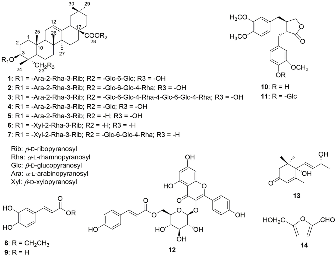
Our current research on phytochemical profile of the folk medicinal plant Anemone chapaensis Gagnep. led to the isolation of a new saponin, chapaenoside (1), along with 13 known compounds (2-14). Their structures were elucidated on the basis of extensive chemical and spectroscopic methods (Nuclear Magnetic Resonance [NMR] spectroscopy and mass spectrometry) as well as comparison with the literature data. The isolated compounds were tested for their cytotoxic activities against 9 cancer cell lines by MTT method. The result indicated that the major saponin prosapogenin CP6 (5) showed selectively strong activity against NCI-N87, RD, Panc-1, and MIA Paca-2 cells with the IC50 values of 5.4, 7.5, 7.5, and 2.7 μM, respectively. The results of phytochemical constituents and principal bioactive saponins from A. chapaensis contributed not only to its phytochemical profile associated with chemotaxonomy but also to biological evidence of the title plant.
Anemone is a genus of more than 150 species of flowering plants in the family Ranunculaceae, native to the temperate zones of northern and southern hemispheres.1 Many Anemone species have been used traditionally in medicine. The plant Anemone chapaensis Gagnep., which was first discovered in 1929, is considered as the most tropical distribution of Anemone species and limitedly distributed in the northern west region of Vietnam surrounding Hoang Lien mountains at 1500 to 2000 m altitude. Many Anemone species have been studied on both phytochemical and pharmacological profiles, which provide various bioactive compounds including mainly phenolics and saponins.2 The roots of A. chapaensis have long been used in folk medicine for the treatment of pain, pharyngitis, rheumatism, tumor, etc.; however, there have been no up-to-date reports on both phytochemical and biological activity.
In our ongoing investigation on the selected Vietnamese medicinal plants,3,4 the current phytochemical study on A. chapaensis for the first time led to the isolation of 1 new saponin, named chapaenoside (1), and 13 known compounds (2-14) (Figure 1). This paper herein reports the isolation and structural identification of the isolated compounds as well as their cytotoxic activity against cancer cell lines.
Structures of the isolated compounds from the aerial parts of Anemone chapaensis (1-14).
Chapaenoside (1) was isolated as a white amorphous powder and gave positive reaction with Libermann-Burchard reagent. Its molecular formula was established as C58H94O26 by high-resolution electrospray-ionization mass spectrometry (HR-ESI-MS) experiment (found at m/z [M+H]+ 1207.6145, calcd. for C58H95O26 1207.6112). Acid hydrolysis of 1 with 1 M HCl liberated l-arabinose, d-glucose, l-rhamnose, and d-ribose as confirmed by gas chromatography (GC) analysis. The 1H NMR spectrum of 1 showed signals of 6 tertiary methyl groups at δ 0.82, 0.96, 0.98, 1.00, 1.09, and 1.25 (each 3H and s), a trisubstituted olefinic proton at δ 5.27 (br s), and a pair of germinal oxymethylene (-CH2-O-) protons at δ 3.68 and 3.86 (each d, J = 12.0 Hz). In addition, the 1H NMR spectrum revealed the presence of l-arabinose, d-glucose, l-rhamnose, and d-ribose units with 5 anomeric protons at δ 4.54 (d, J = 6.0 Hz), 5.37 (d, J = 8.5 Hz), 4.36 (d, J = 8.0 Hz), 5.24 (d, J = 2.0 Hz), and 5.03 (d, J = 4.0 Hz), respectively. The J values of these anomeric protons indicated that the glycosidic linkage of l-arabinose and l-rhamnose were of α-form, and those of d-glucose and d-ribose were belonging to β-form.5
The 13C NMR and Distortionless Enhancement by Polarization Transfer (DEPT) spectra of 1 revealed the presence of 30 carbon resonances accounting for an aglycone including 6 methyl carbons at δ 13.8, 16.6, 17.9, 24.0, 26.4, and 31.5, 2 olefinic signals at δ 123.8 and 144.9, an oxymethine carbon at δ 82.3, an oxymethylene carbon at δ 64.6, and a carboxyl signal at δ 178.1, which were typical of the hederagenin skeleton.6-8 The 13C NMR data also confirmed the presence of 5 sugar units with the anomeric carbons at δ 104.6, 104.6, 104.2, 101.5, and 95.8, respectively.5 The chemical shifts of C-3 (δ 82.3) and C-28 (δ 178.1) were evidence for glycosylation at both C-3 and C-28.8 The sugar sequences at C-3 and C-28 were proposed by the Heteronuclear Multiple Bond Correlation (HMBC) spectrum after assignments of the protons and the carbons by a combination of HMQC and Correlated Spectroscopy (COSY) data, starting from the anomeric proton of each sugar unit (Table 1). Accordingly, H-1 of Ara (δ 4.54) gave an HMBC correlation with the C-3 at δ 82.3, and subsequent cross peaks of H-1 of Rha (δ 5.24)/C-2 of Ara (δ 76.3) and H-1 of Rib(III) (δ 5.03)/C-3 of Rha (δ 76.3) were revealed. Additionally, 2 cross peaks of H-1 of Glc(IV) (δ 5.37)/C-28 (δ 178.1) and H-1 of Glc(V) (δ 4.36)/C-6 of Glc(IV) (δ 69.5) were also observed to fix the sugar chain at C-28 (Figure 2). Regarding the proposed α-l-rhamnose unit, the deduction of α-form was furthermore based on its anomeric carbon signal at δ 101.5 as well as the observed correlation between its anomeric proton signal at 5.24 (d, J = 2.0 Hz, H-1(Ara)) and proton signal at δ 3.77 (1H, H-5(Ara)) in the Nuclear Overhauser Spectroscopy (NOESY) spectrum.9 The overall structure of 1, especially its linkage sites, partial structures, and stereochemistry, was assigned by COSY, Heteronuclear Single Quantum Coherence (HSQC), HMBC, COSY, and NOESY spectra, respectively (Figures 3S-8S, Supplemental Material). The full assignments of the 1H NMR and 13C NMR data of 1 were listed in Table 1. Hence, chapaenoside (1) was unambiguously concluded to be 3-O-[β-d-ribopyranosyl(1→3)-α-l-rhamnopyranosyl](1→2)-α-l-arabinopyranoside hederagenin 28-O-[β-d-glucopyranosyl(1→6)-β-d-glucopyranosyl].
1H-NMR (CD3OD; 500 MHz) and 13C-NMR (CD3OD; 125 MHz) for Chapaenoside (1).
COSY and key HMBC correlations of chapaenoside (1).
The other 13 compounds (2-14), which were isolated from the EtOAc and n-BuOH fractions of the ethanol extract of both aerial parts and roots of A. chapaensis using combined various column chromatography (CC), were identified as huzhangoside D,2,10 hupehensis saponin F,3,7,11 clemastanoside D,4,6 prosapogenin CP6 (5),10 huzhangoside A,6,10 huzhangoside C,7,10 ethyl caffeate,8,12 caffeic acid,9,13 arctigenin,10,14 arctiin,11,15trans-tiliroside,12,16 blumenol A,13,17 and 5-hydroxymethylfurfural14,18 on the basis of spectroscopic data and comparison with the respective literature data. To the best of our knowledge, this is the first chemical investigation of the plant A. chapaensis.
Cytotoxic activity of the isolated compounds was tested on a panel of 9 cancer cell lines (HepG2, A549, MCF7, Ovcar-8, NCI-N87, Hela, RD, Panc-1, and MIA Paca-2) using the 3-(4,5-dimethylthiazol-2-yl)-2,5-diphenyltetrazolium bromide (MTT) assay.3 Among the isolates, only 2 saponins 5 and 6 showed considerable inhibitory effects on some cancer cell lines (Table 2), while the other compounds displayed weak cytotoxicity against all cell lines. The observation of cytotoxic activity of 2 saponins 5 and 6 clearly indicated that the substitution of sugar at the carbonyl group (C-28) significantly reduces the cytotoxicity of these saponins. It was noteworthy that prosapogenin CP6 (5) showed strong activity against NCI-N87, RD, Panc-1, and MIA Paca-2 cells with the IC50 values of 5.4, 7.5, 7.5, and 2.7 µM, respectively, in comparison with doxorubicin as a positive control. However, this compound exhibited less effect on the rest of tested cell lines. Recent studies have indicated that an oleane-type saponin raddeanin, the major compound in the plant Anemone raddeana with COOH group at C-28, showed significant anticancer activities.19-22 Therefore, more studies on anticancer activity of the major saponin prosapogenin CP6 (5) of A. chapaensis could be considered.
Cytotoxicity of Saponins Against 9 Cancer Cell Lines.
Comp
Cytotoxicity (IC50, μM)
HepG2
A549
MCF7
Panc-1
NCI-N87
RD
Hela
Ovcar-8
MIAPaca-2
5
16.7
13.2
24.1
7.5
5.4
7.5
-
11.8
2.7
6
11.3
-
-
19.6
18.2
12.2
-
10.6
-
Dox
3.3
1.9
1.3
1.5
1.9
0.3
0.3
2.4
2.8
-, IC50 >30 μM; Dox, doxorubicin was used as the positive control.
Up-to-date phytochemical investigations of the genus Anemone have revealed various kinds of constituents, including triterpenoids, steroids, saponins, lactones, fat and oils, and alkaloids and saccharides.2 Among them, saponins are not only abundant in this genus but also rich in Clematis, Pulsatilla, and Cimicifugae.2 The isolation of these pentacyclic triterpenoid saponins (1-7) reveals the close genetic relationship between A. chapaensis and the other species of the genus Anemone and some genus in the family Ranunculaceae. Besides, compounds 10 to 13 were isolated from the genus Anemone for the first time, especially arctigenin (10) and arctiin (11) were mainly contented in the family Asteraceae. The major compounds prosapogenin CP6 (5), arctigenin (10), arctiin (11), and trans-tiliroside (12) might serve as chemotaxonomic markers of this plant to differentiate the other species within the genus Anemone.
In conclusion, this is the first report on chemical constituents of A. chapaensis and some of their cytotoxic activities against 9 human cancer cell lines. One new saponin, namely chapaenoside (1), together with 6 known triterpene saponins (2-7), 2 hydroxycinnamic acid derivatives (8-9), 2 lignans (10-11), 1 flavonoid (12), 1 norsesquiterpenoid (13), and 1 furan (14), was isolated from the whole plants of this species. Among them, prosapogenin CP6 (5) showed significant activities against Panc-1, NCI-N87, and RD cells with the IC50 values of 7.50, 5.40, and 7.51 µM, respectively, while displayed moderate-to-weak cytotoxicity against the other tested cell lines. This compound might be a potential candidate for further investigation on the molecular mechanisms of action on specific anticancer targets. This is the first evidence of pharmaceutical evidence of bioactive compounds from the title plant.
Experimental
General Procedure
Column chromatography was performed on silica gel (Kieselgel 60, 70-230 mesh and 230-400 mesh, Merck) and YMC*GEL resins (ODS-A, 12 nm S-150 µm, YMC Co., Ltd.). Analytical thin layer chromatography (TLC) was performed on Kieselgel 60 F254 (Merck) plates (silica gel, 0.25 mm layer thickness) and RP-18 F254 (Merck) plates (0.25 mm layer thickness). Spots were visualized by spraying with 10% aqueous H2SO4 solution, followed by heating for 3 to 5 minutes. Electrospray ionization mass spectra (ESI-MS) were recorded on a Varian Agilent 1100 Liquid Chromatography/Mass Selective Detector (LCMSD) mass spectrometer. High-resolution electrospray-ionization mass spectrometry experiments employed a JEOL AccuTOF LC 1100 mass spectrometer (JEOL, Tokyo, Japan). The NMR spectra were recorded on a Bruker AVANCE III HD 500 spectrometer. Gas chromatography (Shimadzu GC-2010 plus QP2020, Shimadzu Corp., Japan) using a Shimadzu SH-Rxi-5 Sil capillary column (0.25 mm ID × 30 mm) (column temperature 210°C; detector temperature 300°C; injector temperature 270°C; and He gas flow rate 30 mL/min [splitting ratio: 1/20]) was used for sugar analysis.
Plant Materials
The whole plants of A. chapaensis were collected in 1500 to 1800 m height of Hoang Lien mountain, Sapa district, Lao Cai province, Vietnam, in May 2015 and taxonomically identified by one of the authors (P.T.T.) and Dr Do Thi Xuyen, Faculty of Biology, VNU University of Science. The voucher specimens (PQ-01.2015-VDL) were deposited in the Department of Herbal Analysis and Standardization, National Institute of Medicinal Materials, Hanoi, Vietnam.
Extraction and Isolation
The dried roots (2.5 kg) and aerial parts (3.0 kg) of A. chapaensis were separately powdered, extracted with 96% ethanol (15 L × 3), and then concentrated under reduced pressure to yield 235 and 247 g of each crude extracts, respectively. Both of these extracts were successively triturated with n-hexane, ethyl acetate (EtOAc), and n-butanol (BuOH).
The BuOH extract (52 g) of the aerial parts was separated by CC on silica gel 60, eluting with EtOAc/MeOH/H2O (20/1/0.3 → 1/1/0.3, 0/1/0, v/v) to obtain 12 fractions (B1 → B12). Fraction B9 (1.5 g) was further applied to a RP-18 column eluting with MeOH/H2O (2:1, v/v) to yield 1 (36 mg) and 4 (7 mg). Fraction B11 was subjected to a RP-18 column with MeOH/H2O (65/35, v/v) as eluent to yield 2 (57 mg) and 3 (18 mg). The BuOH extract (102 g) obtained from the roots was also fractionated on silica gel CC eluted with EtOAc/MeOH/H2O of increasing polarity (20/1/0.3 → 1/1/0.3, 0/1/0, v/v) to yield 31 fractions (C1 → C31). Fraction C13 was chromatographed on a RP-18 column eluted with a gradient of MeOH/H2O (3:1 → 4:1, v/v) to afford 5 (31 mg) and 6 (60 mg). Fraction C25 was purified by a RP-18 column with MeOH/H2O (70:30, v/v) as eluent to yield 7 (75 mg). The EtOAc extract (126 g) of the aerial parts was fractionated by silica gel 60 CC, eluting with n-hexane/acetone (15:1 → 0:1, v/v), yielding 11 fractions (E1 → E11). The E4 fraction (2.4 g) was submitted to an RP-18 column, eluting with MeOH/H2O (45:55, v/v) to obtain 6 subfractions (E4.1 → E4.6). Fraction E4.1 was then separated using silica gel CC with n-hexane/EtOAc (3:1, v/v) to give 14 (30 mg). Fraction E4.5 was purified by silica gel CC with n-hexane/EtOAc (4:1, v/v) elution solvent to yield 8 (51 mg). The E5 fraction (3.1 g) was subjected to RP-18 CC with MeOH/H2O (2:3, v/v) to give 4 subfractions (E5.1 → E5.4). Fraction E5.3 was purified by silica gel CC with n-hexane/acetone (3:1, v/v) elution solvent to yield 13 (10 mg). Fraction E5.4 was crystallized on addition of water to yield 10 (57 mg). The E6 fraction (1.4 g) was further separated using RP-18 CC with MeOH/H2O (35:65, v/v) elution solvent to yield 4 subfractions (E6.1 → E6.4). Fraction E6.2 was purified by silica gel CC with CH2Cl2/MeOH (30:1, v/v) as eluent to yield 9 (23 mg). The E8 fraction (0.9 g) was further applied to a RP-18 column eluting with MeOH/H2O (1:1) to give 12 (42 mg). Finally, the E10 fraction (1.2 g) was subjected to a RP-18 column eluting with MeOH/H2O (45:55, v/v) to afford 5 subfractions (E10.1 → E10.5). Fraction E10.5 was purified by silica gel CC with EtOAc/MeOH (25:1, v/v) as eluent to yield 11 (53 mg).
Chapaenoside (1)
White amorphous powder.
: +12 (c 0.15, MeOH).
HR-ESI-MS: m/z [M+H]+ 1207.6145
1H NMR (500 MHz, CD3OD) and 13C NMR (100 MHz, CD3OD): see Table 1.
Acid Hydrolysis and Sugar Identification
A solution of the new compound 1 (2.0 mg) in HCl 1.0 M (3.0 mL) was heated under reflux for 2 hours. Then, the reaction mixture was concentrated in vacuum to dryness. The residue was extracted with CHCl3 and H2O (5 mL each, 3 times). Next, the sugar residue obtained by concentration of the water layer was dissolved in dry pyridine (0.1 mL). Then, l-cysteine methyl ester hydrochloride in pyridine (0.06 M, 0.1 mL) was added to the solution. After heating the reaction mixture at 60°C for 2 hours, 0.1 mL of trimethylsilylimidazole was added. Heating at 60°C was continued for further 2 hours, and the mixture was evaporated in vacuum to give a dried product, which was partitioned between n-hexane and H2O. The n-hexane layer was analyzed using the GC procedure (see “General Procedure” section). The peaks of the hydrolysates of the saponin 1 were detected at tR 4.51 minutes (l-rhamnose), tR 8.21 minutes (l-arabinose), tR 8.66 minutes (d-ribose), and tR 14.12 minutes (d-glucose), respectively. The retention times (tR) for the authentic samples (Sigma) after being treated similarly were 4.50 minutes (l-rhamnose), 8.21 minutes (l-arabinose), 8.66 minutes (d-ribose), and 14.11 minutes (d-glucose), respectively. Co-injection of the hydrolysates of the saponin 1 with the standard sugars gave single peaks.
Supporting information - Supplemental material for Chemical Constituents From the Vietnamese Medicinal Plant Anemone chapaensis and Their Cytotoxic Activity
Supplemental material, Supporting information, for Chemical Constituents From the Vietnamese Medicinal Plant Anemone chapaensis and Their Cytotoxic Activity by Ha Thi Thanh Huong, Pham G. Nam, Hoang Van Hung, Phi T. Xuyen, Nguyen M. Khoi, Tran M. Hung, Nguyen H. Tung and Phuong T. Thuong in Natural Product Communications
Footnotes
Declaration of Conflicting Interests
The author(s) declared no potential conflicts of interest with respect to the research, authorship, and/or publication of this article.
Funding
The author(s) disclosed receipt of the following support for the research, authorship, and/or publication of this article: This work was supported by the National Foundation for Science and Technology Development of Vietnam (Grant number 106-YS.05-2014.32).
References
1.
HaoDC.XiaoPG.MaHY.PengY.HeCN. Mining chemodiversity from biodiversity: pharmacophylogeny of medicinal plants of Ranunculaceae. Chin J Nat Med. 2015;13(7):507-520.doi:10.1016/S1875-5364(15)30045-5
2.
HaoDC.GuX.XiaoP. Anemone medicinal plants: ethnopharmacology, phytochemistry and biology. Acta Pharm Sin B. 2017;7(2):146-158.doi:10.1016/j.apsb.2016.12.001
3.
ThuongPT.HungTM.KhoiNMet al. Cytotoxic and anti-tumor activities of lignans from the seeds of Vietnamese nutmeg Myristica fragrans. Arch Pharm Res. 2014;37(3):399-403.doi:10.1007/s12272-013-0185-4
4.
UtoT.TungNH.OhtaTet al. Antiproliferative activity and apoptosis induction by trijuganone C isolated from the root of Salvia miltiorrhiza Bunge (Danshen). Phytother Res. 2018;32(4):657-666.doi:10.1002/ptr.6013
5.
AgrawalPK. NMR spectroscopy in the structural elucidation of oligossacharides and glycosides. Phytochemistry. 1992;31(10):3307-3330.doi:10.1016/0031-9422(92)83678-R
6.
KizuH.ShimanaH.TomimoriT. Studies on the constituents of Clematis species VI. The constituents of Clematis stans Sieb. et Zucc. Chem Pharm Bull. 1995;43(12):2187-2194.doi:10.1248/cpb.43.2187
7.
WangMK.WuFE.ChenYZ. Triterpenoid saponins from Anemone hupehensis. Phytochemistry. 1997;44(2):333-335.doi:10.1016/S0031-9422(96)00394-9
8.
YanLH.XuLZ.LinJ.YangSL.FengYL. Triterpenoid saponins from the stems of Clematis parviloba. J Asian Nat Prod Res. 2009;11(4):332-338.doi:10.1080/10286020902727348
9.
YuS.FuS.LiuB.ZhangY.ZhouG. Two new quercetin glycoside derivatives from the fruits of Gardenia jasminoides var. radicans. Nat Prod Res. 2015;29(14):1336-1341.doi:10.1080/14786419.2014.1001389
10.
MizutaniK.OhtaniK.WeiJX.KasaiR.TanakaO. Saponins from Anemone rivularis. Planta Med. 1984;50(4):327-331.doi:10.1055/s-2007-969722
11.
LiF.LiuX.TangMet al. Structure revision of hupehensis saponin F and G and characterization of new trace triterpenoid saponins from Anemone hupehensis by tandem electrospray ionization mass spectrometry. Carbohydr Res. 2012;353:49-56.doi:10.1016/j.carres.2012.03.020
12.
LimaTC.SouzaRJ.SantosADet al. Evaluation of leishmanicidal and trypanocidal activities of phenolic compounds from Calea uniflora Less. Nat Prod Res. 2016;30(5):551-557.doi:10.1080/14786419.2015.1030740
13.
JangSY.BaeJS.LeeYH.OhKY.ParkKH.BaeYS. Caffeic acid and quercitrin purified from Houttuynia cordata inhibit DNA topoisomerase I activity. Nat Prod Res. 2011;25(3):222-231.doi:10.1080/14786410903339044
14.
RahmanMMA.DewickPM.JacksonDE.LucasJA. Lignans of Forsythia-Intermedia. Phytochemistry. 1990;29(6):1971-1980.doi:10.1016/0031-9422(90)85050-P
15.
HodajE.TsiftsoglouO.AbaziS.Hadjipavlou-LitinaD.LazariD. Lignans and indole alkaloids from the seeds of Centaurea vlachorum Hartvig (Asteraceae), growing wild in Albania and their biological activity. Nat Prod Res. 2017;31(10):1195-1200.doi:10.1080/14786419.2016.1226823
16.
LiuWJ.HouXQ.ChenH.LiangJY.SunJB. Chemical constituents from Agrimonia pilosa Ledeb. and their chemotaxonomic significance. Nat Prod Res. 2016;30(21):2495-2499.doi:10.1080/14786419.2016.1198351
17.
KuangH-xue.YangB-you.XiaY-gang.FengW-sheng.XiaYG.FengWS. Chemical constituents from the flower of Datura metel L. Arch Pharm Res. 2008;31(9):1094-1097.doi:10.1007/s12272-001-1274-6
18.
KangHS.ChoiJH.ChoWK.ParkJC.ChoiJS. A sphingolipid and tyrosinase inhibitors from the fruiting body of Phellinus linteus. Arch Pharm Res. 2004;27(7):742-750.doi:10.1007/BF02980143
19.
GuanYY.LiuHJ.LuanXet al. Raddeanin A, a triterpenoid saponin isolated from Anemone raddeana, suppresses the angiogenesis and growth of human colorectal tumor by inhibiting VEGFR2 signaling. Phytomedicine. 2015;22(1):103-110.doi:10.1016/j.phymed.2014.11.008
20.
GuG.QiH.JiangTet al. Investigation of the cytotoxicity, apoptosis and pharmacokinetics of Raddeanin A. Oncol Lett. 2017;13(3):1365-1369.doi:10.3892/ol.2017.5588
21.
MaB.ZhuJ.ZhaoAet al. Raddeanin A, a natural triterpenoid saponin compound, exerts anticancer effect on human osteosarcoma via the ROS/JNK and NF-κB signal pathway. Toxicol Appl Pharmacol. 2018;353:87-101.doi:10.1016/j.taap.2018.05.025
22.
PengF.WangX.ShuMet al. Raddeanin A suppresses glioblastoma growth by inducing ROS generation and subsequent JNK activation to promote cell apoptosis. Cell Physiol Biochem. 2018;47(3):1108-1121.doi:10.1159/000490187