
Abstract
Lippia alba is a plant widely studied due to both chemical diversity and bioactivities related to its ethnobotanical uses. In this work, the composition of the volatile secondary metabolites (volatile fractions/essential oil, EO) of the flower/leaves of L. alba (from northern region of Colombia) was determined by solid phase micro-extraction/distillation-solvent extraction/microwave-hydrodistillation/gas chromatography-mass spectrometry (MWHD/GC-MS), along with some in vitro biological properties (cytotoxicity and acetylcholinesterase enzyme [AChe] inhibition) from leaf EO. Outstanding results were found: (i) cis-piperitone oxide (~13%-46%), germacrene D (~11%-30%), and limonene (~10%-22%) characterized the volatile secondary metabolites from different parts of the plant; (ii) leaf EO showed a moderate hemolytic activity (HC50: 580 ± 1 µg/mL), a significant cytotoxicity on lymphocytes (LC50: 127 ± 3 µg/mL), a high cytotoxicity on HEp2 cell line (LC50: 38 ± 2 µg/mL), and a moderate inhibitory effect on AChE (IC50: 28 ± 2 µg/mL). Based on these results, a new chemovar of L. alba is reported (represented by cis-piperitone oxide) along with its promising cytotoxic and AChE inhibiting properties.
Lippia alba Mill. is a shrub widely distributed and used in Central and South America for different purposes, eg, ethnomedicine and seasoning. 1 In the northern region of Colombia, this plant is commonly called “oreganito, orégano de cerro, curalotodo, or pronto alivio,” and it is prepared as infusions or tinctures to treat colds and digestive problems, and for fever relief. 2 In accordance with the reviewed scientific literature, there are 32 chemovar (ie, type/subtype) known for the essential oils (EOs) from L. alba, which are represented by one or combination of constituents as bicyclosesquiphellandrene, camphor, carvone, β-caryophyllene, caryophyllene oxide, citral, citrol, dihydrocarvone, estragole, eucalyptol, germacrene D, α-guaiene, limonene, linalool, lippione, myrcene, myrcenone, E/Z-β-ocimene, ocimenone, piperitone, and γ-terpinene, 1,3,4,5,6,7,8,9,10,11,12,13,14,15,16,17,18,19,20,21,22,23,24,25 in relative amounts ranging between ~10% and 91% and EO yields around ~0.1% to 3.2%. For Colombia, 4 chemovar were previously reported. 22 -25 In addition, due to the structural diversity of the L. alba EO, a lot of them have shown an interesting and wide range of bioactivities, eg, antibacterial/antifungal, antiviral, anticancer, antigenotoxic, cytotoxic, neurosedative, analgesic, anti-inflammatory, cardiovascular, antiulcerogenic, anticonvulsant, antioxidant, acaricidal, repellent, and insecticide. 1,18,24,25,26,27,28,29,30,31,32,33,34 In this work, a new compositional analysis by gas chromatography-mass spectrometry (GC-MS) of the volatile fractions (flowers/leaves—simultaneous distillation-extraction, simultaneous distillation-extraction (SDE)/solid phase micro-extraction, SPME) and EO (leaves— microwave-hydrodistillation [MWHD]) from L. alba was established, along with the in vitro cytotoxic effects (on human erythrocytes and lymphocytes, and HEp2 cell line) and the inhibition of acetylcholinesterase enzyme (AChE) by EO.
The botanical sample was identified as L. alba (Mill.) N.E. Br. ex Britton & P. Wilson, and its EO was a transparent liquid, with a weight yield of 0.5% ± 0.2%. Table 1 presents the main molecules (>0.5%) positively identified (~88%-99%) in the volatile fractions (SPME/SDE) of flowers/leaves and EO of leaves, based on the elution order of the total ion current chromatogram. The classification according to the compound families was characterized mostly by p-mentha(e)ne derivatives [~26%-74%, including oxepanes (~8%-47%)] and sesquiterpenoids (~22%-66%). The most important components of the volatile fractions were cis-piperitone oxide (~13%-38%), germacrene D (~11%-30%), and limonene (~10%-15%). Likewise, the composition of the leaf EO remained consistent with that one of the volatile fractions with some difference in the relative amounts, ie, cis-piperitone oxide (~42%-46%), limonene (~14%-22%), and germacrene D (~14%-18%). Additionally, the simple maceration process (SMP) with ethyl acetate carried out on each aerial part of L. alba allowed to verify that the composition of the volatile metabolites was reliable (Table 1), namely, cis-piperitone oxide (~44%-56%), germacrene D (~16%-32%), and limonene (~8%-15%) were the most abundant compounds.
Chemical Composition by GC-MS of Volatile Secondary Metabolites of Flowers and Leaves From Lippia alba Isolated by HS-SPME (PDMS; PA), SDE, SMP, and MWHD.

BP, base peak; EO, essential oil; F, flowers; HS-SPME, headspace-solid phase micro-extraction; L, leaves; MI, molecular ion; NR, not registered; PDMS, polydimethylsiloxane; PA, polyacrilat; R I, retention indices on column Rxi-1ms; SDE, simultaneous distillation-extraction; SMP, simple maceration process; tr, traces.
Values are reported as average ± standard deviation.
Tentatively identified.
When these compositions were compared with the revised literature on the chemistry of L. alba EO, 3 -25 any report mentioning an EO with high content of cis-piperitone oxide (>10%) was not found; this molecule was always reported as an absent or minor/trace component (~0%-1%). Nonetheless, limonene and germacrene D have often been described as abundant (~9%-51%). Therefore, cis-piperitone oxide is reported for the first time as the main constituent of both the EO from L. alba leaves (~42%-46%) and the volatile fractions from flowers/leaves (~13%-38%). It is noteworthy that (i) a report 19 on L. alba EO constituted by piperitenone oxide (>50%), which has a structural similarity with the cis-piperitone oxide, was found; (ii) an EO of another Lippia species (Lippia schaueriana) contained piperitone oxide (~51%) as the main component 35 ; and (iii) cis-piperitone oxide and its stereoisomer (trans-piperitone oxide) have been reported as abundant compounds in some Mentha and Calamintha, C. species, eg, Mentha, M.asiatica [trans-piperitone oxide (~64%)], Mentha, M. longifolia [cis-/trans-piperitone oxides (~17%-78%)], and Mentha, M. spicata [cis-/trans-piperitone oxides (~31%-86%)] 36 -43 ; Calamintha, C. nepeta ssp. glandulosa [piperitone oxide (~25%-74%) and cis-/trans-piperitone oxides (~55%)], Calamintha, C. sylvatica ssp. sylvatica [cis-piperitone oxide (~67%-75%)], and Clinopodium umbrosum [cis-piperitone oxide (~63%)], 44 -47 among others.
Otherwise, the in vitro values of the 50% hemolytic, lethal, and inhibitory concentrations (HC50, LC50, and IC50) on normal/tumor cells (lymphocyte/erythrocyte cells and HEp2 line) together with the effect on AChE by the L. alba EO, as a measure of biological potential, are registered in Table 2. Pursuant to these results, the EO presented a moderate hemolytic activity (>100 and <1000 µg/mL), a significant cytotoxicity (>100 and <250 µg/mL) on lymphocytes, a high cytotoxicity on HEp2 cell line (>10 and <100 µg/mL), and a moderate inhibitory effect on AChE (>10 and <100 µg/mL). In spite of that, the EO never surpassed to the corresponding positive control in all the applied tests. However, it was circa 3 times more active on HEp2 line than on the lymphocyte cells, which could suggest a high degree of selectivity by the tumor line. Meanwhile, the moderate activity of hemolysis by EO may be indicative of slight damage at the cell membrane level.
Effects of Essential Oil From Lippia alba Leaves on Human Cells and AChE a .
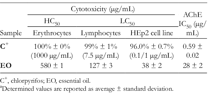
C+, chlorpyrifos; EO, essential oil.
Determined values are reported as average ± standard deviation.
The literature analysis about the cytotoxicity of the EO of different chemo-varieties from L. alba showed that some EO exhibited effects against cell lines, eg, citral and carvone chemotypes were cytotoxic against HeLa line [CC50 (50% cytotoxic concentration, µg/mL): 3.5 ± 0.7 and 74 ± 7, respectively] and Vero cells (LC50: ~31-48 µg/mL); citral chemotype was effective on K562 cells [IC50 (µg/mL): 13.0 ± 0.1-37 ± 1] and Vero cells [CC50 (µg/mL): 65 ± 3-142 ± 1]; carvone, piperitone, ocimenone, and citral chemotypes were active against HepG2 cells [IC50 (µg/mL): ~34 ± 3-412 ± 31], A549 line [IC50 (µg/mL): ~30 ± 3-304 ± 24] and Vero cells [IC50 (µg/mL): ~89 ± 9-592 ± 42]. 48 -51
For comparative purposes, some reports of plant EO containing cis-/trans-piperitone oxides (as major component) and assessment of cytotoxicity were selected. Thus, EO from Mentha, M. mozaffarianii, composed of piperitone oxide (~33% ± 1%-51.5% ± 0.6%), presented high cytotoxicity on MOLT-4 line [IC50 (µg/mL): ~22 ± 5-26 ± 2]; M. longifolia EO constituted by piperitone oxide (~40% ± 1%-65% ± 2%) were active against MCF-7 [IC50 (µg/mL): ~45 ± 2-51 ± 2] and LN-CaP cells [IC50 (µg/mL): ~44 ± 2-52 ± 3]; Dysphania ambrosioides EO rich in cis-piperitone oxide (~35%) reduced the viability of GM07492-A line (IC50: 207 ± 4 µg/mL). Besides, Minthostachys mollis EO (~30% cis-piperitone oxide) was cytotoxic against Vero cells (CC50 < 25 µg/mL). 52 -55 This effect of EO against tumor cells could be attributed to p-mentha(e)ne derivatives (eg, epoxides), which have demonstrated different degrees of effectiveness [%GI (percentage grown inhibition, 25 µg/mL): ~5% ± 2%-99.9% ± 0.2%] on some tested cell lines (HCT-116, OVCAR-8, and SF295). 56
Regarding the moderate inhibitory effect presented by L. alba EO on AChE, it was corresponding with the study by Miyazawa et al, 57 who reported that the EO from M. arvensis (Tosa-hakka), which contained 33% of piperitone oxide, showed an IC50 value of 49 µg/mL on the AChE. In the same work, the authors found that EOs rich (~33%-84%) in p-mentha(e)ne derivatives (eg, menthone, piperitone, pulegone, carvone, and piperitenone epoxide) were, in one way or the other, active on AChE (IC50: ~28-88 µg/mL). In addition, Miyazawa et al 58 studied the effect on AChE of a group of p-mentha(e)ne molecules and found that p-mentha(e)ne ketones were the most active molecules with IC50 values between ~0.9 and 1.8 mM. Accordingly, it could be attributed that p-mentha(e)ne derivatives would be responsible for the inhibitory effect of the EO on the enzyme.
Experimental
Plant Material
Samples (fresh leaves and flowers) from L. alba were collected in San Jacinto town (Departamento de Bolivar) in July 2015/2017. Taxonomic identification (No. Voucher COL588910) was carried out by the Instituto de Ciencias Naturales (Universidad Nacional de Colombia). The plant collection was made under Resolution No. 739 of July 8, 2014, conferred by the Agencia Nacional de Licencias Ambientales (ANLA).
Volatile Fractions
For the isolation of volatile fractions from plant flowers/leaves, 2 methods were used: SDE and headspace-solid phase micro-extraction (HS-SPME).
Simultaneous Distillation-Extraction
The volatile fractions of the fresh parts (5-10 g) from L. alba were obtained by means of the Likens & Nickerson microscale apparatus, modified by Godefroot et al, 59 using CH2Cl2 (2 mL) as extraction solvent for 2 hours. The extracts were dehydrated with anhydrous sodium sulfate, and 1 µL of each of them (individually) was analyzed by GC-MS.
Headspace-Solid Phase Micro-Extraction
The constituents of vapor phase of the flowers/leaves from plant were trapped using SPME device (Supelco), with two coated fibers (polydimethylsiloxane [PDMS], 100 µm; polyacrilate, 65 µm), sampling the headspace. Plant samples (0.5-1 g) were thermally preconditioned (50°C) for 10 minutes and then each fiber was exposed to the headspace of each sample (separately) at 50°C for 30 minutes. Once the sampling was completed, the SPME fiber was desorbed (5 minutes, 25°C) with the analytes inside GC-MS inlet port.
Essential Oil Isolation
Essential oil was obtained of fresh plant leaves (200 g) through hydrodistillation (400 mL of water) assisted by microwave radiation (MWHD) with Clevenger-type apparatus modified with Dean-Stark reservoir. 22 The heating was executed in a domestic microwave oven (Whirpool) operated at 700 W, during 1 hour in 4 cycles of 15 minutes. The EO was decanted, dehydrated with anhydrous sodium sulfate, and prepared [50 µL of each EO with CH2Cl2 (1 mL)] for the corresponding analysis by GC-MS.
Simple Maceration Process
Total extracts from L. alba flowers/leaves were obtained by maceration using ethyl acetate as solvent. The vegetable material (0.5-1 g) was in contact with the solvent (5 mL) under stirring for 7 days at 25°C. The extracts were concentrated to 1 mL, dehydrated with anhydrous sodium sulfate, and analyzed by GC-MS.
Chemical Analysis
The chemical analysis of the volatile secondary metabolites was carried out in a Trace 1310 gas chromatograph coupled to ISQ Series mass spectrometer (Thermo Fisher Scientific), with split/splitless inlet (split ratio, 10:1), liquid autosampler (AI/AS 1310 Series, Thermo Fisher Scientific), or manual injection (SPME). A column Rxi-1 ms (crossbond PDMS, 30 m × 0.25 mm ID × 0.5 µm df, Restek) was useful for the separation. Temperature programming of GC oven was executed as described by Muñoz-Acevedo et al. 60 Chromatographic and spectroscopic data were processed by using Thermo Xcalibur (Version 2.2 SP1.48, Thermo Fisher Scientific) and Automated Mass Spectral Deconvolution and Identification System (AMDIS, Build 130.53, Version 2.70) software.
Linear temperature-programmed retention indices were calculated from a homologous series of C7-C35 aliphatic hydrocarbons and analyzed under the same conditions. The constituents were identified by comparing its mass spectra with those of the available databases (NIST11, NIST Retention Index, and Wiley9), and with linear retention indices reported and consulted in the existing literature. 61 -63
In vitro Biological Activities
All experiments were performed in quintuplicate, with their positive/negative controls and the statistical treatment of data.
Cytotoxicity Assays
On human erythrocytes
The hemolytic activity was carried out based on the methodology by Yang et al 64 modified. The erythrocytes red blood cells (RBC) were isolated by centrifugation at 2500 rpm for 10 minutes, from peripheral blood of healthy donors (18-30 years). Next, a suspension of 4% RBC was prepared in phosphate-buffered saline (PBS) (pH 7.4), which was subjected to different concentrations (100-1000 μg/mL) of EO, for 30 minutes at 37°C. Then, the supernatant liquid was taken from the centrifuged solution and measured at 540 nm in a 96-well plate-reader (FLUOstar Omega).
On human lymphocytes
The cytotoxicity of EO on lymphocytes was assessed using the trypan blue (TB) exclusion test. 65 The isolation of monocytes was performed according to Márquez et al and Calderón-Segura et al 66,67 using peripheral blood from healthy donors. Cell suspension (circa 3000 cell/µL, in Roswell park memorial institute [RPMI]1640 medium) was subjected to different concentrations of EO (100-1000 μg/mL in Dimethylsulfoxide [DMSO]), and incubated for 24 hours at 37°C (5% CO2). Then, an aliquot of sample was mixed with 0.4% TB solution (1:1). The percentage of cell viability was established by a Neubauer camera and a ZEIS contrast microscope (Nikon Microscope, 40×).
On HEp2 cell line
HEp-2 cells (ATCC CCL-23) were cultured (1 × 10⁴ cell/well) and incubated (37°C, 5% CO2, 24 hours) in 96-well plates with RPMI1640 medium (supplemented with 10% fetal bovine serum (FBS) and 2% antibiotic mixture). Subsequently, the EO was added and the mixture was incubated again (24 hours). 68,69 Then, the medium was discarded, and 100 µL of 500 µg/mL, 3-(4,5-dimethylthiazolyl-2)-2,5-diphenyltetrazolium bromide solution in RPMI medium (without FBS) was added; it was incubated (3 hours) and centrifuged (3000 rpm, 15 minutes). The reaction mixture was discarded and DMSO (100 µL) was added. The plate was analyzed in a 96-well reader at 570 nm. 70 The percentage of cell viability in each treatment was calculated using the absorbances obtained in control 0 (100% viability) as a reference.
Acetylcholinesterase Enzyme Inhibition
The in vitro inhibition of the EO/chlorpyrifos (C+) on the AChE was measured pursuant to the method by Ellman et al 71 modified for which the samples (EO/C+, 50 µL of each), prepared at 4-125 µg/mL (C+, 0.3-4.8 µg/mL), were placed to react in a 96-well plate with the AChE (50 µL—0.25 U/mL) for 30 minutes at 25°C. Afterward, the substrate [5,5´-dithiobis-(2-nitrobenzoic acid) (100 µL, 0.2 mM), AChI (0.24 mM), and Na2HPO4 (0.04 M)] was added to each well and the final mixture was incubated at 37°C for 6 minutes and analyzed in a plate-reader at 412 nm. All solutions of samples/enzyme were prepared in PBS buffer. The IC50 value was obtained from the graph of percent inhibition (at 6 minutes) vs concentration of the evaluated substance.
Footnotes
Acknowledgments
The authors thank Dr Homero San Juan for providing the HEp2 cell line, and Colciencias and Departamento del Atlántico [“Jóvenes Investigadores (M.C.G., 2015)” and “Formación de Capital Humano (J.D.R./Y.S.D-M., Convocatoria No. 673, 2014)].”
The author(s) declared no potential conflicts of interest with respect to the research, authorship, and/or publication of this article.
Declaration of Conflicting Interests
The author(s) declared no potential conflicts of interest with respect to the research, authorship, and/or publication of this article.
Funding
The author(s) disclosed receipt of the following financial support for the research, authorship, and/or publication of this article: Universidad del Norte (Proyecto 2013-DI0024—Área Estratégica en Biodiversidad).