
Abstract
Alpinia pricei Hayata (Zingiberaceae) is a spicy plant endemic to Taiwan. In this study, several behavioral analyses were used to evaluate the neuropharmacological activity of A. pricei in mice. Oral administration of 100, 300, and 500 mg/kg of A. pricei extract (APE) significantly prolonged pentobarbital-induced sleeping time in mice by 24.5%, 74.7%, and 79.0%, respectively. Also, the antidepressant effect of APE was evaluated using suspended tail and forced swimming tests. The immobility periods of mice in the suspended tail and forced swimming tests were reduced after the administration of APE. Further, an elevated plus-maze test was used to study the anxiolytic activity of APE. After treatment with 500 mg/kg of APE the time the mice spent in the open arms (31.55 ± 13.65 seconds) and the number of times they entered the open arms (51.75 ± 16.51 times) (P < 0.05) of the plus-maze increased significantly compared to a saline-treated group. Our results also revealed that APE showed potent analgesic activity in the tail-flick test; all dosages of APE prolonged the tail-flick time for up to 90 minutes. In conclusion, APE had a potent effect on the neuropharmacological activities of mice. Finally, the main compounds of APE were separated, and spectral analysis was conducted. The major constituents of APE were characterized as 5,7-dimethoxyflavanone (
Keywords
Alpinia plants (Zingiberaceae) are well-known medicinal herbs; they have been reported to possess multiple activities, including antioxidant, anti-inflammatory, anticancer, immunostimulatory, hepatoprotective, and antinociceptive. 1–7 Alpinia pricei Hayata is a perennial rhizomatous spicy plant endemic to Taiwan. Traditionally, the leaves of A. pricei are used to make zongzi (glutinous rice dumplings); the aromatic rhizome of A. pricei is thought to dispel abdominal distension and increase stomach secretion and peristalsis. Over the past decades, our research group has studied the phytochemistry and bioactivities of A. pricei. First, we used a high concentration of sugar to induce metabolic syndrome in C57BL/6J mice; we found that visceral adiposity and insulin resistance induced by a chow-based hypercholesterolemic diet could be alleviated by A. pricei supplementation. 8 It was also confirmed using the animal model that the ethanol extract of A. pricei could regulate blood lipids and avoid lipid peroxidation. 9 Our study also found that the ethanol extract of A. pricei could regulate the cell cycle by protein expression, causing cells to stagnate at the G2/M phase, and at the same time promote the release of reactive oxygen species (ROS), mitochondrial membrane potential decline, and polyadenosine diphosphateribose polymerase (PARP) lysis, which in turn caused tumor cell apoptosis. 10 Phytochemical studies showed that the major components of A. pricei were flavonoids, phenolic acids, sterols, styrylpyrone, and fatty acids. 11,12 We also demonstrated that the colorectal cancer cell line (HCT116) produced large amounts of ROS with flavokawain B treatment, leading to the activation of GADD153, eventually resulting in granulocyte pathways in the form of cell apoptosis. 13 In addition to the chalcone skeleton flavonoid activity, our results obtained from cell and animal studies demonstrated that the styrylpyrone-class compound, desmethoxyyangonin, had significant inhibitory activity against fulminant hepatitis through anti-inflammatory activity. 14 To understand the effect of A. pricei on the central nervous system (CNS), several animal behavioral analyses were performed to evaluate the neuropharmacological activities of A. pricei. The major compounds of A. pricei were also characterized in this study.
In the open field test, as shown in Table 1, the total pathway for vehicle control was 3622.3 ± 582.9 cm, and the speed of movement was 12.93 ± 1.59 cm/s. When the mice received 100, 300, and 500 mg/kg of A. pricei extract (APE), the distances of movement were reduced to 2897.4 ± 790.0, 2886.5 ± 394.1, and 2920.0 ± 452.9 cm, respectively. The speed of movement for the experimental groups was also decreased to 9.98 to 10.18 cm/s in a dose-dependent manner. However, oral administration of APE did not have significant effects on the time spent immobile.
Effect of APE on Locomotor Activity of Mice in an Open Field Test.

APE, Alpinia pricei extract.
Data are presented as mean ± SE from 10 animals. Observations were made 60 minutes following the oral administration of vehicle (control) or APE.
*P < 0.05 compared with vehicle-treated controls.
The effect of oral administration of A. pricei and desmethoxyyangonin (one of the major components of APE) on pentobarbital-induced sleeping of mice is shown in Table 2. When treated with APE (100, 300, and 500 mg/kg), the pentobarbital-induced sleeping time for mice was dose-dependently prolonged to 24.5%, 74.7%, and 79.0%, respectively. Mice administrated with desmethoxyyangonin (75 mg/kg) prolonged 51.6% sleeping time in the pentobarbital-induced sleeping model. In this study, zolpidem hemitartrate was used as the positive control; it prolonged the pentobarbital-induced sleeping time from 2229.8 ± 172.2 to 2804.8 ± 203.6 seconds at the dosage of 0.3 mg/kg.
Effect of APE and Desmethoxyyangonin on Pentobarbital-induced Sleeping Time in Mice.

APE, Alpinia pricei extract.
All animals were treated with pentobarbitone sodium (45 mg/kg, IP) 60 minutes after oral administration of vehicle, APE, and desmethoxyyangonin. *P < 0.05, **P < 0.01 compared with vehicle-treated controls.
As shown in Figure 1, the maximal antidepressant activity of APE was obtained at a dosage of 500 mg/kg, resulting in 30.8% immobility reduction; the immobility time of the control group was 188.61 ± 45.07 seconds; the immobility time of APE treatment at the dosages of 100, 300, and 500 mg/kg were 158.71 ± 83.29, 138.12 ± 49.16, and 130.51 ± 49.02 seconds, respectively. Fluoxetine hydrochloride, an antidepressant drug, was used as a positive control; and the immobility time was reduced to 91.47 ± 25.59 seconds at a dosage of 10 mg/kg.

Effect of APE (100, 300, and 500 mg/kg) and fluoxetine hydrochloride (10 mg/kg) on the suspended tail test. Each bar represents values as mean ± SE (n = 10). *P < 0.05, **P < 0.01 compared with vehicle-treated controls. APE, Alpinia pricei extract.
The elevated plus-maze test was performed to evaluate the anxiolytic activity of APE in this study. Trazodone hydrochloride was used as a reference compound. Table 3 shows the effect of APE and trazodone hydrochloride on mouse behavior in the elevated plus-maze test. The time spent in the open arms was 12.68 ± 8.67 seconds, and the number of entries into the open arms was 18.88 ± 11.74 for vehicle control (treated animals with saline). Independent t-test revealed that APE administration (500 mg/kg) significantly increased the time spent in the open arms (31.55 ± 13.65 seconds) and the number of entries into the open arms (51.75 ± 16.51 times) (P < 0.05). The time spent in the open arms for trazodone hydrochloride treatment (10 mg/kg; orally) was 28.86 ± 10.31 seconds and the number of entries into the open arms was 25.63 ± 14.91 times.
Effects of APE and Trazodone Hydrochloride on Behavior in the Elevated Plus-Maze Test.
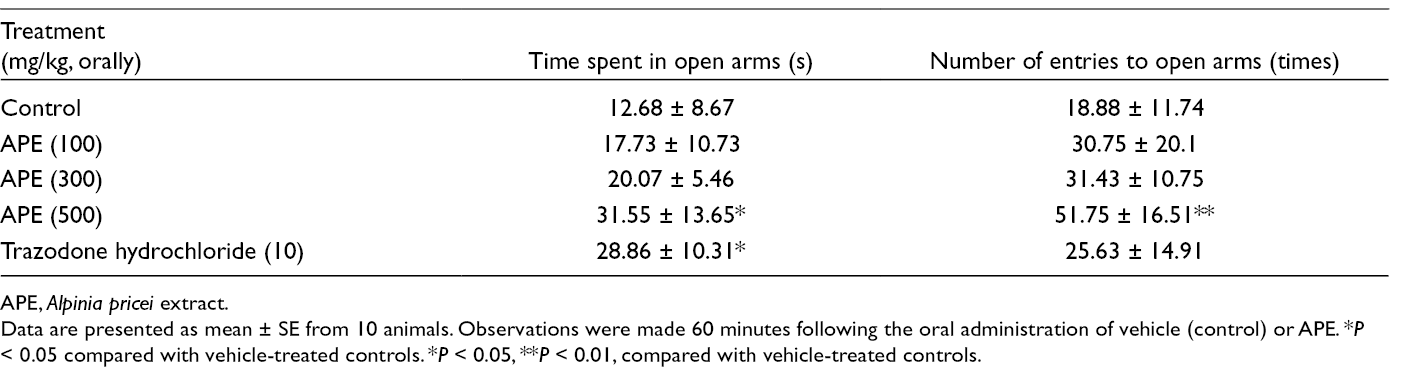
APE, Alpinia pricei extract.
Data are presented as mean ± SE from 10 animals. Observations were made 60 minutes following the oral administration of vehicle (control) or APE. *P < 0.05 compared with vehicle-treated controls. *P < 0.05, **P < 0.01, compared with vehicle-treated controls.
Figure 2 shows the results of the forced swimming test. In the groups of APE treatments, the time immobile was significantly reduced. At a dosage of 500 mg/kg given by the oral route, a significant decrease in the duration of immobility time in comparison to the control group was seen, with values changing from 127.3 to 45.5 seconds. Fluoxetine hydrochloride (dosage = 10 mg/kg) reduced the immobility time from 127.3 to 90.0 seconds.
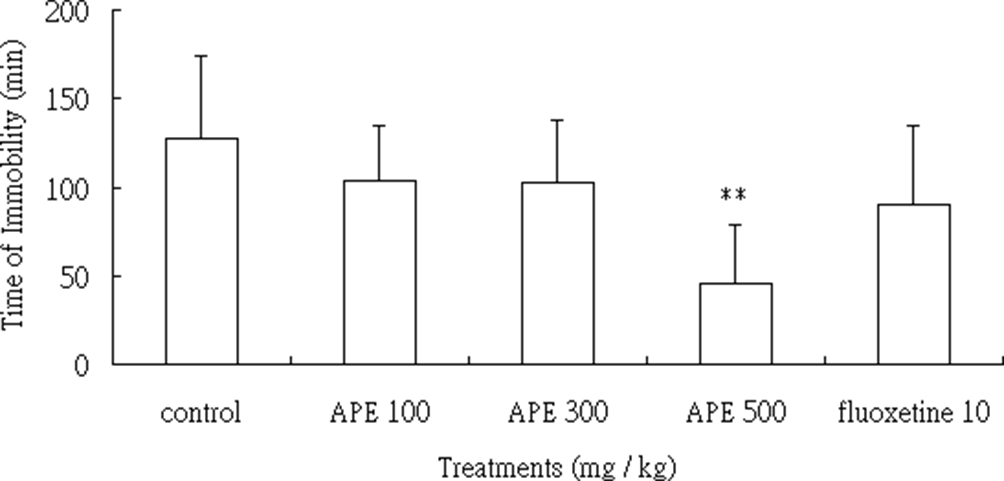
Effect of APE (100, 300, and 500 mg/kg) and fluoxetine hydrochloride (10 mg/kg) on the forced swimming test. Each bar represents values as mean ± SE (n = 10). **P < 0.01 compared with vehicle-treated controls. APE, Alpinia pricei extract.
In the tail-flick test, Figure 3 shows significant prolongation of the reaction time at all dosages at 30 minutes posttreatment. All dosages of APE administration significantly prolonged (P < 0.05) the tail-flick time for up to 90 minutes. However, the maximal analgesic activities were reached at 60 minutes for each dosage; the activity of the positive control group, acetaminophen (90 mg/kg), could be maintained to 90 minutes.

Effects of oral administration of APE and AAP (60 mg/kg) on analgesia measured by the hot tail-flick method in mice. Each bar represents values as mean ± SE (n = 10). *P < 0.05, **P < 0.01, ***P < 0.001 compared with vehicle-treated controls. APE, Alpinia pricei extract; AAP, acetaminophen.
Since APE revealed potent neuropharmacological activities for Institute of Cancer Research (ICR) mice, we further characterized the major compounds of APE by using chromatography followed by spectral analysis. The metabolite profiling of APE by liquid chromatography/mass spectrometry is shown in Figure 4. The major compounds
Characterization of Phenolic Compounds in APE Using LC-MS.

APE, Alpinia pricei extract; LC/MS, liquid chromatography/mass spectrometry.

Metabolite profiling of APE. 1, 5,7-Dimethoxy-flavanone; 2, desmethoxyyangonin; 3, 2′,4′,6′-trimethoxychalcone; 4, cardamonin; 5, trans/cis-3,5-dimethoxystilbene; 6, flavokawain B. APE, Alpinia pricei extract.
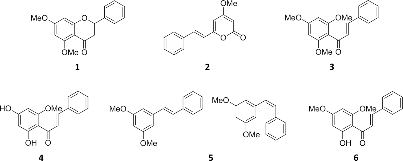
The structures of the main compounds in APE. 1, 5,7-Dimethoxyflavanone; 2, desmethoxyyangonin; 3, 2′,4′,6′-trimethoxychalcone; 4, cardamonin; 5, trans/cis-3,5-dimethoxystilbene; 6, flavokawain B. APE, Alpinia pricei extract.
The neuropharmacological effect of APE on mice was studied. Our results indicated that APE reduced the locomotor activity of mice; however, the time immobile did not increase in mice after treatment with APE in the open field test. And pretreating the mice with APE noticeably prolonged the pentobarbital-induced sleeping time in a dose-dependent manner. In the antianxiety effect assay (elevated plus-maze test), APE apparently increased the time spent and number of entries into the open arms. Moreover, APE exhibited antinociceptive activity according to the results obtained in the tail-flick test. It is interesting that the neuropharmacological effect of APE was similar to the activity of kava-kava (Piper methysticum G. Forster), which is a traditional psychoactive beverage used in the South Pacific.
19
Extensive literature exists on the pharmacologically active constituents of kava-kava, which are mainly a group of lactone molecules, named kavalactones, which had a strong effect on the CNS in an animal trial.
20
The main compounds of APE seen in this study, including desmethoxyyangonin, cardamonin, and flavokawain B, were previously identified in the western Pacific plant kava-kava. It is, therefore, reasonable to infer that the mechanisms of the neuropharmacological activities exhibited by A. pricei might be similar those of kava-kava. In this study the effect of APE on the neuropharmacological activities was evaluated in mice. According to our data, APE has a measurable effect on the neuropharmacological activities, suggesting the possible application of APE as a functional food and/or pharmaceutical to aid CNS regulation. Furthermore, we also characterized the composition of APE: the main compounds were 5,7-dimethoxyflavanone (
Experimental
Plant Material and Extract Preparation
The rhizomes of A. pricei were collected from Ping-tung County in southern Taiwan in March 2014 and were identified by Dr Yen-Hsueh Tseng (National Chung-Hsin University). The voucher specimen was deposited in the herbarium of the same university. The air-dried roots (2 kg) of A. pricei were extracted with 10 L 70% ethanol (EtOH) in H2O at room temperature. The total crude extract was evaporated to yield the EtOH extract (APE) (166 g).
Animal Experiments
The male ICR mice (4 weeks old, 25-28 g) were purchased from BioLasco (Yilan, Taiwan). APE was emulsified with 1% carboxymethylcellulose sodium salt and dissolved in distilled water. Animals were treated with APE at doses of 100, 300, and 500 mg/kg and desmethoxyyangonin 75 mg/kg (orally), 60 minutes before the experiments. Controls were received vehicle at the same volume (10 mL/kg) and were administered by the same route as the treated groups. Zolpidem hemitartrate (0.3 mg/kg), fluoxetine (10 mg/kg), trazodone hydrochloride (10 mg/kg), and acetaminophen (60 mg/kg) used as reference compounds were treated (10 mL/kg orally) after dissolving in distilled water. The experiments were performed after the approval of the protocol by the local Institutional Ethics Committee. All experiments were carried out following the current guidelines for the care of laboratory animals and the ethical guidelines for investigations of experimental pain in conscious animals. 21 All the actions of mice in each model were image recorded for visual and automated quantitative analysis by the same person. The behaviors of mice were recorded using a DSP CCD camera (Model: KMS-63F4) connected to a computer with Noldus software (Ethovision version 4.0, Noldus Information Technology, Wageningen, Netherlands) for data acquisition. Behavioral analyses were conducted open field test, pentobarbital-induced sleeping time, tail suspension test, forced swimming test, elevated plus-maze test, and tail-flick test. 22–26
Extraction and Purification
The major compounds in the APE were obtained at retention times of 18.4, 20.6, 21.5, 22.8, 26.0, and 27.3 minutes by semi-preparative high performance liquid chromatography. A luna silica (Luna C18) column (250 × 10 mm, Phenomenex, Torrance, California, US) was conducted with two solvent systems, H2O (A) and 100% acetonitrile (B). The gradient elution profile was as follows: 0 to 3 minutes, 80% A to B; 3 to 60 minutes, 80-0% A to B (linear gradient); 60 to 80 minutes, 0% A to B; the flow rate was 2.5 mL/min and the detector wavelength was set at 280 nm. The structures of compounds were then elucidated using spectroscopic analyses. UV spectra of test compounds were recorded with a Jasco V-550 spectrometer (Halifax, Nova Scotia, Canada) and IR spectra obtained from a Bio-Rad FTS-40 spectrophotometer (Hercules, California, US). Electron-impact mass spectrometry (EIMS) and high-resolution EIMS data were collected with a Finnigan MAT-958 mass spectrometer (Waltham, Massachusetts, US) and NMR spectra recorded with Bruker Avance 500 and 300 MHz FT-NMR spectrometers (Billerica, Massachusetts, US), at 500 MHz (1H) and 75 MHz (13C).
Metabolite Profiling of APE
The APE profiling was performed by ultra-high performance liquid chromatography linked to electrospray ionization -ion trap mass spectrometer. A Waters RP-18 column (100 × 2.1 mm, 1.7 µm, Milford, Massachusetts, US) was employed with two solvents systems, H2O with 0.1% formic acid (A) and 100% acetonitrile (B) in this study. The gradient elution profile was as follows: 0 to 2 minutes at 80% A, 2 to 6 minutes 80% A to 40% A, 6 to 16 minutes 40% A to 0% A, 16 to 19 minutes at 100% B; the flow rate was 0.3 mL/min for application to the mass spectrometer. The ESI was used in both the positive and negative modes. The MS operating conditions were as follows: capillary voltage, 4.5 kV; end plate offset voltage, 500 V; nebulizer, 60 psi; dry gas, 10 psi; dry temperature, 300°C; full scan from m/z 100 to 800.
Statistical Analysis
Data are expressed as means ± SD. Statistical comparisons of the results were made using analysis of variance. Significant differences (*P < 0.05 and **P < 0.01) between the control (untreated) and treated cells were analyzed by Dunnett’s test.
Footnotes
Declaration of Conflicting Interests
The author(s) declared no potential conflicts of interest with respect to the research, authorship, and/or publication of this article.
Funding
The author(s) disclosed receipt of the following financial support for the research, authorship, and/or publication of this article: This study was supported by the Ministry of Science and Technology, Taiwan, (106-2313-B-005-012-MY3).