
Abstract

Vision loss has been highlighted as a major public health concern; approximately 90 million Americans older than 40 years have vision or eye problems. 1 By 2050, the Centers for Disease Control and Prevention (CDC) estimates the prevalence of diabetic retinopathy (DR) and age-related macular degeneration (AMD) to increase by 72% and 100%, respectively. 1 The CDC has identified DR as the most common cause of vision loss and blindness among working-age individuals, and AMD is the most common cause of permanent vision loss among those aged 65 years or older. 2
Community retinal screenings increase access to eye care in underserved populations. We report the utility of optical coherence tomography (OCT)-fundus photography combined imaging for community-based retinal screenings. We reviewed records from two tele-ophthalmology community screenings held during the pandemic period which utilized the gold standard non-mydriatic retinal photography for tele-ophthalmology 3 alongside OCT to identify additional macular pathology. Thirty-nine patient records from two community screenings in Newark, New Jersey, were evaluated, where both a Canon CR-2 PLUS AF Digital Non-Mydriatic Retinal Camera (Tokyo, Japan) and the Optovue RT-Vue (Fremont, CA) OCT unit were deployed. Imaging was reviewed by a board-certified ophthalmologist to identify retinal disease and referral determination. The ophthalmologist first reviewed the fundus photos of each case to diagnose any retinal pathology that needed an ophthalmic referral. Then, OCT-B images of the macula were provided to the examiner to see if OCT findings changed the referral pattern. Changes in referral pattern based on OCT-B imaging were recorded.
Of the records reviewed, 47% of patients were male, with an average age and body mass index of 55 years and 30.0, respectively. Of the eyes screened, referral for in-person retinal examinations by a comprehensive ophthalmologist were recommended for 18 eyes (25%) based on fundus photography alone. This included five eyes for DR, 15 for AMD, and two for both DR and AMD (Table 1). The 18 eyes belonged to 12 individuals: four people referred for DR evaluation and 10 for AMD evaluation, including two people for both DR and AMD evaluation (Table 1). Of these 18 eyes, eight eyes (seven individuals) showed evidence of macular edema on OCT-B images. Therefore, the referral for these eight eyes was adjusted as an urgent referral directly to a retina service. The results of OCT-B imaging allowed for 44% of the eyes with retinal pathology (11% of the total eyes) to be referred directly for subspecialty care (Table 1).
Reasons for Referral for In-Person Retinal Evaluation.
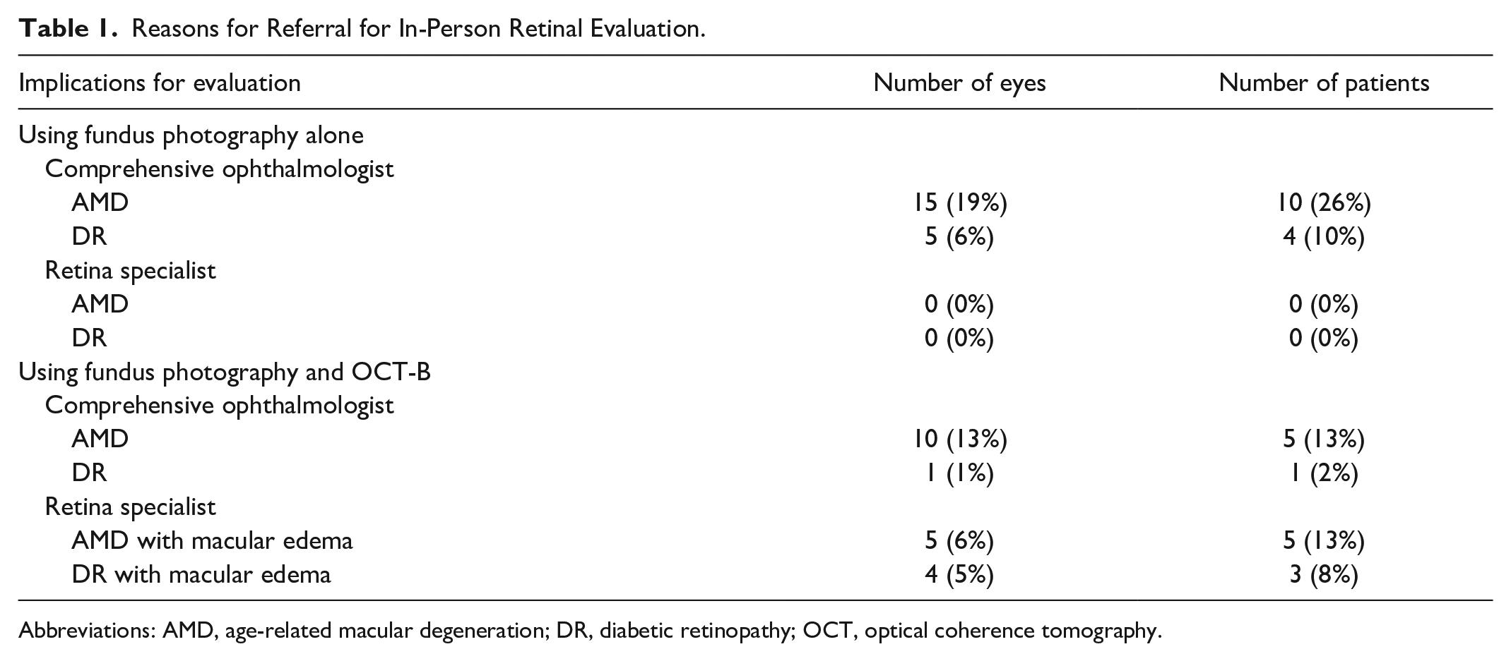
Abbreviations: AMD, age-related macular degeneration; DR, diabetic retinopathy; OCT, optical coherence tomography.
In this study, we demonstrated that the dual use of fundus photography and OCT-B imaging in at-risk communities can allow for more accurate assessment of macular disease, which is especially critical in populations with limited access to ophthalmic care. The addition of OCT-B in this setting allowed identification of eyes with severe disease for triage to a retina specialist. Streamlining patients to the necessary clinic reduces obstacles to health care access. Future studies may assess the utility of OCT-B imaging with fundus photography in diabetics in endocrinology clinics to identify urgent pathology.
Footnotes
Abbreviations
AMD, age-related macular degeneration; CDC, Centers for Disease Control and Prevention; DR, diabetic retinopathy; OCT, optical coherence tomography.
Declaration of Conflicting Interests
The author(s) declared no potential conflicts of interest with respect to the research, authorship, and/or publication of this article.
Funding
The author(s) received no financial support for the research, authorship, and/or publication of this article.