
Abstract
Importance
Patients who undergo total laryngectomy lose the voice function permanently. It is important to reconstruct the voice function of the patients after total laryngectomy.
Objective
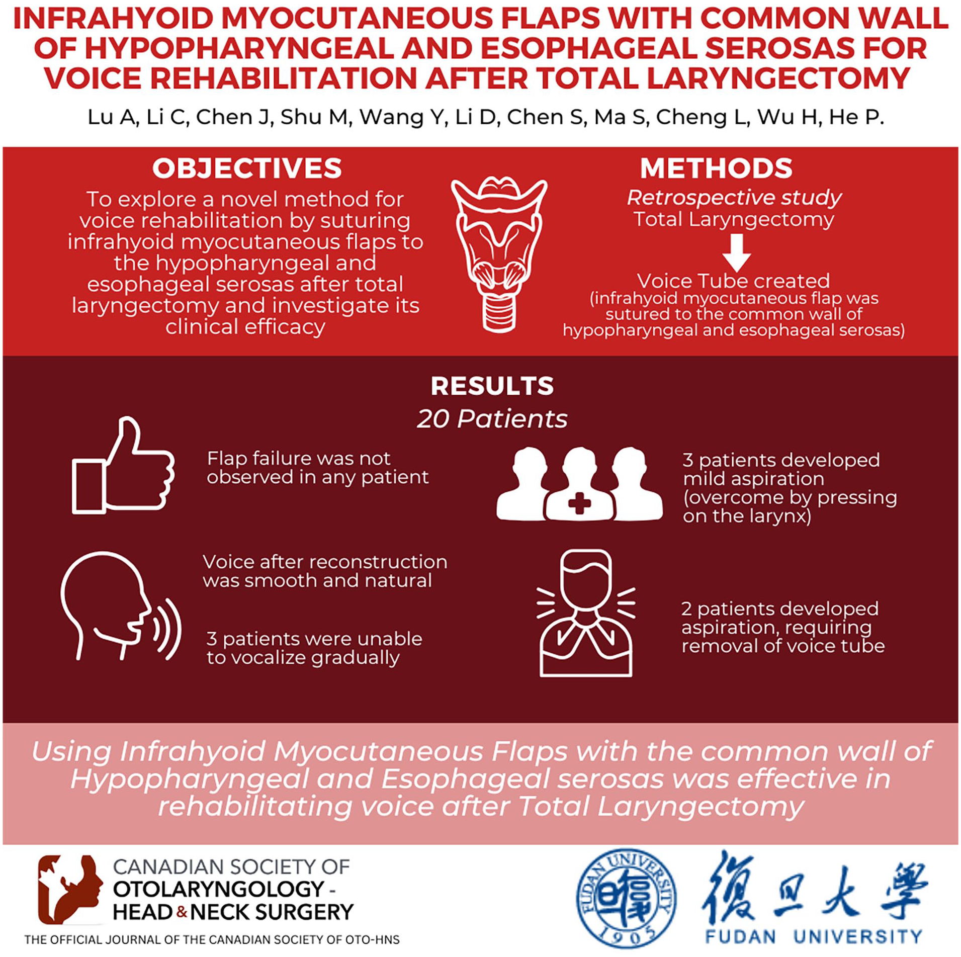
To explore a novel method for voice rehabilitation by suturing infrahyoid myocutaneous flaps to the hypopharyngeal and esophageal serosas after total laryngectomy and investigate its clinical efficacy.
Design
Clinical study (retrospective study).
Participants
Twenty patients with advanced laryngocarcinoma who underwent total laryngectomy.
Methods
Following total laryngectomy, the pharyngeal cavity was formed via layer-by-layer suturing, and the infrahyoid myocutaneous flap was sutured to the common wall of hypopharyngeal and esophageal serosas to create a voice tube.
Results
Flap failure was not observed in any patient. All patients were able to vocalize after surgery. The voice after the reconstruction was smooth and natural. However, 3 patients were unable to vocalize gradually. Two patients experienced aspiration, requiring the removal of the voice tube, while 3 patients exhibited mild aspiration, which could be overcome by pressing the larynx on the voice tube surface.
Conclusions
Using infrahyoid myocutaneous flaps with the common wall of the hypopharyngeal and esophageal serosas was effective in rehabilitating voice after total laryngectomy. Future clinical studies are necessary to validate the effectiveness of this technology for voice rehabilitation.
Introduction
Laryngocarcinoma is usually treated with surgery, chemoradiotherapy, immunotherapy, and so on, with surgery being the primary treatment. The rationale is to preserve or rebuild laryngeal function while removing the tumor as radically as possible. Patients who have undergone total laryngectomy experience a complete loss of pronunciation, and swallowing protection and breathing may be significantly affected. Rehabilitating laryngeal function and improving the living quality of patients after an operation have been challenging for ear, nose, and throat (ENT) surgeons.1,2 Nonsurgical methods of voice rehabilitation include electrolarynx, pneumatic artificial larynx, and esophageal speech. Electrolarynx and pneumatic artificial larynx have been criticized for unnatural sounds or social stigma. Although esophageal speech has the advantages of good sound and low cost, the strenuous training process and low success rate make it difficult to apply to every patient. 3 Among surgical methods, tracheoesophageal puncture (TEP) is highly regarded for speech restoration. In particular, TEP with prosthesis remains the “gold standard” and the primary choice in developed countries. 2 However, the high cost and frequent need to replace the prosthesis restrict it as a primary treatment in developing countries.
Recently, there has been an increasing use of flaps as voice tubes to rehabilitate the function of pronunciation.3,4 The infrahyoid myocutaneous flap is soft, easy to harvest, and has abundant blood supply. It has been widely used in repairing pharyngeal defects. 5 However, this type of myocutaneous flap has never been applied to voice tube reconstruction. Our team marks a pioneering approach in utilizing infrahyoid myocutaneous flaps to reconstruct pharyngotracheal voice tube, 6 achieving a satisfactory effect. This study sought to improve this technique by suturing the infrahyoid myocutaneous flap to the common wall of the hypopharyngeal and esophageal serosas. This study discussed the effectiveness of this new technique on patients after total laryngectomy.
Materials and Methods
Patients
This study aimed to investigate the pronunciation of patients who underwent total laryngectomy and reconstruction of voice tubes at the Eye, Ear, Nose and Throat Hospital of Fudan University between July 2022 and October 2023. This study encompassed 20 cases, including 1 female and 19 males. All patients were diagnosed with primary laryngeal cancer (T3-T4) conforming to the indications of total laryngectomy, most of whom had squamous carcinoma, except 3 cases with carcinosarcoma, verrucous carcinoma, and small cell carcinoma. None of the patients received chemoradiotherapy preoperatively. Six patients developed lymph node metastases as per the postoperative pathological report (metastatic squamous cell carcinoma, N1-N2). All patients experienced symptoms of hoarseness. Two patients complained of dysphagia.
Surgical Methods
Voice tube reconstruction was performed simultaneously with total laryngectomy. To harvest satisfactory skin flaps, total laryngectomy and neck lymph node dissection were performed through a longitudinal incision in front of the neck. Total laryngectomy using traditional methods necessitates ensuring the blood supply to the sternohyoid and sternothyroid muscles. The pharyngeal cavity was closed in 3 layers: mucosa, submucosa, and serosa. The infrahyoid myocutaneous flap was generally harvested from the opposite side of the lymph node dissection, with flap sizes ranging from 1 × 6 to 2 × 8 cm2. If lymph node dissection was performed on both sides, the side where the surgeon was accustomed to harvesting the skin flap was chosen. The skin flap was sutured downward to the blind end of the trachea and upward to the pharyngeal cavity. The middle part of the skin flap was sutured with the pharyngeal and esophageal serosal layers to form a voice tube (Figure 1). If necessary, a contralateral skin flap of 1 × 2 cm2 size could be taken to facilitate the formation of an opening at the tracheal end of the voice tube. In patients who retained the epiglottis, after folding the epiglottis downward and adjusting it, the upper end of the flap was sutured with the epiglottic mucosa so that the epiglottis would cover the opening of the voice tube to prevent accidental aspiration due to water ingress. For patients who aspirated after voice tube reconstruction, Ω-shaped epiglottoplasty could be performed. The mucosa around the pharyngeal opening of the voice tube was incised in a nearly-circular manner, allowing partial detachment of the voice tube from the surrounding soft tissue. Then, the mucosa on both sides of the pharyngeal opening of the voice tube was folded and sutured to form an Ω-shaped epiglottic structure, help preventing aspiration (Figure 2). A urinary catheter could be inserted and left in the lumen of the voice tube, aiding in dilation and drainage to prevent stenosis or occlusion of the voice tube. Finally, the incision was closed by subcutaneous and skin suturing.

Surgical procedure. (a) Flap design. (b) Total laryngectomy. (c) Flap (red arrow) sutured to the hypopharyngeal and esophageal serosas (yellow arrow). Blue arrow indicates tracheostoma. (d) Completion of flap inset. (e) Insertion of a urinary catheter (black arrow) for dilation. (f) Postoperative appearance. Blue arrow indicates tracheostoma.

Ω-Shaped epiglottoplasty. (a) Preoperative view (the suction tube was inserted into the pharyngeal opening). (b) Incision of the mucosa around the pharyngeal opening. (c) Suturing of the mucosal flap to constrict entrance. (d) Reconstructed epiglottis.
Assessment Methods
The grade, roughness, breathiness, asthenia, and strain (GRBAS) scale was utilized to evaluate the voice objectively. The GRBAS scale is widely used for perceptual evaluation of voice quality and grades voice on a 4-point scale (0 = normal to 3 = severe). 7 The grade (G) score is the most reliable in the GRBAS scale and was thus used to assess the voice function.
The deglutition function was evaluated using the water swallow test. During the test, the patients are instructed to sit upright and drink 30 ml of warm water. Deglutition function is classified into 5 grades by observing the time patients use to drink the water and the aspiration situation. The following are the 5 grades assigned: Grade Ⅰ (excellent): the patient successfully swallows 30 ml of water in 1 gulp; Grade Ⅱ (good): The patient finishes drinking water in more than 2 gulps without aspiration; Grade Ⅲ (medium): The patient swallows 30 ml of water in 1 gulp with aspiration; Grade Ⅳ (available): The patient swallows 30 ml of water in more than 2 gulps with aspiration; Grade Ⅴ (poor): The patient repeatedly aspirates and is unable to swallow all the 30 ml of water. 8 The assessment was performed 3 months after voice tube reconstruction.
Results
Twenty patients recovered their pronunciation after surgery by blocking the tracheostoma with their fingers. However, 2 of them gradually became unable to speak after radiotherapy (9 patients received radiotherapy after the operation), and 1 patient experienced voice tube atresia after the removal of the catheter. The remaining 17 patients (85%) exhibited good pronunciation and restored some sociabilities. Three patients experienced strenuous pronouncing, but the speech was satisfactory in intelligibility and consistent in language. In these patients, the voice tube was unobstructed, and the airflow was observed from the pharyngeal opening of the voice tube under a laryngoscope (Figure 3). Catheters placed to prevent stenosis and atresia are usually removed 3 to 4 weeks after operation. Postoperative feeding was achieved through the nasogastric feeding tube, with the recovery of oral feeding time being generally 2 weeks after the operation and sometimes 3 to 4 weeks. There was no aspiration before the operation. After the operation, 2 patients (10%) aspirated and were unwilling to keep the voice tube. Due to their older ages and risk of pneumonia, the voice tube was excised. But the pronunciation remained good before excision. Three patients (15%) experienced mild aspiration while drinking, but it was overcome by pressing the skin on the surface of the voice tube. Patients who underwent Ω-shaped epiglottic reconstruction did not experience aspiration. Pharyngocutaneous fistula occurred in 4 patients (20%) after operation, which improved after debridement and antibiotic treatment. There was no flap failure or local necrosis throughout the study (Table 1).

(a) Voice tube (arrow) and cavum pharyngis (asterisk) under CT. (b) Voice tube (red arrow) and introitus esophagi (black arrow) under a laryngoscope. The pharyngeal opening dilated, and the air flowed out when pronouncing. (c) The pharyngeal opening closed when not pronouncing. CT, computed tomography.
Laryngeal Function and Adverse Outcome in 20 Patients.

Three patients who experienced aphonia did not participate in the G score evaluation. They had normal voices before they exhibited aphonia.
Two patients requested to remove the voice tube due to aspiration.
Discussion
Total laryngectomy is one of the most effective treatments for advanced laryngeal cancers. However, removing the larynx can result in permanent loss of speech function, which causes substantial damage to the patient’s social contact and quality of life. 9 Researchers have made several efforts to rehabilitate voice. However, each technique has its strengths and limitations. Esophageal speech is the most economical and convenient way of pronunciation rehabilitation, which can be achieved without surgery or auxiliary equipment. However, its success rate is low, approximately 50% to 70%,3,10 the volume and continuity of pronunciation are poor, and it is prone to abdominal pain, stomach distension, acid reflux, and other gastrointestinal symptoms. The electrolarynx is simple to use, but the pronunciation is relatively monotonous, and the intelligibility is low. 11 The pneumatic artificial larynx affects patients’ social interaction and negatively impacts their psychology. Laryngeal transplant is a less feasible option. 3 TEP prosthesis is the preferred method of voice rehabilitation because of its natural pronunciation, low training demand, and relatively-simple operation. However, it is overly reliant on doctors and too expensive to be widely used in developing countries. Additionally, there are complications such as granulation tissue, obstruction, displacement, leakage, and difficulty in replacement and cleaning.12,13
Since the concept of the voice tube was proposed, diverse flaps have been used for voice rehabilitation. Infrahyoid myocutaneous flap is mainly utilized for oral defect repair, oropharyngeal repair, and hypopharyngeal repair. Its role in voice rehabilitation is often easily overlooked. 5 As seen with other voice tube surgeries, the infrahyoid myocutaneous flap may be utilized in the primary stage with total laryngectomy. TEP prosthesis is generally chosen for a secondary stage, which can also be performed in a primary stage with an increased rate of stenosis. 14 The infrahyoid myocutaneous flap offers several advantages when considering the anatomical structure of strap muscles. The rich blood supply of infrahyoid myocutaneous flaps offers a high survival rate. It is wide enough for easy access. It is thin and pliable, with a high similarity to the original regional tissue, which explains its better speech function. In this study, all patients were able to pronounce after surgery. Some researchers have utilized infrahyoid myocutaneous flaps to reconstruct the voice tube. The regained sound was fluent and natural.6,15 In this study, most cases (85%) recovered pronunciation finally. Intelligibility and loudness were satisfactory, and no flap failure occurred.
When the flaps come from regions outside of the neck, donor-site morbidity cannot be ignored. The free visceral flaps represented by the jejunum can often lead to complications, including diarrhea, intestinal leakage, abscess, or hernia, which could be serious and result in high perioperative mortality. 16 The incidence and risk of free fascial flaps, such as radial forearm flaps, are not higher than those of free visceral flaps. However, free fascial flaps pose additional risks of decreased motor capacity and numbness in the forearm.17-19 Meanwhile, the operation of nonlocal flaps often cannot be independently completed by the ENT doctor, with the change in the surgical field increasing the operation time. As a local skin flap, the donor-site morbidity of infrahyoid myocutaneous flaps is low.5,20,21 It does not require transferring to a different surgical field, making the operation time shorter and increasing the tolerance of patients. These features are especially beneficial to the elderly or frail patients.
Preserving or restoring the swallowing protection function is an important part of laryngeal function reconstruction. As the voice tube lies close to the skin, pressing the superficial skin can effectively prevent aspiration. Surgeons preserved the epiglottis or folded the flap into an epiglottic shape to prevent aspiration.3,15 In our experience, preserving the epiglottis alone may not protect the voice tube from food inflow, because the distance between the epiglottis and the pharyngeal opening of the voice tube may change. Therefore, even if the epiglottis is preserved, it needs to be redesigned carefully. The problem of aspiration was solved in patients who underwent Ω-shaped epiglottic reconstruction. Compared to sliding epiglottis reconstruction,22,23 Ω-shaped epiglottic reconstruction can be performed endoscopically and has a lower impact on the surrounding tissues. Also, when compared to other endoscopic epiglottoplasty techniques, 24 Ω-shaped epiglottic reconstruction does not increase the technical difficulty and is more suitable for patients with voice tubes.
Nine patients received chemotherapy postoperatively, among whom 2 patients experienced aphonia gradually, and the laryngoscope showed a narrow pharyngeal opening of the voice tube, which may be due to tissue edema and scarring. Radiotherapy is associated with flap necrosis and stricture. 21 Studies on repairing the pharyngeal cavity reported previous cervical radiotherapy as a relative contraindication.5,20 Our previous study did not observe any significant effect of radiotherapy on speech function. 6 Thus, future studies are necessary to confirm radiotherapy’s effect on the infrahyoid myocutaneous flap and its mechanism.
The voice tube’s length and diameter should be carefully designed to adapt to the patient’s situation. Long and narrow voice tubes may increase airflow resistance, negatively impacting the sound. Conversely, a flap that is too short or wide can also cause air leakage or aspiration.25,26 The diameter of the tube is also related to aspiration and pronunciation. This area can provide an island of skin of about 7 × 4 cm2, reaching a maximum of 10 × 5 cm2. 27 This study reports a pioneering approach to suture the flap with the serous layer of the hypopharynx and esophagus, making the operation less demanding on the flap size. A 1 cm wide skin flap can meet the surgical requirements, which not only reduces skin tension but also reduces the local necrosis rate of the flap. There was no necrosis either in the overall flap or at anastomotic site. Dubsky et al covered the free flap of the jejunum with a pectoral major myocutaneous flap, making double flaps. 28 Under suitable conditions, it is advisable to reinforce the voice tube with the opposite strap muscle to reduce the probability of air leakage and pharyngocutaneous fistula. This also prevents the flap from being overstretched during the suture.
As infrahyoid myocutaneous flap is a local flap, the extension of the T4 primary cancer to the infrahyoid muscle is an evident contraindication, limiting its use. These flaps are also not available to patients who have had previous thyroid surgery, resulting in damaged blood vessels.5,20 However, its advantages make it a reliable alternative for natural pronunciation, mild injuries, no implant, light postoperative reaction, and quick recovery. Though there exist certain technical requirements, training can help ENT doctors master the technique, and it is not difficult to popularize. This study posed limitations, such as the small sample size and short follow-up time. More and larger-scale prospective studies are required to demonstrate the effectiveness of the novel technology. In conclusion, using infrahyoid myocutaneous flaps is a suitable option for voice rehabilitation after total laryngectomy.
Conclusion
This study demonstrated the effectiveness of the infrahyoid myocutaneous flap in reconstructing the voice in patients who underwent total laryngectomy, with the voice improved after suturing the flap with the common wall of hypopharyngeal and esophageal serosas. Although this technique cannot completely overcome the problems such as accidental aspiration, its good pronunciation effect and minor complications make it a viable alternative for voice rehabilitation.
Supplemental Material
sj-jpg-1-ohn-10.1177_19160216241301327 – Supplemental material for Infrahyoid Myocutaneous Flaps with Common Wall of Hypopharyngeal and Esophageal Serosas for Voice Rehabilitation After Total Laryngectomy
Supplemental material, sj-jpg-1-ohn-10.1177_19160216241301327 for Infrahyoid Myocutaneous Flaps with Common Wall of Hypopharyngeal and Esophageal Serosas for Voice Rehabilitation After Total Laryngectomy by Zhiyan Lu, Changjiang Li, Jian Chen, Min Shu, Yimiao Wang, Dan Li, Siyi Chen, Shuaichi Ma, Lei Cheng, Haitao Wu and Peijie He in Journal of Otolaryngology - Head & Neck Surgery
Supplemental Material
sj-jpg-2-ohn-10.1177_19160216241301327 – Supplemental material for Infrahyoid Myocutaneous Flaps with Common Wall of Hypopharyngeal and Esophageal Serosas for Voice Rehabilitation After Total Laryngectomy
Supplemental material, sj-jpg-2-ohn-10.1177_19160216241301327 for Infrahyoid Myocutaneous Flaps with Common Wall of Hypopharyngeal and Esophageal Serosas for Voice Rehabilitation After Total Laryngectomy by Zhiyan Lu, Changjiang Li, Jian Chen, Min Shu, Yimiao Wang, Dan Li, Siyi Chen, Shuaichi Ma, Lei Cheng, Haitao Wu and Peijie He in Journal of Otolaryngology - Head & Neck Surgery
Supplemental Material
sj-jpg-3-ohn-10.1177_19160216241301327 – Supplemental material for Infrahyoid Myocutaneous Flaps with Common Wall of Hypopharyngeal and Esophageal Serosas for Voice Rehabilitation After Total Laryngectomy
Supplemental material, sj-jpg-3-ohn-10.1177_19160216241301327 for Infrahyoid Myocutaneous Flaps with Common Wall of Hypopharyngeal and Esophageal Serosas for Voice Rehabilitation After Total Laryngectomy by Zhiyan Lu, Changjiang Li, Jian Chen, Min Shu, Yimiao Wang, Dan Li, Siyi Chen, Shuaichi Ma, Lei Cheng, Haitao Wu and Peijie He in Journal of Otolaryngology - Head & Neck Surgery
Supplemental Material
sj-jpg-4-ohn-10.1177_19160216241301327 – Supplemental material for Infrahyoid Myocutaneous Flaps with Common Wall of Hypopharyngeal and Esophageal Serosas for Voice Rehabilitation After Total Laryngectomy
Supplemental material, sj-jpg-4-ohn-10.1177_19160216241301327 for Infrahyoid Myocutaneous Flaps with Common Wall of Hypopharyngeal and Esophageal Serosas for Voice Rehabilitation After Total Laryngectomy by Zhiyan Lu, Changjiang Li, Jian Chen, Min Shu, Yimiao Wang, Dan Li, Siyi Chen, Shuaichi Ma, Lei Cheng, Haitao Wu and Peijie He in Journal of Otolaryngology - Head & Neck Surgery
Footnotes
Declaration of Conflicting Interests
The author(s) declared no potential conflicts of interest with respect to the research, authorship, and/or publication of this article.
Funding
The author(s) disclosed receipt of the following financial support for the research, authorship, and/or publication of this article: The present study was supported by the Project of Shanghai Municipal Health Commission (grant no. 202340219) and the National Natural Science Foundation of China (grant no. 81870710).
Supplemental material
Additional supporting information is available in the online version of the article.
References
Supplementary Material
Please find the following supplemental material available below.
For Open Access articles published under a Creative Commons License, all supplemental material carries the same license as the article it is associated with.
For non-Open Access articles published, all supplemental material carries a non-exclusive license, and permission requests for re-use of supplemental material or any part of supplemental material shall be sent directly to the copyright owner as specified in the copyright notice associated with the article.