
Abstract
An immunosensor was developed on an interdigitated electrode (IDE) by voltammetry sensing for the early identification of the autoimmune disease ‘rheumatoid arthritis (RA)’ by detecting the biomarker anti-cyclic citrullinated peptide antibody (anti-CCP). Higher immobilization of cyclic citrullinated peptide (CCP) as a probe was achieved by using green synthesized iron oxide nanoparticles (IONPs). Field-emission scanning electron microscopy and field-emission transmission electron microscopy observations revealed that the polydispersed material displayed multifaceted features. X-ray photoelectron spectroscopy analysis confirms the occurrence of Fe, O, and C groups on the synthesized IONPs. IONPs were immobilized with a probe on IDE through bifunctional aldehyde-amine linkers. Due to the elevated occupancy of CCP and the highly efficient electric transfer from IONPs, higher changes in current are observed upon binding of anti-CCP with CCP. In the linear range from 8 to 250 pg/mL, the sensitivity and detection limit of anti-CCP were 8 and 15 pg/mL, respectively, with a regression coefficient of y = 1E−06x−3E−07; R2 = 0.9637. Control experiments with nonimmune antibody and anti-carcinoembryonic antigen indicate the specific detection of anti-CCP. Furthermore, spiking of anti-CCP in human serum does not interfere, representing the specific detection of anti-CCP. This CCP-immobilized IDE through IONP helps to quantify anti-CCP levels in the biological fluid for diagnosing RA.
Introduction
Rheumatoid arthritis (RA) is an inflammatory autoimmune disease generally diagnosed based on common joint inflammation that affects 1% of the population worldwide. 1 Primarily, it causes stiffness and swelling in the joint area of the feet and hands. Under serious conditions, it causes extra-articular manifestations in blood vessels, joints, skin, and other internal organs. Identification of RA at its later stage affects quality of life with the ultimate increment in the rate of mortality and morbidity. 2 Thus, diagnosing RA at its earlier stage is extremely necessary to provide suitable medication to prevent irreversible damage to the joints. Generally, the presence of blood-based biomarkers in the serum helps to diagnose various diseases, including RA. 3 According to the criteria of EULAR 2010, anti-cyclic citrullinated peptide antibody (anti-CCP) and rheumatoid factor are currently used as biomarkers to diagnose RA and its condition.4–6 It has been proven that anti-CCP is an efficient biomarker with 67% sensitivity and 94% specificity. Furthermore, quantifying the level of anti-CCP helps in the early diagnosis of RA.7–9 At present, enzyme-linked immunosorbent assays are used to quantify the anti-CCP level with higher specificity and sensitivity. Apart from that, various sensing strategies, such as RAMAN scattering, optical biosensors, and electrochemical sensors, have been utilized to quantify the level of anti-CCP.8,10,11 Most sensors use cyclic citrullinated peptide (CCP) as the probe to detect anti-CCP in patient serum. Researchers are still focusing on improving the performance of biosensors for diagnosing RA by quantifying the level of anti-CCP. This research targets anti-CCP detection by CCP interaction on an iron oxide nanoparticle-modified interdigitated electrode (IDE) sensor.
Nanoparticles have a size in nanometers and show great interest in the fields of diagnosis, medicine, manufacturing, environment, and electronics.12–16 Nanomaterials, such as gold, silver, silica, graphene, carbon, and copper, have been synthesized from 5 to 100 nm for different application purposes on sensing surfaces.17–21 Due to the excellent biocompatibility of nanoparticles with biological molecules and greater surface area, various surface functionalization can be performed to attach an analyte/target on the sensor surfaces. Since the sensitivity of biomolecules highly depends on the interaction of the target and analyte, probe immobilization through nanoparticles improves the limit of detection of the target molecule.22,23 Various researchers have proved that nanoparticle-modified capture molecules attached to sensing surfaces with higher numbers attract an increased number of target molecules. In particular, it increases the current flow during the target-analyte interaction process.24–26 The current research prefers to use iron oxide nanoparticles (IONPs) to immobilize the probe CCP on IDE for the detection of anti-CCP. The application of IONPs with biosensors brings out positive features, including biocompatibility and high electroconductivity, and improves the stabilized probe molecule to obtain the detection signal with amplification.27–29 Moreover, IONPs act as electrode materials to improve signal amplification. In particular, immunoassays on electrochemical sensors in the presence of IONPs bring significant advantages, such as low cost, high sensitivity, and easier functionalization for biocompatibility. 29 Considering the above characteristics, IONPs were synthesized by a greener method using plant extract as the reducing agent.
Synthesizing nanoparticles by a greener method brings out several advantages, such as environmental friendliness, stability, ease of preparation, and cost-effectiveness. 30 In particular, plant extracts have been used as the capping, reducing, and stabilizing agent during the synthesis process. Polyphenol bioactive compounds extracted from flowers, roots, stems, fruits, seeds, and gum have been utilized for nanoparticle production. 31 The Durian extract used in this study is mainly composed of carbohydrates, natural polysaccharides that can be used as both reducing and stabilizing agents for the synthesis of IONPs without involving other chemicals/reagents. 32 Nanoparticles synthesized by using a greener method show higher stability with antimicrobial activity and can be applied for various purposes, including biosensors.33–35 Herein, amine-modified IONPs were attached to IDE, and then a glutaraldehyde chemical linker was used to immobilize CCP on IDE. This method of probe attachment improves the number of molecules on the sensing electrodes and increases the current flow upon interaction with anti-CCP.
An IDE sensor developed with aluminum as the electrode and silicon dioxide (SiO2) as the base was utilized here to detect and quantify the level of anti-CCP. Surface functionalization with IONPs improves the effect of cation exchange properties and gives a quick response of current change upon binding of biomolecules on the sensing surfaces. Amine-aldehyde surface chemistries were conducted to immobilize IONPs with CCP on the sensor surface. The detection limit of anti-CCP was determined by titrating dose-dependent anti-CCP binding on immobilized CCP. Additionally, anti-CCP was spiked in human serum and detected by CCP to identify the anti-CCP in real-life situations. Overall, this experimental procedure helps to quantify the level of anti-CCP at higher sensitivity, which would help to diagnose rheumatoid arthritis and its condition during the period of treatment. Further, this study can be a model system to be followed in other sensing surfaces/systems and suitable for different disease biomarker detections. Optimal analysis with different IONP sizes and with an alternate metallic particle will provide several ways to expand this research.
Materials and methods
Materials
Human CCP antibody, glutaraldehyde, bovine serum albumin (BSA), anti-CCP, human serum, CCP and FeSO4 were purchased from Sigma–Aldrich (St Louis, MO, USA). (3-Aminopropyl)triethoxysilane (APTES) was received from Merck (Kenilworth, NJ, USA). Field-emission scanning electron microscopy (FESEM; Hitachi, Japan), field-emission transmission electron microscopy (FETEM; JEOL, Japan) and X-ray photoelectron spectroscopy (XPS; Thermo Scientific, K-Alpha, UK) were utilized to characterize the nanoparticles by following the procedures outlined earlier. 36
Biosynthesis of iron oxide nanoparticles
Ferrous sulfate solution (0.05
Interdigitated electrode sensing surface fabrication
The traditional wet etching technique was followed to fabricate IDEs. This preparation of the electrode mainly follows the following major steps.37,38 Before starting the process, AutoCAD software was utilized to design the sensing surface with the same size of electrodes with various gap sizes, and then on the photomask, the designed sensing electrode was printed. (1) A silicon wafer base substrate was washed with the Radio Corporation of America washing solutions (RCA1 and RCA2); (2) to form a layer of SiO2, thermal oxidation was conducted at 500°C (1 h); (3) a thermal evaporator and an aluminum (Al) coil was used to deposit the Al on the oxidized layer; (4) an Al-layer was covered with positive photoresist by utilizing the spin-coater at the speed of 3000 rpm and soft-baked for 1 min (90°C); (5) the desired pattern was transferred by using UV-light and then unexposed area was cleared by dipping the substrate in the developing solution followed by hard-baking for 1 min (110°C); (6) the unexposed area of Al was removed by dipping the substrate in Al-etching solution; and (7) finally, the prepared IDE was washed by acetone followed by distilled water.
Preparation of amine-modified iron oxide nanoparticles
IONPs were modified into amines with APTES to attach to the fabricated IDE surface. To prepare amine-modified IONPs, 50 μL of IONPs was diluted in 450 μL of 100% ethanol. This mixture was added to 1% diluted APTES in 30% ethanol and kept in a shaker for 2 h. The APTES-modified IONPs were recovered by centrifugation and washed with 25% ethanol. Finally, the APTES-IONPs were dried at room temperature for further use.
Surface preparation of iron oxide nanoparticles-cyclic citrullinated peptide on interdigitated electrode
CCP was coated on a fabricated IDE to identify the anti-CCP. Before being processed, the surface of IDE was treated with 1% potassium hydroxide for 10 min. Then, 1 mg/mL dispersed IONP-APTES was added to the potassium hydroxide-treated surface and kept for 2 h. After 2 h, the unbound IONP-APTES was eliminated by washing the surface with 25% ethanol. Then, 2% of diluted glutaraldehyde in Phosphate-buffered saline (PBS) was dropped and rested for 2 h followed by washing to eliminate the unbound glutaraldehyde. Finally, 200 nM CCP was dropped on the aldehyde-modified electrode and rested for 1 h. This CCP immobilized electrode was utilized to quantify the anti-CCP level.
Quantification of anti-cyclic citrullinated peptide on cyclic citrullinated peptide immobilized surface
Anti-CCP antibody was quantified on the IONP-CCP-modified IDE surface. To minimize the signal-to-noise ratio, the electrode was covered with 20 μl of BSA (0.5 mg/mL) before the identification of anti-CCP and then washed with PBS to clear the additional BSA. After the blocking step, anti-CCP at concentrations of 4–250 pg/mL was dropped independently on the surface to test the interaction with immobilized CCP. The current changes were calculated before and after the interaction to determine the detection limit of CCP.
Identification of spiked anti-cyclic citrullinated peptide in human serum
A selective anti-CCP antibody identification experiment was conducted by spiking anti-CCP in 1:100 diluted human serum. Anti-CCP at 8, 15, 30, 60, 125, and 250 pg/mL was spiked in diluted human serum and dropped on CCP-modified IDE, and the current changes were recorded. To determine the specific detection of anti-CCP, the following experiments were performed: (1) nonimmune antibody-1; (2) nonimmune antibody-2; and (3) control protein (interleukin-6) instead of specific antibody and its protein. The results were compared with the specific experiments for CCP detection with and without IONP.
All the measurements were carried out by ammeter connected with dual probe station and the current from 0 to 2 V was supplied. The output readings were recorded with 0.1 V intervals at room temperature. Before being each measurement three washings were done by 10 m
Results and discussion
The findings delineate immunosensing rheumatoid arthritis biomarkers by IONP-seeded voltammetry sensors, primarily to determine the extra-articular manifestations in blood vessels, joints, skin, and other internal organs (Figure 1(a)). Figure 1(b) shows the representation of anti-CCP antibody determination on an IONP-modified sensing electrode. For this detection, an IDE sensing surface was used and measured by a dual probe station (Figure 1(c)). The fabricated IDE surface was initially tested under a high-power microscope to confirm the uniformity of the gaps and fingers (Figure 1(d)). To fabricate the IDE surface, the conventional steps involved were followed, and several devices were prepared in a single batch (Figure 2). Amine-aldehyde chemical modifications were conducted to immobilize CCP on the sensor surface. First, the surface was covered with amine-IONPs, and then glutaraldehyde was attached to the amine-modified IONPs. Then, CCP was allowed to attach to the immobilized glutaraldehyde followed by covering the surface with the blocking agent ethanolamine. Finally, anti-CCP was quantified on the CCP-modified surfaces. The sensing electrode was modified into IONPs, which enhanced the electric current flow upon attaching the biomolecules and aided in immobilizing more CCP attachment on the electrode. This electrode will interact with a higher number of anti-CCP antibodies and help to quantify the lower level of anti-CCP antibodies. In this study, the current-volt analysis was carried out at the lower optimal range of 0–2 V with the desired intervals (0.1 V). The desired lower range was due to the mechanism (dipole moment) happens on the sensing surface upon biomolecular/molecular attachment and their interactions (Figure 1(b)). The measurement with dipole moment works well at lower current ranges and it became marginal when the current increased to higher levels.22,23,25 (a) Progress with rheumatoid arthritis development. (b) IDE surface profile and molecular attachment. Representation of anti-cyclic citrullinated peptide detection on CCP-modified IDE. CCP was attached to IDE through iron oxide nanoparticles immobilization by the chemical linkers (3-Aminopropyl) triethoxysilane and glutaraldehyde. The involvement of dipole moment upon molecular attachment or interaction is shown by arrows. (c) Dual probe station for measurement. Polarities are shown. (d) Observation of the surface electrode by high-power microscopy. Image with electrode, gaps, and fingers on IDE is shown. IDE: interdigitated electrode; CCP: cyclic citrullinated peptide. Steps involved in the fabrication of interdigitated electrode. Eight major steps in the fabrication process are displayed.

Characterization of iron oxide nanoparticles
The morphology of the synthesized IONPs was verified by FESEM and FETEM analyses (Figures 3 and 4). As shown in Figure 3(a) and (b), the synthesized nanoparticles were polydispersed and multifaceted. The energy dispersive X-ray spectroscopy (EDX) results in Figure 3(c) indicate the presence of Fe (74.3%), O (19.67%), and C (6.03%) with the synthesized nanoparticles. The FETEM images in Figure 4(a) and (b) also represent the FETEM results. The EDX results from FETEM analyses in Figure 4(c) confirm the occurrence of Fe, O, and C in the synthesized IONPs. These results confirmed that the synthesized IONPs had the proper characteristics. Furthermore, selected area electron diffraction analysis with FETEM was carried out to prove the crystalline nature of the prepared IONPs by showing bright spots (Figure 4(d)). (a) FESEM image of IONPs at a magnification of 50k. (b) FESEM image of IONP at the magnification of 180k. Nanoparticles are distributed as polydisperse and multifaceted. (c) EDX analysis of IONP. The occurrence of Fe, O, and C confirms the synthesized IONPs. FESEM: Field-emission scanning electron microscopy; IONP: iron oxide nanoparticles. (a) FETEM image of IONPs at a scale bar of 100 nm. (b) FETEM image of IONP at the scale bar of 50 nm. Nanoparticles are distributed as polydisperse and multifaceted. (c) EDX analysis of IONP. The occurrence of Fe, O, and C confirms the synthesized IONPs. (d) selected area electron diffraction analysis on IONP with FETEM. FETEM: field-emission transmission electron microscopy; IONP: iron oxide nanoparticles.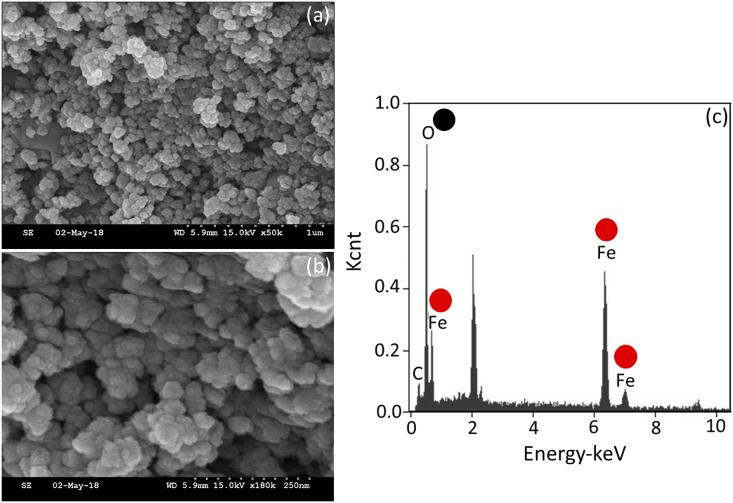

The chemical composition and the surface electronic states of the synthesized IONPs were analyzed by XPS analysis (Figure 5(a)–(c)). The XPS graph confirms the presence of Fe, O, and C with the binding energies of Fe2p3/2 and Fe2p1/2 states located at 710.79 and 725.07, respectively. These peaks indicated the structural configuration of FeO(OH) and Fe3O4. These peaks show that “Fe” is divalent, and the peak at 725.07 eV is found as the transition to Fe2p1/2, associated with the main peak of Fe 2p3/2 at 710.79 (Figure 5(a) and (b)). The O1 s state binding energy at 530.18 is attributed to the occurrence of O-H and R-O and Fe(OH)3 and FeO(OH) (Figure 5(c)). The peak at 284.6 eV confirms the occurrence of C-O in the synthesized IONPs.
39
On the IONP surface, the carbon atoms might form sp2 and sp3 hybridizations and bond to oxygen groups [carbonyl (C=O), hydroxyl (C–OH), and carboxyl (C–OOH)] resulting from oxygen adsorption. On the other hand, functionalization of IONPs is expected to provide an increased number of carbon groups from biomolecules. These carbon oxygen bonds in the O 1 s spectrum are due to the oxidation of the carbon layer and surface functionalization. The C 1 s and O 1 s spectra indicate the carbon and oxygen states of the components with the adsorbed molecular layers. O− and OH− species may adsorb as molecular forms and receive a negative charge on the surface, which occurs during the oxidation of Fe.
40
X-ray photoelectron spectroscopy analysis of IONP. (a) Survey scan of IONP. Fe2p, O1 s, and C1 s peaks were identified. (b) Profile for Iron peaks. Peaks at 710 eV and 725 eV confirm the occurrence of Fe2p. (c) Profile for oxygen peak. Peak at 530 eV confirms the presence of Oxygen. (d) X-ray diffraction analysis diffraction peaks analysis. The predominant peaks are at (104), (018), and (214). IONP: iron oxide nanoparticles.
Further confirmation was carried out by X-ray diffraction analysis to reveal the crystalline peaks. Based on the analysis, three major peaks were noticed at planes (104), (018), and (214). In general, IONPs display five diffraction peaks to elucidate the crystallinity, and the current study has proven three peaks (Figure 5(d)).
Cyclic citrullinated peptide immobilization process through iron oxide nanoparticles
CCP was attached to IDE through IONPs with amine-aldehyde chemical interactions. Figure 6(a) shows the current flow for the CCP attachment process on the IDE electrode. As displayed in the figure, when amine-IONPs were dropped on the OH-treated electrode, the current response was noted as 9.85E-12 A, and after linking with glutaraldehyde, it increased to 3.49E-11 A. This current increment confirms the linker formation of amine with aldehyde, and then with further addition of CCP, the current was drastically increased to 1.06E-10 A (Figure 6(b)). This current enhancement was due to the binding of CCP on the immobilized glutaraldehyde. Since a higher number of APTES was attached through IONPs, it attracted higher glutaraldehyde immobilization followed by CCP. Furthermore, it was proven by various researchers that probe immobilization with nanoparticles gives a proper orientation of probe molecules on sensing surfaces, facilitating the binding of a higher number of target molecules.41–43 Here, IONPs help to attach CCP on the IDE electrode, which indeed aids in quantifying the lower level of anti-CCP antibody. (a) Surface immobilization process of CCP on interdigitated electrode. Current changes for each immobilization step were recorded. Increasing current was noted after each immobilization. (b) Difference in current between each immobilization step. A higher current was noted after immobilizing the CCP on glutaraldehyde. CCP: cyclic citrullinated peptide.
Determination of anti-cyclic citrullinated peptide
Anti-CCP antibody was quantified on the IONP-CCP-modified IDE electrode. Before identification, the chemically modified sensing electrode was covered with the blocking agent ethanolamine to reduce the signal-to-noise ratio. After dropping BSA, the current was slightly increased from 1.06E-10 to 1.11E-10 A, and then different concentrations of anti-CCP were dropped (Figure 7(a) and (b)). As shown in Figure 7(a), after dropping 4 pg/mL anti-CCP, no significant changes in current were noticed. Furthermore, upon adding 8 pg/mL, the current was increased from 1.06E-10 to 1.18E-10 A, and then with increasing anti-CCP concentrations to 15, 30, 60, 125, and 250 pg/mL, the current responses were increased to 1.43E-10, 1.6E-10, 1.82E-10, 2.19E-10, and 2.89E-10 A, respectively. Clearly, it was noted that with increasing concentrations of anti-CCP, the current responses also gradually increased (Figure 7(b)). The differences in current were plotted in Excel, and the limit of detection was calculated. As shown in Figure 7(b) (inset figure), the detection limit of the anti-CCP interaction with CCP was calculated to be 15 pg/mL with a regression coefficient (R2) of 0.9637. (a) Titrating anti-CCP on CCP-modified surfaces. Anti-CCP concentrations from 4 to 250 pg/mL were allowed to interact with CCP. Clear current changes were noted for each CCP concentration. Current levels for the interaction of anti-CCP with CCP. With increasing anti-CCP, current levels gradually increased. (b) Difference in current levels for the interaction of anti-CCP with CCP. Plotted in an Excel sheet and calculated the limit of detection as 15 pg/mL (inset). CCP: cyclic citrullinated peptide; anti-CCP: anti-cyclic citrullinated peptide.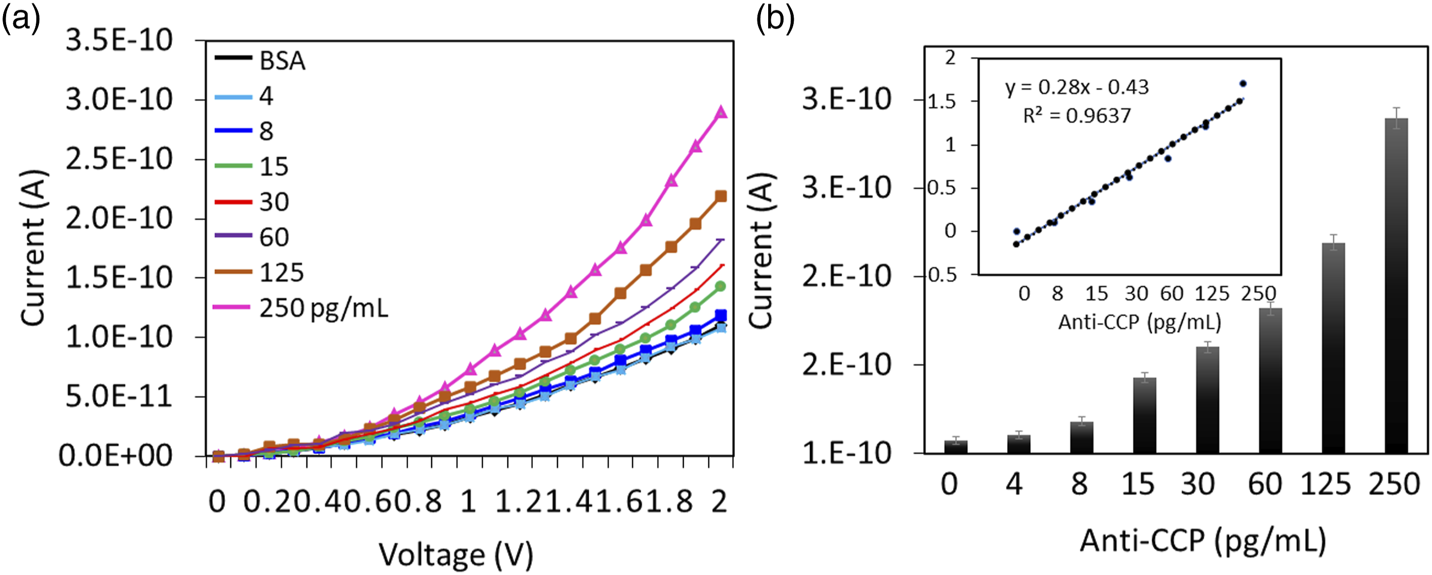
Anti-cyclic citrullinated peptide and cyclic citrullinated peptide interaction in the presence of human serum
Anti-CCP was spiked in diluted human serum to identify the performance of the detection strategy in real biological samples. As shown in Figure 8(a), with all concentrations of anti-CCP, current responses were increased when the anti-CCP levels were increased. At the same time, control experiments with nonspecific proteins and nonimmune antibodies failed to interact with CCP, confirming the specific interaction of CCP with anti-CCP (Figure 8(b)). Furthermore, it was noticed that the current response of anti-CCP was much higher with IONPs than with the surface without IONPs due to the higher immobilization of anti-CCP followed by CCP interaction on the sensing electrode (Figure 8(b)). In the presence of IONP two-fold increment in the output signal was noticed due to higher number of molecular assemblies with an enhanced surface area. The variations with different measurements are displayed by the averaged error values. The reproduced data on three different electrode surfaces were displayed in Figure 9 and no significant changes were noticed with all surface modifications or molecular interactions. The device designed is cheaper, and the surface can be reused depending on the probe utilized. Based on our preliminary measurements, if antibody can be removed by a denaturing agent, which will not affect the surface, such as mild urea treatment, the surface can be reused at least three times. The reproducibility test with the surface modifications/interactions was carried out with three devices fabricated from the same batch and averaged (n = 3). (a) Selective detection of anti-CCP. Neither the nonimmune antibody nor the control protein showed current changes. (b) Spiking with detection of anti-CCP. Anti-CCP was spiked in human serum. Spiked anti-CCP did not affect the detection, indicating the selective detection of anti-CCP. Furthermore, with iron oxide nanoparticles, the current response was noted for the specific anti-CCP and cyclic citrullinated peptide interaction. Anti-CCP: anti-cyclic citrullinated peptide. Reproducibility on sensing surface. Three different electrode surfaces from the same batch of fabrication were tested. No significant variations were found with all the surface changes.

Conclusions
Rheumatoid arthritis is an inflammatory autoimmune disease that damages the joint, causes pain and disability, and affects other physical functions. This work was focused on detecting anti-CCP antibody (anti-CCP), which has been found to be an established biomarker for rheumatoid arthritis. CCP was utilized as the probe and immobilized on the IDE sensing surface through IONPs, which were synthesized by a greener method. Images of IONPs were captured by FESEM and FETEM analyses, and the chemical structures of Fe, O, and C were confirmed by XPS analysis. CCP was attached to IDE through IONP by using the chemical linkers APTES and glutaraldehyde. Anti-CCP was detected on CCP-modified surfaces with good sensitivity and limits of detection of 8 and 15 pg/mL, respectively. Control experiments revealed the selective detection of anti-CCP and by spiking of anti-CCP in human serum. This method of anti-CCP quantification helps to diagnose the early stages of rheumatoid arthritis.
Footnotes
Declaration of conflicting interests
The author(s) declared no potential conflicts of interest with respect to the research, authorship, and/or publication of this article.
Funding
The author(s) received no financial support for the research, authorship, and/or publication of this article.