
Abstract
Polymerized polypeptide nanomicelles have attracted much attention as novel drug carriers because of their good biocompatibility and degradability. To prepare doxorubicin (DOX)-loaded nanomicelles, an amphiphilic peptide, FFHFFH-KKGRGD (P12), was synthesized by solid-phase synthesis, and the physicochemical and drug-release properties, as well as the cytotoxicity of the nanomicelles, were evaluated in vitro. The P12-DOX polymer micelles were prepared by dialysis. The morphology and particle size were characterized by transmission electron microscopy and dynamic light scattering. The critical micelle concentration (CMC) of the polymer was determined by the probe method, and the drug-release characteristics of the micelles were studied by dynamic dialysis. The cytotoxicity and uptake of the P12-DOX micelles were evaluated against mouse breast cancer cells (4T1) and human umbilical vein endothelial cells. The peptide polymer micelles containing DOX were uniformly sized and had a spherical core–shell structure with an average particle size of 128.6 nm. The CMC of the polymer was low (0.0357 mg/mL). The in vitro release of DOX from the micelles is slow and is consistent with first-order kinetics. The copolymer micelles of the P12 polypeptide and DOX can be used as nanoscale spherical carriers of hydrophobic drugs and have broad applicability.
Introduction
The application of nanoparticle drug-delivery systems has attracted widespread attention in the field of tumor diagnosis and treatment. With their unique function and design, there have been significant advances in the application of nano-drug-delivery systems in eliminating malignant tumors. Many drug-delivery systems with high tumor-targeting ability, sensitivity to the tumor microenvironment, and high efficacy have been developed in the field of tumor nanomedicine. 1 A variety of nanocarriers have been developed and approved for antitumor drug targeting. These nanocarriers, namely liposomes, micelles, nanotubes, dendrimers, and peptides, have a wide range of advantages, such as high selectivity, versatility, specificity, biocompatibility, and precisely controlled drug release. 1 Polypeptides, as recently developed carrier materials, have attracted great interest in nanoscience and materials science because of their good biocompatibility and degradability. 2 –4 Scientific research shows that compared with inorganic metal materials, peptide-coated palladium nanoparticles have significantly improved their stability and water-solubility characteristics and can be used for highly sensitive biological analysis of trypsin in human urine samples. 5 Gold nanoparticles and quantum dots modified with pentapeptides Cys-Ala-Leu-Asn-Asn as ligands also have very good stability and can be used for a variety of bioanalyses. 6,7 In particular, as one of the many types of polypeptides, amphiphilic polypeptides have excellent self-assembly properties and can self-assemble into “core–shell” nanomicelles with hydrophilic shells and hydrophobic cores in aqueous solutions. 8 –10 Because of the hydrophobic cores, micelles can contain water-insoluble drugs and significantly improve their bioavailability. 9 In addition, micelles can significantly delay drug release, improve drug stability, and reduce toxicity under normal physiological conditions. 9,11 –13 Studies have shown that self-assembled peptide nanomaterials have a wide range of applications, including in gene delivery, bioimaging, drug transportation, and hemostasis. 7,14 –16 Since polypeptides are biodegradable in vivo and have been shown to have no toxic or immunogenic effects at lesion sites, research on drug-delivery systems based on amphiphilic polypeptides has received increasing attention. 14,17 For example, Gelain et al. studied amphiphilic polypeptides, such as A6D and V6D, composed entirely of amino acid residues, and the results showed that these small peptides could self-assemble into nanotubes and vesicles. 18 Liang et al. synthesized three amphiphilic peptides and studied their self-assembly behaviors and encapsulation performances. 19 Their results showed that these peptides could self-assemble into drug-encapsulating nanospheres. 20
Doxorubicin (DOX) is an antitumor antibiotic that inhibits the synthesis of RNA and DNA. 21,22 Its strongest inhibitory effect is against RNA, and it has a broad spectrum of antitumor activity, making it effective against a variety of tumors. 23,24 DOX is a periodic nonspecific drug that causes tumor cell death during various stages of growth. 25 In clinical practice, DOX is mainly used for the treatment of breast cancer, lung cancer, liver cancer, and stomach cancer, but DOX is chemically unstable, poorly soluble in water and prone to hydrolysis, photolysis, and other changes, which seriously limit its clinical application and treatment efficacy. 26 In this article, DOX was selected as the model drug, and self-assembled amphiphilic peptides (N-Phe-Phe-His-Phe-Phe-His-Lys-Lys-Gly-Arg-Asp-C, P12) were synthesized by solid-phase synthesis. Then, DOX-loaded polymer micelles were prepared by dialysis. The physicochemical and drug-release properties of these polymer micelles were investigated, and their antitumor effects in vitro were evaluated.
Experimental
Materials and main reagents
DOX hydrochloride (analytical-reagent grade): Nanjing Dilger Medical Technology Co. Ltd, China; amino acids and 2-chlorotrityl chloride resin: Hefei Guopeptide Biotechnology Co. Ltd, China; methanol, piperidine, ninhydrin, 1-hydroxybenzotriazole (HOBt), N,N-diisopropyl ethylamine (DIEA), 4′,6-diamidine 2-phenylindole (DAPI), N,N-diisopropylcarbodiimide (DIC), dichloromethane (DCM), dimethylformamide (DMF), ethyl ether, trifluoroacetic (TFA), and triethylamine (analytical-reagent grade): Sinopharm Chemical Reagent Co. Ltd, China; and CCK-8 cell proliferation and cytotoxicity detection kits: Saimo Feishier Technology Co. Ltd, China.
The synthesis of the linear amphiphilic peptide P12
The peptide P12 was prepared using an Fmoc solid-phase peptide synthesis method. 27 First, 2-chlorotrityl chloride resin was added to 15 mL of DCM and allowed to swell for 3 min. Then, 15 mL of a pre-prepared coupling solution containing 0.55 g of Fmoc-Phe-OH, 0.45 g of HOBt, 1.5 mL of DIEA, and 1.5 mL of DIC was added to the swollen 2-chlorotrityl chloride resin for 1.5 h. A Kaiser test was then performed to confirm that the amino acid and resin were completely coupled. The unreacted active chloride groups were reacted with methanol and DCM for 30 min. After washing the resin with DMF several times, the Fmoc group at the amino end of the peptide was removed by treatment with 20% piperidine for 20 min. Then, a Kaiser test was carried out to confirm the exposure of the –NH2 groups. The resin was washed with 15 mL of DMF several times. After the resin was thoroughly washed, the next amino acid was condensed onto the resin until all the desired amino acids had been installed; the last amino acid of P12 was Fmoc-Asp(OtBu)-OH. The Fmoc protecting groups on the coupled compound were cleaved by treatment with 20% piperidine for 30 min, and the coupling reagent was removed by washing with 20 mL of CH3OH for 5 min. Finally, the resin was treated with TFA solution (volume ratio of TFA:water is 95:5) with shaking for 2 h, which simultaneously released the polypeptide from the resin and deprotected the side chains. The insoluble crude peptide product was obtained after cleaning with ice-cold ethyl ether.
Purification of peptide P12 by HPLC
The crude peptide product was dissolved in water, filtered through a 0.45-µm filter membrane, and purified by RP-HPLC. The chromatographic conditions were as follows: Sephadex G-100, 5 μm, 1 × 18 cm column, Eluant A: 0.1% TFA/H2O, Eluant B: 0.1% TFA/80% acetonitrile-H2O (v/v), gradient elution 20 → 80% B, 20 min, flow rate 1.0 mL/min, and detection wavelength λ = 214 nm.
Mass spectrometric and NMR analysis of peptide P12
Liquid chromatography-mass spectrometry (LC-MS) was used to determine the molecular weight of peptide P12. The spectrometry conditions were as follows: ESI(+) ion source, target product scanned with an SIM pattern, scanning range (m/z) of 800–600, interface temperature of 300°C, DL temperature of 250°C, heat block temperature of 200°C, drying gas flow rate of 15 L/min, capillary voltage of 4500 V, and detection voltage of 1960 V.
An Avance Neo NMR spectrometer from Bruker (Germany) was used to acquire the proton nuclear magnetic resonance ( 1 H NMR) and carbon nuclear magnetic resonance ( 13 C NMR) spectra of the P12 peptides. The NMR analytical conditions were as follows: the sample was dissolved in D2O. The height of the solvent in the NMR tube was approximately 3–4 cm. The amount of sample required for the acquisition of the hydrogen spectrum was approximately 5–10 mg and that for the acquisition of the carbon spectrum was more than 20 mg. The sweeping range of NMR was set at 1 H: −2 to 13 ppm, 13 C: −20 to 230 ppm, Spectrometer frequency (1H): 600 MHz, and Spectrometer frequency (13C): 150 MHz.
Determination of the CMC of peptide self-assembly
A 0.125 mg/L solution of pyrene in acetone and a 0.4 g/L solution of peptide P12 in double-distilled H2O (ddH2O) were prepared as stock solutions. Then, 50 μL of the pyrene solution was added into 8 25-mL conical flasks. After the acetone was completely evaporated, 10 mL of peptide P12 dispersions at concentrations ranging from 0.00625 g/L to 0.2 g/L in ddH2O was added to the conical flasks. The mixtures were ultrasonicated for 30 min, incubated in a 60°C water bath for 30 min, and finally incubated at a constant temperature of 40°C for 12 h, all while being shielded from light. Then, with the longest ultraviolet absorption wavelength of P12 peptide (258 nm) as the excitation wavelength, the fluorescence spectra ranging from 260 nm to 320 nm were recorded to detect micelle formation under different peptide concentrations using a fluorescence spectrophotometer (RF-5301PC, Shimadzu, Japan). The ratios of the fluorescence intensities at 260 and 262 nm were calculated (I 262/I 260), and a plot of the fluorescence intensity ratios as a function of the logarithm of the copolymer concentration was prepared. The intersect was the critical micelle concentration (CMC) value, and the ratio of I 262/I 260 obviously increased with concentration.
Preparation of the P12-DOX micelles
The DOX-loaded P12 micelles were prepared via dialysis. 28 Under ultrasonication, 30 mg of P12 and 5 mg of DOX were dissolved in 3 mL of dimethyl sulfoxide and dialyzed against ddH2O in a MWCO of 8 kDa dialysis bag at 37°C for 12 h to remove the organic solvent and unloaded DOX, forming P12-DOX micelles. The micelles were collected for subsequent analysis by centrifugation.
Investigation of the encapsulation efficiency and drug-loading ratio of P12-DOX
The encapsulation efficiency and drug-loading ratio of P12-DOX were calculated using the following equations 29 :


where W e is the amount of drug in the micelles, W t is the initial amount of drug, and W m is the total mass of micelles.
Detection of the morphology and particle size of the P12-DOX micelles
The P12-DOX micelles were distributed onto the copper mesh and dyed with 1% phosphotungstic acid solution for 15 min. Then, the morphology of the P12-DOX micelles was observed by transmission electron microscopy (TEM; Hitachi 770, Japan), with an accelerating voltage of 120 kV. The particle size and distribution of the P12-DOX micelles were determined at a wavelength of 633 nm and a temperature of 25°C by a Zetasizer Nano-ZS 90 (UK).
Detection of the release profile of the P12-DOX micelles in vitro
The in vitro release profile of the P12-DOX micelles was assessed by the dialysis bag diffusion method. 20 A series of 1 mg/mL solutions of P12-DOX were prepared in phosphate buffer at pH 1.2, 4.5, and 6.8, and then 2 mL of each solution was transferred to a dialysis bag (MWCO = 8000 Da), which was then placed in a flask with 20 mL of phosphate-buffered saline (PBS) at pH 1.2, 4.5, and 6.8, respectively. Then, the flasks were placed on a shaker at 37°C and 100 r/min, and 2 mL of the PBS outside the dialysis bag was removed at different intervals and immediately replaced with 2 mL of fresh dissolution medium. An ultraviolet spectrophotometer (754 PC; APL, Shanghai, China) was used to measure the absorbance of the 2-mL aliquot of dialysate at 485 nm. The intensity was calculated based on the equation of its standard curve y = 0.00879x + 0.023333 with R2 0.99877. Then, the amount of drug released from the P12-DOX micelles in different pH environments was plotted.
Cytotoxicity assessment of the P12-DOX micelles
The cytotoxicity of the P12-DOX micelles was determined using a CCK-8 assay against mouse breast carcinoma cells (4T1 cells) and against human umbilical vein endothelial cells (HUVECs). The two kinds of cryogenic cells were removed from a liquid nitrogen container and placed in a 37°C sink to allow them to thaw. Then, the cells were suspended and washed with 2 mL of complete medium containing RPMI-1640, 10% fetal bovine serum, and 1% penicillin–streptomycin. The settled cells were resuspended in complete medium, placed onto a tissue culture dish and cultured in a CO2 culture box at 37°C with 5% CO2. The following experiments were carried out when the cells were in excellent condition. The 4T1 cells and HUVECs were incubated until 90% of the cells adhered and then digested with 1 mL of 0.25% trypsin in a culture box for 2 min. The suspended cells were collected by centrifugation at 800 r/min for 5 min, and then the collected cells were resuspended in complete medium and counted with a hemocytometer. The tested cells were diluted to 1 × 10⁵ cells/mL with complete medium. Then, 100 μL of the tested cell suspension was transferred onto a 96-well plate (approximately 10,000 cells/well) and cultured in a CO2 culture box until the cells adhered. After the supernatant was removed, P12, P12-DOX, and DOX solutions at six concentrations (50, 25, 12.5, 6.25, 3.125, and 0 μg/mL) and 100 μL of fresh complete medium were added to the 96-well plate. The samples were cultured in a CO2 culture box for 24 h. Then, the supernatant was removed again, and another 100 μL of fresh complete medium containing 0.10% CCK-8 was added to the 96-well plate, and the samples were cultured in a CO2 culture box at 37°C for another 4 h. Then, the absorbance of each well at 450 nm was detected with a full-featured microplate reader (Synergy NEO, USA), and the cell survival rates under different treatment conditions were calculated.
Cellular uptake experiments with the P12-DOX micelles
The cellular uptake of P12-DOX micelles was observed by laser scanning confocal microscopy. The tested 4T1 cells and HUVECs were inoculated into a 24-well plate (2 × 10⁴ cells/well) and cultured in a CO2 culture box at 37°C and 5% CO2 for 24 h until the cells adhered. Then, the supernatant was removed, and the adhered cells were washed with PBS twice to remove cells that were dead or not tightly attached. Then, the P12-DOX micelles were added to the test cells and cultured in a constant-temperature CO2 culture box for 4 h. After culturing, the supernatant was removed, and the cells were washed twice with PBS and then fixed with 10% formalin for 15 min. Subsequently, the tested cells were washed again with PBS and dyed with DAPI (to stain the nuclei) for 15 min. Laser scanning confocal microscopy (FV1200; OLYMPUS, Japan) was used to determine uptake.
Results and discussion
The purification and analysis of the P12 peptide by HPLC, LC-MS, and NMR spectroscopy
HPLC was used to purify the target peptide (P12). The retention time of P12 was 10.8 min (Figure 1), and its purity was approximately 96.46%. Its mass spectrum showed a peak for [M+H]+ at 1523.75 (Figure 2), which was consistent with the theoretical mass of 1522.75 g/mol. Its diagnostic fragment ions are m/z 508.55 and 762.4. Therefore, the target peptide (P12) was synthesized.

HPLC purity assessment of the synthesized P12 peptide.
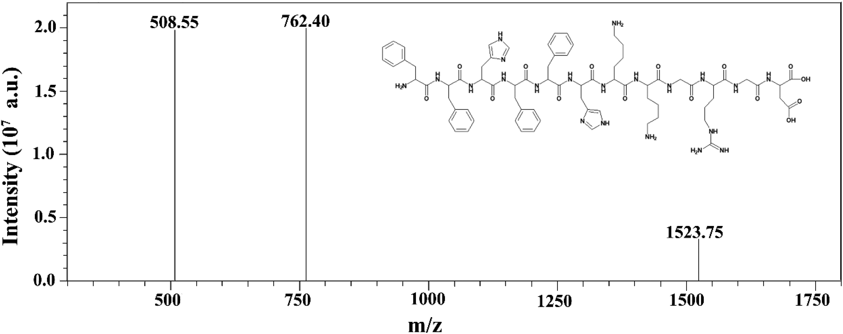
Mass spectrum of the P12 peptide and its molecular structure.
The chemical formula of the peptide is C74H99N21O15, and its theoretical molecular weight is 1522.75 g/mol. The peptide contains 74 carbon atoms and 99 hydrogen atoms, including 25 active hydrogen atoms. NMR spectroscopy indicated the presence of another 74 hydrogen atoms. The 74 hydrogen atoms were divided into 5 types. According to the order of their chemical shifts, they were divided into Ar-
1 H (600 MHz) NMR data for the P12 peptide in D2O.a
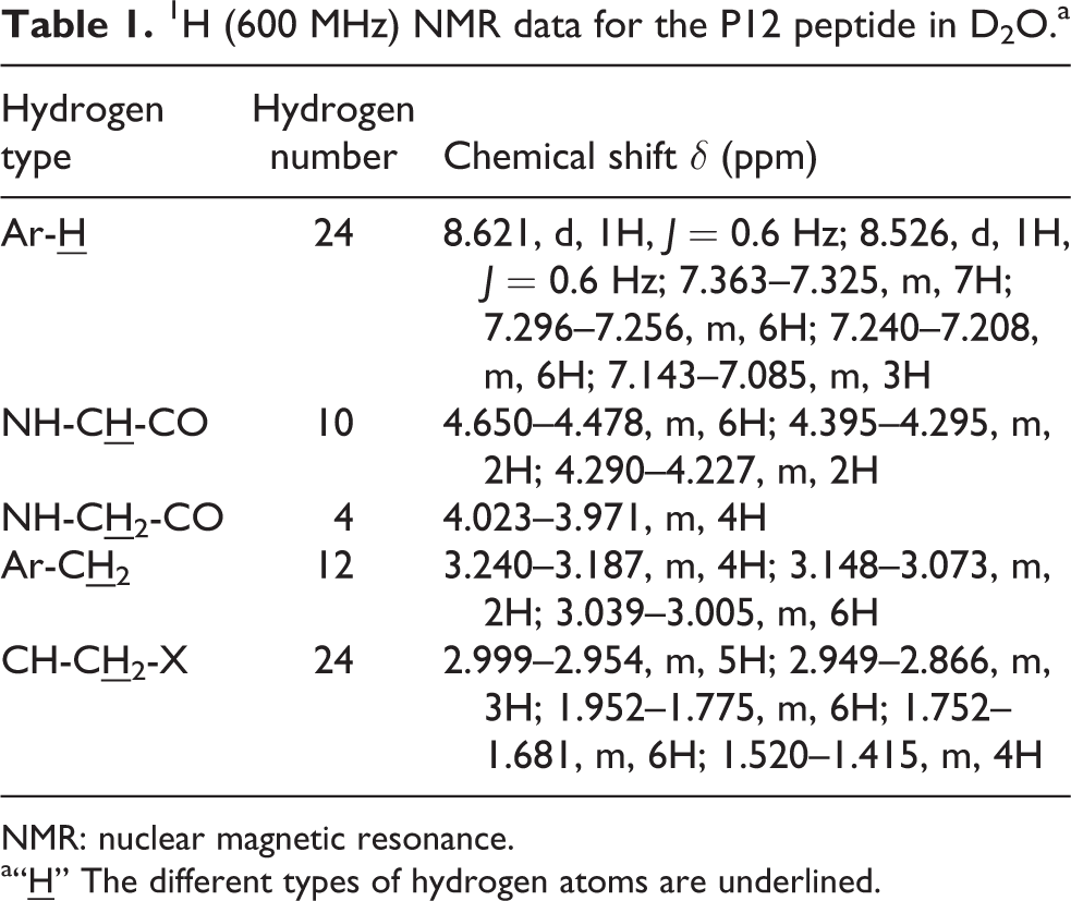
NMR: nuclear magnetic resonance.
a“
13C (150 MHz) NMR data for the P12 peptide in D2O.

NMR: nuclear magnetic resonance.
Detection of the CMC of the P12 peptide
The P12 peptide was composed of a hydrophobic A6 block and a hydrophilic A6 containing an RGD block, allowing it to self-assemble into micelles in aqueous solutions. The CMC value of the micelles of the P12 peptide was determined by fluorescence spectrometry with pyrene as the hydrophobic probe. 30 As shown in Figure 3(a), the intensities of the fluorescence emission peaks increased as the concentration of the P12 peptide solution increased. The increase in intensity was accompanied by a slight redshift, and the most intense peak shifted from 260 nm to 262 nm. The above phenomenon confirmed the formation of the micelle structure of the P12 peptide because the hydrophobic probe (pyrene) entered the hydrophobic core of the micelles from the external hydrophilic medium. As shown in Figure 3(b), the x-coordinates were the logarithm of the peptide concentration in Figure 3(a), and the corresponding y-coordinates were the ratio of I262/I260, and the I262/I260 ratio changed slightly at low concentrations. When the concentration of the peptide P12 increased above a certain point (0.0357 mg/mL), the ratio of I262/I260 increased dramatically, indicating that pyrene had entered the hydrophobic core of the P12 micelles. The CMC value was calculated based on the intersection of the extrapolated fitting lines to be approximately 36 mg/L, corresponding to a molar concentration of approximately 22 μM, which is less than the CMC of 45 μM for the self-assembly of amphiphilic antimicrobial peptides reported in the literature. 31 This result indicates that the P12 micelles can potentially serve as a delivery vehicle for hydrophobic drugs.

Fluorescence emission spectra of pyrene with increasing concentrations of the peptide (a) and CMC (b). CMC: critical micelle concentration.
Detection of the drug-loading ratio and encapsulation ratio of the P12-DOX micelles
To quantitatively analyze DOX during the experiment, a standard curve of DOX was prepared using a series of solutions with different concentrations of DOX at 485 nm. There was a good linear relationship between the concentration and the UV absorption spectra of DOX over a certain concentration range. As shown in Figure 4(a), the equation of the standard curve was y = 0.00879x + 0.023333 with R2 0.99877.

The standard curve of DOX (a) and the effect of different feed ratios on the encapsulation performance of the peptide (b). DOX: doxorubicin.
As shown in Figure 4(b), as the concentration of P12 peptide increased, the encapsulation efficiency of DOX increased gradually. However, when the ratio of P12 peptide to DOX exceeded 6:1, the encapsulation efficiency of DOX began to decrease, probably due to the low proportion of DOX encapsulated by the micelles. The drug-loading ratio first increased and then decreased with increasing P12 peptide content. When the ratio of P12 peptide to DOX was 4:1, the drug loading of micelles reached 31.00 ± 1.58%, and the encapsulation efficiency was approximately 32.41 ± 1.56%. This result is approximately three times the drug-loading ratio of the amphiphilic peptide drug carriers reported in the literature of 10%. 5
Investigation of the morphology of P12 and P12-DOX nanomicelles
The morphology of the P12 and P12-DOX nanomicelles was observed by TEM under neutral conditions (pH 7.0) with a P12 peptide concentration of 0.5 mg/mL. The P12 peptide self-assembles through hydrophobic interactions to form P12 nanomicelles (Figure 5(a) and (b)) and P12-DOX nanomicelles loaded with DOX (Figure 5(c) and (d)). Both the particles of P12 and the P12-DOX nanomicelles are relatively uniform in size, and the average diameters of both P12 and the P12-DOX nanomicelles indicated by TEM are smaller than the sizes determined by DLS (105.2 nm and a PDI of 0.213 for P12 and 128.6 nm and a PDI of 0.257 for P12-DOX). The slight difference between the TEM observations and the DLS measurements may be due to the material being in different states for these two measurements. The former measures the micelle powder (solid state), and the latter determines the particle size of the micelles in solution. In addition, the P12 and P12-DOX nanomicelles may stick together or aggregate in solution, resulting in an uneven distribution of nanomicelle diameters and an increase in the observed particle size.

TEM images of P12 and P12-DOX nanomicelles (a) and (c) and particle sizes of the nanomicelles by DLS (b) and (d), respectively. TEM: transmission electron microscopy; DOX: doxorubicin.
Analysis of the drug-release properties of P12-DOX in vitro
The drug-release characteristics are a key parameter in the preparation of drug carriers. The World Health Organization has clear definitions, standards, and methods for evaluating drug release or dissolution in drug-delivery systems. 32 DOX is a hydrophobic drug that occupies the hydrophobic core of the P12 nanomicelles. We simulated the DOX release characteristics of the P12-DOX nanomicelles in three different pH environments: gastric juice (pH 1.2), intestinal juice (pH 6.8), and tumor environment (pH 4.6). 8 As shown in Figure 6, the DOX release was dependent on the pH of the environment. The P12-DOX nanomicelles show a sudden release phenomenon at the beginning of drug release. The DOX in the nanomicelles may not be evenly distributed and some may be partially adsorbed on the surface of the microspheres and are thus easily released. Slow release began after approximately 20 h, and the cumulative release percentages at 60 h were 81.4% (pH 1.2), 62.0% (pH 4.6), and 37.9% (pH 6.8), indicating that the nanomicelles can facilitate sustained release of the drug. More acidic conditions resulted in greater cumulative release of DOX over the same period, indicating that the P12-DOX nanomicelles are somewhat sensitive to acid, which is consistent with the acidic internal environment of tumor cells. 33

The release profiles of the P12-DOX nanomicelles at different pH values. DOX: doxorubicin.
The in vitro cytotoxicity and cellular uptake
To evaluate the feasibility of the P12-DOX nanomicelles as a drug-delivery vehicle for the treatment of tumors, a CCK-8 assay was used to evaluate their cytotoxicity. Using P12 and DOX as experimental controls, we studied the toxic effects of P12, DOX, and P12-DOX nanomicelles at different concentrations on 4T1 cells and HUVECs. As shown in Figure 7(a) and (b), P12 had almost no effect on the growth of either tumor cells or normal cells, indicating that the P12 peptide had good biocompatibility, providing a theoretical basis for its further application. At the same drug concentration, the survival rate of cells treated with P12-DOX micelles was higher than that of cells exposed to DOX alone, indicating that the P12-DOX nanomicelles reduced the cytotoxicity of DOX, especially its cytotoxicity to HUVECs at a concentration of 50 μg/mL. In addition, the drug has a concentration-dependent therapeutic effect on tumors. The survival rate of 4T1 cells exposed to P12-DOX micelles was lower than that of HUVECs, indicating that the system can target tumor cells.

Cell viability of 4T1 cells (a) and HUVECs (b) after incubation with P12, DOX, and P12-DOX. DOX: doxorubicin; HUVEC: human umbilical vein endothelial cell.
To further investigate the function of the P12-DOX nanomicelles as potential drug carriers, we observed the uptake of P12-DOX nanomicelles by tumor cells and normal cells by laser confocal microscopy. 8 As shown in Figure 8, the normal cells and tumor cells showed different degrees of uptake of P12-DOX, but due to the specific binding ability of the RGD sequence in the P12 polypeptide to the alpha-v beta-3 receptor on tumor cell membranes, the tumor cells are better able to take up P12-DOX and more of the drug enters the cell.

Uptake of P12-DOX nanomicelles by HUVECs (a) and 4T1 cells (b) (the blue signals correspond to nuclei and the green signals correspond to DOX). DOX: doxorubicin; HUVEC: human umbilical vein endothelial cell.
Conclusion
The amphiphilic peptide P12 was successfully synthesized by solid-phase synthesis. The peptide can self-assemble to form nanomicelles in aqueous solutions and encapsulate the hydrophobic drug DOX, suggesting its potential as an antitumor drug-delivery system to improve the therapeutic effect of the drug on tumor cells. Furthermore, P12-DOX nanomicelles allowed the sustained and controlled release of DOX and showed a certain degree of pH-responsive release.
As a potential drug-delivery vehicle, P12 had a suitable CMC and was taken up by tumor cells through specific binding to tumor cells. This result indicated that P12-DOX has practical significance, as it could be used to improve the curative effects of existing drugs on tumor cells, making it broadly applicable. Further studies on the ability of P12-DOX micelles to target tumor cells in animals are underway.
Footnotes
Author contributions
These authors contributed equally to this work: Ping Song, Wanzheng Li.
Declaration of conflicting interests
The author(s) declared no potential conflicts of interest with respect to the research, authorship, and/or publication of this article.
Funding
The author(s) disclosed receipt of the following financial support for the research, authorship, and/or publication of this article: This work was supported by the National Natural Science Foundation of China (NSFC) [grant number 31671797], Anhui Natural Science Foundation [grant number 1808085QC85], Natural Science Foundation of Anhui University [grant numbers KJ2017A123, KJ2018A0117, and TSKJ2017B15], Anhui Province Major Scientific and Technological Projects [grant number 17030701014], and Youth Talent Support Program of Anhui Polytechnic University [grant number 2016BJRRC006].