
Abstract
Rheumatoid arthritis (RA) is a systemic autoimmune disease affecting 0.5–1% of the worldwide population. Whilst predominantly causing chronic pain and inflammation in synovial joints, it is also associated with significant extra-articular manifestations in a large proportion of patients. Among the various pulmonary manifestations, interstitial lung disease (ILD), a progressive fibrotic disease of the lung parenchyma, is the commonest and most important, contributing significantly to increased morbidity and mortality. The most frequent patterns of RA-associated ILD (RA-ILD) are usual interstitial pneumonia and nonspecific interstitial pneumonia. New insights during the past several years have highlighted the epidemiological impact of RA-ILD and have begun to identify factors contributing to its pathogenesis. Risk factors include smoking, male sex, human leukocyte antigen haplotype, rheumatoid factor and anticyclic citrullinated protein antibodies (ACPAs). Combined with clinical information, chest examination and pulmonary function testing, high-resolution computed tomography of the chest forms the basis of investigation and allows assessment of subtype and disease extent. The management of RA-ILD is a challenge. Several therapeutic agents have been suggested in the literature but as yet no large randomized controlled trials have been undertaken to guide clinical management. Therapy is further complicated by commonly prescribed drugs of proven articular benefit such as methotrexate, leflunomide (LEF) and anti-tumour necrosis factor α agents having been implicated in both ex novo occurrence and acceleration of existing ILD. Agents that offer promise include immunomodulators such as mycophenolate and rituximab as well as newly studied antifibrotic agents. In this review, we discuss the current literature to evaluate recommendations for the management of RA-ILD and discuss key gaps in our knowledge of this important disease.
Introduction
Interstitial lung disease (ILD) is a progressive fibrotic disease of the lung parenchyma. Occurring in association with several connective tissue diseases, it is the commonest and most important pulmonary manifestation of rheumatoid arthritis (RA) [Brown 2007; O’Dwyer et al. 2013]. RA-associated ILD (RA-ILD) may be a consequence of the chronic immune activation and inflammation that occurs in RA and which subsequently promotes aberrant fibroproliferation, or can be due to drug-related or infectious precipitants [O’Dwyer et al. 2013]. RA-ILD contributes significantly to decreased quality of life, progressive chronic disability, high utilization of healthcare resources and poorer mortality, with mean survival under 3 years [Kelly et al. 2014]. The management of ILD in patients with RA is a challenge. Several therapeutic agents have been suggested in the literature but as yet no large randomized controlled trials have been undertaken to guide clinical management. In this perspective, we discuss the current literature to evaluate recommendations for the management of RA-ILD and discuss key gaps in our knowledge of this important disease.
Histopathological and radiographic classification
RA-ILD has well described subtypes that are shared with the idiopathic interstitial pneumonias (IIPs). The four major histopathological and high-resolution computed tomography (HRCT) patterns of RA-ILD are shown in Table 1.
The four major histopathological and HRCT patterns of RA-ILD.
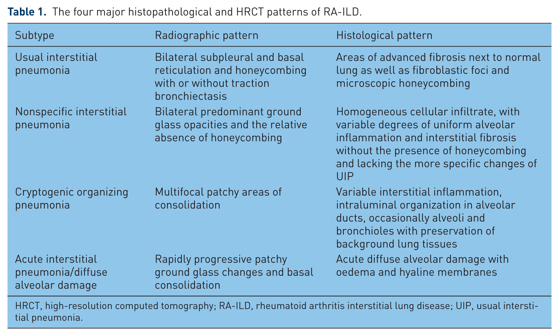
HRCT, high-resolution computed tomography; RA-ILD, rheumatoid arthritis interstitial lung disease; UIP, usual interstitial pneumonia.
The most common patterns found are usual interstitial pneumonia (UIP), accounting for 44–66% and nonspecific interstitial pneumonia (NSIP) (24–44%), followed by mixed disease (0–12%). Cryptogenic organizing pneumonia (COP) and acute interstitial pneumonia (AIP/diffuse alveolar damage (DAD)) are uncommon (0–11%), while lymphocytic interstitial pneumonia and desquamative interstitial pneumonia are rare [Tanaka et al. 2004; Lee et al. 2005; Yoshinouchi et al. 2005; Kelly et al. 2014].
A simple staging system for the extent of systemic sclerosis related ILD was proposed [Goh et al. 2008] and this has been shown to also correlate well with RA-ILD [Sathi et al. 2011]. Goh and colleagues’ system defines the extent of disease as limited if less than 20% of the lung parenchyma is affected and as extensive otherwise. Intermediate values (10–30%) are categorized by the forced vital capacity (FVC), with FVC values greater than 70% indicative of limited disease and vice versa [Goh et al. 2008].
Epidemiology
Incidence and prevalence
The reported prevalence of RA-ILD varies depending on the characteristics of the population selected, the definitions and criteria for diagnosis used, as well as the diagnostic tools employed. Prevalence has been reported in the range from 3.6% to 60% [Ayhan-Ardic et al. 2006; Bongartz et al. 2010; Richman et al. 2013; Kelly et al. 2014].
Annual incidence of RA-ILD is reportedly as high as 4.1 per 1000 people [Koduri et al. 2010]. The estimated lifetime risk of developing ILD for patients with RA is approximately 10% [Bongartz et al. 2010]. Interestingly, the incidence of other extra-articular manifestations (EAMs) for example vasculitis has decreased in the last decade but there has been an apparent increase in reported cases of RA-ILD [Bartels et al. 2010]. However, as O’Dwyer and colleagues argue, over the last decade improved clinical awareness of RA-ILD along with improved survival times (particularly as advancing years is a risk factor for its development) may have contributed to an increase in ILD incidence [O’Dwyer et al. 2013].
Mortality and prognosis
In addition to the increased mortality associated with RA itself, RA-ILD is a significant cause of mortality. The median survival of patients with untreated RA-ILD is approximately only 3 years [Bongartz et al. 2010; Koduri et al. 2010]. Compared with the general population, ILD accounts for 6–13% of the excess mortality of patients with RA and is the second most common cause of premature death after cardiovascular disease [Young et al. 2007; Bongartz et al. 2010]. The increased mortality can be mainly ascribed to respiratory failure due to ILD progression and infective complications of RA.
ILD associated with connective tissue diseases including RA purportedly has a lower mortality than idiopathic ILD [Agusti et al. 1992; Flaherty et al. 2003; Lee et al. 2005; Rajasekaran et al. 2006; Park et al. 2007; Song et al. 2013]. However, others have found no difference in prognosis [Hubbard and Venn, 2002; Kocheril et al. 2005; Kim et al. 2009, 2010; Bongartz et al. 2010]. These discrepancies may be a result of subtle undetected histologic differences, immunosuppressive RA drugs or the systemic effects of chronic autoimmune activation associated with RA [Cavagna et al. 2013].
The major determinants of prognosis appear to be RA-ILD subtype and disease extent. Studies have suggested UIP carries a worse outlook compared with NSIP, COP and overlap syndromes [Lee et al. 2005; Park et al. 2007; Kim et al. 2010; Nakamura et al. 2012; Kelly et al. 2013]. The acute DAD histological pattern has the highest mortality, with median survival time only 0.2 years [Tsuchiya et al. 2011]. More extensive fibrosis or worsening of the extent of disease on HRCT also appears to predict survival, with extensive disease (>20% of lung affected on HRCT) associated with twice the relative risk (RR) of dying compared with limited disease [Sathi et al. 2011; Kelly et al. 2013].
Pathogenesis
The aetiopathogenesis of RA-ILD remains as yet unclarified. Though RA itself is a risk factor for the development of fibrotic lung disease, only a subset of patients with RA develop ILD. A number of multifactorial components may contribute to RA-ILD development. Associated risk factors encompass environmental, serologic, clinical, genetic and drug related.
Demographic factors associated with developing ILD in RA include older age, male sex and longstanding disease [Gabbay et al. 1997; Bongartz et al. 2010; Koduri et al. 2010; Mori et al. 2012; Kelly et al. 2014]. Occupational exposure, for example silica inhalation, contributes to the development of ILD through chronic lung inflammation [Schwarz and King, 2010]. Other risk factors reported include high RA disease activity, high-grade functional impairment and the presence of other systemic features, for example articular erosions and rheumatoid nodules [Bongartz et al. 2010; Koduri et al. 2010; Habib et al. 2011].
A relationship between smoking and an increase in the prevalence of ILD has been identified in several studies [Gochuico et al. 2008; Kelly et al. 2014], with current or previous smoking history reported to confer an increased risk for ILD with an odds ratio (OR) of 3.8 for over 25 pack years [Saag et al. 1996]. However, other reports have found no association [Biederer et al. 2004]. A recent study of 356 patients with RA and pulmonary disease (ILD or airway disease) found the strong association observed in the univariate analysis between smoking history and ILD was not maintained after multinomial logistic regression analysis; this suggests factors other than smoking may trigger ILD occurrence in RA [Mori et al. 2012]. It is important to note RA-ILD can occur in nonsmokers [Ayhan-Ardic et al. 2006].
There is an association between positive rheumatoid factor (RF) and RA-ILD and evidence of a similar link with antibodies to cyclic citrullinated peptides (ACPA) has been found [Turesson et al. 2007; Inui et al. 2008; Aubart et al. 2011; Habib et al. 2011; Mori et al. 2012; Giles et al. 2014; Kelly et al. 2014; Yin et al. 2014; Doyle et al. 2015]. The association is inconsistent with some authors disputing these findings [Bongartz et al. 2007; Inui et al. 2008]; however a recent meta-analysis by Zhu and colleagues has concluded serum ACPA positivity is highly associated with the risk of RA-ILD and idiopathic pulmonary fibrosis among patients with RA, with an OR of 4.7 [95% confidence interval (CI) 2.071–10.572, p < 0.001) [Zhu et al. 2014].
The interaction between smoking, the lungs and RA is intriguing. Citrullinated proteins have been observed in lung tissue obtained from RA-ILD [Bongartz et al. 2007] and in the bronchoalveolar lavage (BAL) fluid of smokers but not in nonsmokers [Klareskog et al. 2006; Makrygiannakis et al. 2008]. Interestingly, both RF and ACPA have been detected in patients with ILD without clinical evidence of RA or to predate subsequent RA, particularly in smokers [Gizinski et al. 2009; Fischer et al. 2012].
This has led to the postulation that site-specific citrullination in the lungs leads to the generation of ACPA, which then promote immune dysregulation and lung abnormalities in patients with RA early in the rheumatoid process, especially in smokers. Cigarette smoke can potentially stimulate protein citrullination in the lung due to increased activity of pulmonary peptidylarginine deaminase, thus triggering human leukocyte antigen (HLA)-DR (shared epitope) restricted immune reactions to autoantigens. These data therefore suggest an aetiology involving an integration of environmental exposures, autoimmunity and genetic susceptibility may result in the development of not only ILD but also RA [Klareskog et al. 2006; Makrygiannakis et al. 2008; Chan et al. 2013]. Clearly, this is not the whole answer as RA-ILD can occur in nonsmokers [Ayhan-Ardic et al. 2006].
It appears likely that genetic predisposition plays a role. Michalski and colleagues observed patients with RA-ILD are more likely to possess specific α1-antitrypsin variants, particularly non-M1M1 α1-antitrypsin phenotypes [Michalski et al. 1986]. Several HLA alleles are associated with an increased susceptibility for RA-ILD, such as HLA-DR4, HLA-B40 and HLA-B54 [Charles et al. 1991; Sugiyama et al. 1994; Mori et al. 2012]; carriage of HLA-DRB1*1501 and *1502 alleles has been implicated with a RR ratio for RA-ILD of 4.02 (p = 0.013) [Migita et al. 2010; Mori et al. 2012]. Similarly, Furukawa and colleagues found DR2 serology (DRB1*15 and *16 alleles) appeared to impart increased risk for ILD in Japanese patients with RA, while HLA-DRB1 ‘shared epitope’ was associated with reduced risk [Furukawa et al. 2012]. However, these associations need to be confirmed by further studies.
Diagnosis
Diagnosis is based on clinical presentation, physical examination and investigations including sputum and blood tests, pulmonary function tests (PFTs), HRCT scan, occasionally BAL and more rarely a lung biopsy. Investigations are crucial for determining the type and severity of lung disease and its change over time. Specific subtypes of IIP have significantly different prognoses and treatment associated with them and therefore an accurate diagnosis is extremely important for optimum management; however the relevance of determining the subtype in selecting therapy for patients with RA-ILD is still to be determined.
Clinical presentation
Whilst some patients with RA-ILD can be asymptomatic, the majority present with progressive exertional dyspnoea, usually with an associated dry cough. On examination, tachypnoea and fine bibasal chest crackles are often evident, with finger clubbing a relative rarity.
Pulmonary function tests
PFTs are a sensitive but relatively nonspecific measure of RA-ILD. The majority of patients with ILD demonstrate physiological abnormalities in both lung volumes and gas transfer, namely a restrictive defect with low total lung capacity (TLC) and low forced vital capacity (FVC), low transfer factor of the lung for carbon monoxide (TLCO) and oxygen desaturation at rest or on exertion [American Thoracic Society/European Respiratory Society, 2002]. Decreased TLCO is reported to be the most sensitive test for predicting the presence of ILD, as well as the extent of disease [Dawson et al. 2001; Hamblin and Horton, 2011]. TLCO also has prognostic value with a low TLCO (<54% of the expected value) predictive of disease progression [Dawson et al. 2002]. However, a recent study comparing baseline vital capacity (VC) and TLCO at presentation with RA-ILD in extensive versus limited disease found that baseline VC was relatively maintained in limited disease but significantly reduced in patients with extensive disease whilst TLCO was reduced in both limited and extensive disease; therefore lung volumes may have greater utility than gas transfer in calculating disease extent at baseline [Chan et al. 2013; Kelly et al. 2013]. In interstitial pulmonary fibrosis (IPF), worsening desaturation (< 88%) is also associated with a poorer prognosis and is useful for guiding oxygen therapy and transplantation [Wells and Ward, 2014]. It should be noted that both TLCO and desaturation with walking can be influenced by the coexistence of emphysema or pulmonary hypertension [Wells and Ward, 2014].
Importantly, PFTs are very valuable for monitoring disease change over time, especially as it is safer and more sensitive than repeat HRCT. Patients with progressive decline in lung volume or gas transfer are likely to have worsening disease while patients with static disease will have correspondingly stable test results. Recommendations for monitoring are 3–6 monthly, then every 6–12 months if stable. Suggested parameters for significant decline are a 15% decrease in carbon monoxide diffusion factor (DLCO) and 10% decrease in FVC from baseline values [American Thoracic Society/European Respiratory Society, 2002].
Bronchoalveolar lavage
BAL is not routinely used as BAL patterns are relatively nonspecific. However, BAL may be a useful adjunct to clinical and radiographic evaluation for patients who lack an assured UIP pattern on thoracic HRCT. BAL fluid in patients with RA-ILD shows predominantly inflammatory cytology with increased lymphocytes, eosinophils, macrophages and neutrophils [Lee et al. 2005]. The results of BAL analysis may have a modest ability to distinguish between different subtypes of RA-ILD; a slight neutrophilia (>4%) may be more frequent in UIP while lymphocytosis (>18%) is more common in NSIP and organising pneumonia (OP) [Cavagna et al. 2013]. BAL results may also have prognostic implications as neutrophilia is linked to more advanced disease and attenuated therapeutic response [Cavagna et al. 2013]. BAL can perhaps be most usefully employed in excluding infectious processes or differential diagnoses such as sarcoidosis.
Imaging
HRCT has become the standard noninvasive method of diagnosing and defining ILD in patients with RA. In the setting of IPF, the diagnostic accuracy of HRCT compared with histological diagnosis for UIP and NSIP has been reported to be approximately 70% in various studies; disagreement occurs in up to one third of cases, particularly in NSIP which can have a varying radiological picture [Sharyn et al. 2001; Flaherty et al. 2003; Silva et al. 2008], in which case surgical lung biopsy (SLB) may be necessary to reach an accurate diagnosis [Johkoh et al. 1999; Raghu et al. 1991; Hunninghake et al. 2001; Sharyn et al. 2001; Flaherty et al. 2003; Mink and Maycher, 2012; American Thoracic Society, 2002]. The predominant feature of honeycombing on HRCT yields a specificity of approximately 95% and sensitivity of approximately 40% for UIP; whereas a predominance of ground glass opacities gives a sensitivity of approximately 95% and specificity of approximately 40% for NSIP [Mink and Maycher, 2012].
Studies have suggested these findings for IPF are similar in RA-ILD, with a high level of correlation between HRCT features and underlying histopathological pattern of UIP [Tanaka et al. 2004; Lee et al. 2005; Kim et al. 2009; Assayag et al. 2014]. With typical HRCT findings, UIP can be diagnosed without SLB among patients with RA-ILD as in IPF [Lee et al. 2005]. However, as with IPF [Sharyn et al. 2001], HRCT findings of the NSIP pattern can be diverse in RA [Lee et al. 2005], therefore SLB may be helpful for diagnosis when the HRCT findings are not typical for UIP. However, larger studies assessing the correlation between HRCT and histopathological patterns of RA-ILD are needed as studies to date have been small. HRCT patterns of common RA-ILD subtypes are outlined in Table 1.
Other imaging modalities, for example radioisotope scans, have no utility. Plain chest radiography can reveal reticular and fine nodular opacities in the lower zones but it has a low sensitivity and can be normal until relatively advanced [Cavagna et al. 2013]. Old HRCTs and X-rays (including abdominal films, the upper slices of which may have encompassed lung bases) can be very helpful in assessing duration of disease [Lake and Proudman, 2014].
Lung biopsy
Surgical lung biopsy is the gold standard for establishing a histopathological diagnosis. However, due to the potential risks associated, most patients are diagnosed without pathological confirmation. As discussed above, HRCT imaging has a high level of correlation with histopathological features of UIP, thus unless the HRCT findings are indeterminate, a biopsy is rarely undertaken.
If a biopsy is undertaken, the preferred option is video-assisted thoracoscopic surgery rather than open-lung biopsy [Cavagna et al. 2013]. Transbronchial biopsies are inadequate for diagnosis with the exception of diagnosing COP, AIP or fungal infection [American Thoracic Society/European Respiratory Society, 2002].
Ultrasound
In addition to standard radiological examination with HRCT, ultrasound of the lung could provide an inexpensive, safe, noninvasive and radiation-free tool for screening for and monitoring nascent structural changes. Corroborating findings of studies in other connective tissue disease (CTD) [Sperandeo et al. 2009; Delle Sedie et al. 2010; Tardella et al. 2012; Barskova et al. 2013], recent research demonstrates lung sonography, either with standard or pocket-size devices, has a high sensitivity (92–100%) and specificity (55–56%) for detecting underlying ILD in patients with RA as determined by HRCT [Cogliati et al. 2014; Moazedi-Fuerst et al. 2014, 2015].
Differential diagnosis
Patients with RA with onset of acute respiratory symptoms may have AIP, an exacerbation of preexisting ILD (either known or previously unknown), infection (particularly as many RA drugs lead to immunosuppression), pulmonary embolism, a drug reaction or a combination of any of these.
It is now well recognized that ILD can be the presenting feature of RA in up to 10% of cases [Chan et al. 2013], therefore patients presenting with an IIP should be investigated for an underlying occult CTD, particularly RA. However, ILD more often arises in patients with established long-term RA [Chan et al. 2013]. It is important to distinguish between primary ILD and secondary diffuse lung disease as a result of indirect complications. Such complications include iatrogenic, for example adverse effects of disease-modifying anti-rheumatic drugs (DMARDs), infection and lymphoproliferative disease.
The ability to distinguish between primary RA-ILD and secondary or indirect complications can be challenging and is principally based on clinician judgment, clinical context, a temporal link of disease development with a particular therapy and response to withdrawal of the suspected agent [Doyle et al. 2014].
Biomarkers
Standardized algorithms for identification of patients with RA at risk of developing clinically significant ILD are lacking. Therefore, discovering unique markers of RA-ILD that can contribute to disease detection could have a significant impact.
Alongside clinical risk factors including age, male sex and smoking history, investigators have attempted to identify serologic proteins associated with RA-ILD. With available evidence suggesting a key role for dysregulated B-cell immunity and citrullinated proteins, the possibility that ACPA might therefore predict the later development of ILD in patients with RA, particularly active smokers, merits consideration. Zhu and colleagues have reported serum ACPA positivity is highly associated with the risk of RA-ILD with an OR of 4.7 (95% CI 2.071–10.572, p < 0.001) [Zhu et al. 2014].
Heat shock proteins can distinguish RA-ILD from RA without ILD with a sensitivity and specificity of 29% and 96% respectively [Harlow et al. 2013]. In CTD-ILD a range of surfactant proteins and cytokines have been identified [Bonella and Costabel, 2014]; increased matrix metalloproteinases have been shown to correlate with IPF [Rosas et al. 2008]. These could all have relevance to RA-ILD [Doyle et al. 2015; Chen et al. 2015]. Other markers of potential relevance for ILD include Krebs von den Lungen 6 [Kinoshita et al. 2004], cytokine and metalloproteinase (MMP) [Oka et al. 2013] and platelet-derived growth factor [Gochuico et al. 2008]. None of these have yet been shown to be of clinical value.
A large UK collaborative study is currently ongoing to assess the potential prognostic value of serological and genetic markers in patients with RA-ILD, CTD-ILD and IIP. It is anticipated that the results will guide subsequent therapeutic trials in patients with IIP (personal communication).
Treatment
The treatment of RA-ILD can be divided into supportive measures and treatment against the inflammatory processes that are putatively responsible for its pathogenesis. To date, there is no specific treatment and combined with the lack of robust evidence to support recommendations, there is no contemporary consensus for the treatment of RA ILD. We evaluate the best available evidence to inform a treatment rationale.
Conservative therapy
For patients with mild disease or contraindications to pharmacological treatments, for example multiple comorbidity, advanced age or frailty, conservative treatment may be advisable. Nonpharmacological treatment may encompass education, psychosocial support and exercise rehabilitation.
The potential of pulmonary rehabilitation in RA-ILD is as yet undefined, but it has shown beneficial short-term effects on dyspnoea, functional exercise capacity and quality of life in IPF [Wells and Hirani, 2008; O’Dwyer et al. 2013]. However, the utility of pulmonary rehabilitation in RA is likely to be limited due to the functional limits articular disease can impose on patients [O’Dwyer et al. 2013]. Consideration may need to be given to the development of RA-specific pulmonary rehabilitation protocols.
With cigarette smoke implicated in not only the pathogenesis of RA-ILD, but also in inducing and worsening both the severity of articular disease and lung damage, encouraging and supporting patients with RA-ILD who smoke in quitting should be a priority. Smoking cessation counselling and support as well as nicotine replacement therapy should be available for all patients with RA.
Supplemental oxygen therapy can be important in attenuating severe symptoms in palliative disease, though its effect on the disease course, if any, remains uncertain [O’Dwyer et al. 2013]. Appropriate treatment for acute and coexisting infections should be given. Finally, annual influenza vaccination and regular pneumococcal vaccination are recommended for all patients with RA-ILD, particularly as RA treatment utilizes many immunosuppressive drugs.
Immunosuppressive agents
Traditionally treatment initiated for RA-ILD was empirical and typically corticosteroids were used as first-line agents based on limited evidence from the IIPs, often with highly variable results. Certain ILD subtypes, in particular NSIP and OP, can be steroid responsive and may be managed with aggressive corticosteroid therapy and frequent follow up [O’Dwyer et al. 2013]. Patients who responded to steroids often had immunosuppressive drugs such as azathioprine added, again based on evidence from IPF when it was felt that steroids and azathioprine resulted in improved survival compared with steroids alone [Raghu et al. 1991]. However, the recent PANTHER-IPF study (Prednisone, Azathioprine, N-acetylcysteine: A Study That Evaluates Response in Idiopathic Pulmonary Fibrosis) contradicted this protocol, having been prematurely terminated as a consequence of increased morbidity and mortality among the azathioprine and prednisone treatment arm [The Idiopathic Pulmonary Fibrosis Clinical Research Network, 2014]. Much of the excess mortality was due to pulmonary infection; it is estimated that corticosteroids increase the risk of serious infection fourfold [Dixon et al. 2012]. The mechanism for this is uncertain but may relate to the observation that modest doses of steroid drop immunoglobulin G levels which may precipitate infection in RA, especially in patients treated with biologics. Corticosteroids may be combined with other immunosuppressive agents such as cyclosporine and cyclophosphamide. However, there is a lack of evidence from controlled studies regarding their efficacy in RA-ILD and recommendations are often extrapolated from IPF and other CTD-associated ILDs.
The roles of free radical scavenger N-acetylcysteine and low-dose warfarin, both of which initially showed promise in IPF [Demedts et al. 2005; Kubo et al. 2005], have also been challenged as a result of recent disappointing trial results [Noth et al. 2012; Raghu et al. 2012].
Cyclophosphamide is an immunosuppressive drug commonly used to treat patients with ILD. However, studies have shown no benefit of cyclophosphamide in IPF and although initially it was thought to have benefit in systemic sclerosis ILD (SSc-ILD) [Tashkin et al. 2006, 2007], a 2008 meta-analysis concluded cyclophosphamide treatment did not result in a clinically significant improvement of pulmonary function (PF) in patients with SSc-ILD [Nannini et al. 2008]. However, when combined with methylprednisolone pulses, cyclophosphamide therapy has demonstrated some modest success in patients with systemic sclerosis associated pulmonary disease, stabilizing or improving lung function parameters [Airo et al. 2007; Yiannopoulos et al. 2007]. No randomized control trial has been performed for its use in RA-ILD but it is utilized in extensive UIP in RA, albeit with limited efficacy data [Chan et al. 2013]. Cyclophosphamide may be useful in extensive or rapidly progressive ILD with high inflammatory activity [Chan et al. 2013] and also have a role in the treatment of steroid-unresponsive suspected drug-induced pneumonitis [Suwa et al. 1998].
Mycophenolate mofetil is an immunosuppressive agent that has potential utility given its additional action on nonimmune cells such as fibroblasts, endothelial cells and smooth muscle cells. Mycophenolate has been shown to stabilize or improve PF in CTD-ILD, mainly scleroderma [Swigris et al. 2006; Tzouvelekis et al. 2012; Fischer et al. 2013]. A meta-analysis of mycophenolate in SSc-ILD demonstrated it had potential for disease stabilization, preventing further FVC decline [Tzouvelekis et al. 2012]. Studies in a mixed CTD cohort with a small number of patients with RA-ILD found mycophenolate helped patients achieve symptomatic improvement and stabilization or improvement in PFTs and imaging, as well as significant tapering of steroids [Saketkoo and Espinoza, 2008; 2009]. Some authors advocate mycophenolate as a first-line agent over cyclophosphamide or a maintenance therapy after cyclophosphamide therapy [Saketkoo and Espinoza, 2009]. However, these data are preliminary and there are no controlled studies directly comparing cyclophosphamide and mycophenolate in ILD-RA. In the treatment of RA-ILD, it appears to be effective at doses of 1–2 g per day in patients with early or limited disease [Swigris et al. 2006; Kelly and Saravanan, 2008; Saketkoo and Espinoza, 2008]; however, it does not appear to be entirely effective in ameliorating the articular manifestations of the disease, necessitating concomitant DMARD prescription [Kelly and Saravanan, 2008]. Overall mycophenolate appears to combine reasonable efficacy with relatively low toxicity, conferring a modest improvement in lung function, although stabilization of lung function is a more typical response. However, the published evidence for its use in RA-ILD remains very small, though existing studies do provide supportive evidence to justify larger clinical trials.
Disease-modifying antirheumatic drugs
The treatment of RA-ILD is complicated due to the majority of traditional DMARDs of proven articular benefit having been implicated in the development of drug-related pneumonitis or accelerated respiratory failure in the setting of existing ILD. The nonbiologic DMARDs most frequently linked to ILD-promoting effects (of which the most common manifestation is acute interstitial hypersensitivity pneumonitis) are methotrexate (MTX) and LEF.
The recommended first-line DMARD for RA [National Institute for Health and Care Excellence, 2009], MTX is known to confer an increased risk of pneumonitis, with a recent meta-analysis by Conway and colleagues discovering a RR of 7.81 (95% CI 1.76−34.72) [Conway et al. 2014]. While relatively rare, affecting between 0.43% and 1% of treated patients [Salliot and Van Der Heijde, 2009; Sathi et al. 2012], it has a mortality rate of around 20% [Chikura et al. 2008]. Patients with reduced baseline PF are at increased risk and are most likely to develop it within the first 6 months of therapy [Chikura et al. 2008; Sathi et al. 2012]. Hence baseline PF tests are advised. Importantly MTX has not been conclusively shown to induce or exacerbate underlying RA-ILD [Gaffo and Alarcón, 2008; Bongartz et al. 2010] or lead to a greater risk of pulmonary death than patients not taking MTX [Conway et al. 2014]. However, the increased risk of pneumonitis means that it may be prudent to avoid first-line use of MTX in patients with a previous diagnosis of ILD. In those patients with RA who develop acute or subacute breathlessness within 6 months of commencing MTX, we recommend suspension of therapy and repeat PF tests. If these show a reduction of more than 10% from baseline values, HRCT of the chest is recommended to help define whether pneumonitis has occurred. Folinic acid rescue therapy with high-dose steroids should be initiated in these cases.
Similarly, LEF has been reported to have associations with potentially fatal rapid onset pneumonitis and ILD [Kamata et al. 2004; Savage et al. 2006; Martin et al. 2007; Inokuma, 2011]. With regards to ILD, approximately 1% of patients treated with LEF develop new or worsening ILD [Hyeon Ju et al. 2007; Sawada et al. 2009] and patients treated with LEF have an almost twofold increase in risk of ILD compared with those not receiving LEF [Suissa et al. 2006]. However, there is no excess risk of developing LEF-induced ILD unless there is history of ILD or MTX use [Suissa et al. 2006].
The main pathogenetic mechanism of both MTX- and LEF-induced pneumonitis is attributed to hypersensitivity reactions, particularly in the case of established lung disease. Susceptibility is linked to genetic factors. The HLA-A*31:01 allele has recently been reported to be a possible predictor of MTX-induced ILD in Japanese patients [Furukawa et al. 2013]. The prevalence of this particular genetic subset in the Japanese population is more than double that in the white population (8.7% versus 3.9%), possibly explaining the increased risk of this drug side effect in Japan [Furukawa et al. 2013]. Similarly, LEF-associated pneumonitis appears to occur much more frequently in specific ethnic populations (namely Japanese and Korean) [Hyeon Ju et al. 200; Sawada et al. 2009], whereas in Western populations pneumonitis only occurs as a complication of LEF therapy at a rate of less than 0.1% [Chan et al. 2013; Roubille and Haraoui, 2014].
Biologic drugs
Antitumour necrosis factor agents
Antitumour necrosis factor (anti-TNF) agents have shown great efficacy in improving symptoms and articular disease progression. However, their expanding use has led to concern regarding potential pulmonary toxicity. New-onset or exacerbation of existing ILD has been reported following the use of all anti-TNF agents approved for RA: infliximab [Mori et al. 2006; Ostor et al. 2006; Takeuchi et al. 2008; Taki et al. 2009], etanercept [Lindsay et al. 2006; Hagiwara et al. 2007; Tournadre et al. 2008; Koike et al. 2009] and adalimumab [Schoe et al. 2006; Dascalu et al. 2010; Yamazaki et al. 2010; Koike et al. 2012; Dias et al. 2014], as well as newer agents certolizumab [Pearce et al. 2012; Glaspole et al. 2013; Lager et al. 2013] and golimumab [Hadjinicolaou et al. 2011].
In a case series of 226 patients taking anti-TNF agents (of whom 83% had RA), the authors observed that 10% of patients developed ILD after initiation of anti-TNF therapy [Ramos-Casals et al. 2007]. Another review in 2011 declared 144 cases of new onset or exacerbations of ILD reported to date in patients with RA treated with anti-TNFs [Panopoulos and Sfikakis, 2011]. In a retrospective study of 122 patients, 89% of whom had RA, Perez-Alvarez and colleagues found worsening or new ILD developing after commencement of anti-TNF agents [Perez-Alvarez et al. 2011]. Nakashita and colleagues’ study of 163 patients with (n = 58) or without established ILD (n = 105) found anti-TNF drug treatment, but not tocilizumab or abatacept, resulted in risk of emergence or progression of ILD, particularly for patients with prexisting ILD [Nakashita et al. 2014]. Strikingly, anti-TNF-induced ILD tends to be rapidly progressive and often fatal with a high mortality of one third of patients, rising to two thirds in those with preexisting ILD [Ramos-Casals et al. 2007; Perez-Alvarez et al. 2011; Roubille and Haraoui, 2014]. It has been recommended that patients with pre-existing RA-ILD receive anti-TNF treatment with caution. Other risk factors for death include older age (>65 years), later onset of ILD and greater immunosuppression [Perez-Alvarez et al. 2011].
With around one third of patients receiving concomitant MTX it has been questioned whether the effect is due to anti-TNF potentiation of the pulmonary toxicity of MTX; however available data argue in favour of an independent and specific potential pulmonary toxicity of anti-TNF drugs [Roubille and Haraoui, 2014].
In contrast, other studies argue against any association between anti-TNF and ILD in RA. A 2010 report from the British Society for Rheumatology Biologics Register on patients with RA-ILD taking either anti-TNF agents or traditional DMARDs found no difference in mortality [Dixon et al. 2010]. However, death from RA-ILD was higher in patients treated with anti-TNF therapy (21%) compared with those on DMARDs (7%), although reporting bias may have contributed [Dixon et al. 2010]. Herrinton and colleagues’ cohort study of over 8000 patients with RA found no evidence that anti-TNF therapy was associated with ILD compared with nonbiologic therapies [Herrinton et al. 2013], though this excluded subjects with preexisting pulmonary disease, therefore a potential role in exacerbation cannot be excluded. Jani and colleagues’ recent systematic review cautioned vigilance, concluding that although a clinically meaningful increased risk of poor prognosis with anti-TNF agents could not be clearly established yet neither could it be excluded [Jani et al. 2014].
However, anti-TNF drugs have also shown potential for pulmonary disease stabilization or improvement [Vassallo et al. 2002; Bargagli et al. 2004; Antoniou et al. 2007; Wang et al. 2011; Horai et al. 2012]. Experimental studies show TNFα may have both profibrotic and antifibrotic effects, suggesting an imbalance between these two roles may either trigger fibrosis or conversely stabilize existing ILD in predisposed individuals, though further work is needed to confirm this hypothesis [Roubille and Haraoui, 2014; Olivas-Flores et al. 2015]. The paradoxical effects of anti-TNF have been highlighted by a report from Komiya and colleagues where adalimumab therapy simultaneously improved RA-associated lung disease and was implicated in the development of drug-induced interstitial disease [Komiya et al. 2011].
Rituximab
Rituximab (RTX) is a monoclonal antibody against the B-cell marker CD20, licensed for the treatment of RA in anti-TNF nonresponders. The demonstration of follicular B-cell hyperplasia and interstitial plasma cell infiltrates in patients with RA-ILD has suggested potential B-cell involvement in its pathogenesis and increased interest in the use of RTX for this indication [Atkins et al. 2006].
The safety and efficacy of RTX are currently under investigation for patients with RA-ILD but studies have been small and results inconclusive. Four small observational studies assessing the safety of RTX in patients with RA with concomitant ILD suggested it was relatively safe and did not lead to progression of ILD [Dass et al. 2011; Becerra and Cambridge, 2013; Romero et al. 2013; Kabia et al. 2015]; it also showed some success as a rescue therapy for severe RA-ILD [Braun-Moscovici et al. 2013] and in an isolated case report [Hartung et al. 2012]. Conversely one 48-week study assessing the efficacy and safety of two courses of RTX in in a small number of patients with progressive RA-ILD did not find any consistent improvement [Matteson et al. 2012]. Data from the British rheumatoid interstitial lung (BRILL) network has been amalgamated with that from the BSRBR database to allow comparison of patients with RA-ILD receiving RTX versus anti-TNF therapy as first-line biologic treatment. Analysis shows significant survival advantages for those treated with RTX (personal communication).
RTX has shown promise in ILD associated with other CTD, such as scleroderma and systemic lupus erythematosus, with improvement or stabilization of lung function [McGonagle et al. 2008; Daoussis et al. 2010, 2012; Haroon et al. 2011; Yoo, 2012; Sumida et al. 2014; Bosello et al. 2015]. However, studies are again either small or single case reports and some reporting bias may exist.
As with other biologic therapies, there have been concerns about potential pulmonary toxicity with RTX. Identified cases come largely from lymphoproliferative disorders [Lioté et al. 2010] but there are a few reports in patients with RA [Soubrier et al. 2008; Panopoulos and Sfikakis, 2011]. RTX-associated ILD is reportedly rare, with estimated incidence of 0.01–0.03% [Hadjinicolaou et al. 2012].
Robust clinical trials are urgently necessary to inform decision making in the use of RTX for this indication. Current ongoing trials include comparing RTX with cyclophosphamide in CTD-ILD [ClinicalTrials.gov identifier: NCT01862926]; RTX for IPF [ClinicalTrials.gov identifier: NCT01969409] and a phase I/II trial assessing the combination of RTX, plasma exchanges and corticosteroids in patients with acute exacerbations of IPF [ClinicalTrials.gov identifier: NCT01266317].
Other agents
There is a paucity of data regarding newer biological therapies such as the T-cell costimulation blocker abatacept or interleukin 6 (IL-6) receptor monoclonal antibody tocilizumab. Apart from one case study of possible deterioration of RA-related ILD after the administration of abatacept (which also had other confounding factors that may account for this occurrence) [Wada et al. 2012], abatacept has not been shown to have any noninfectious pulmonary toxicity [Hadjinicolaou et al. 2011] though Jani and colleagues suggest current data are insufficient to draw firm conclusions [Jani et al. 2014]. There are also few data with regards to efficacy, apart from a small observational study of four patients which demonstrated no adverse pulmonary effects and no deterioration in lung function over the course of treatment [Mera-Varela and Perez-Pampin, 2014]. A single case report showed improvement of RA-ILD with tocilizumab [Mohr and Jacobi, 2011] whereas others have noted ILD occurrence or exacerbation following tocilizumab use [Hadjinicolaou et al. 2011; Wendling et al. 2013; Jani et al. 2014; Roubille and Haraoui, 2014] including one fatality [Kawashiri et al. 2012].
The IL-1 receptor antagonist anakinra has no literature indicative of noninfective pulmonary complications with its use [Roubille and Haraoui, 2014] but also little to suggest potential therapeutic benefit or a potential role in ILD. Contemporary interest has been directed on a final category of biologic agent, the tyrosine kinase inhibitors (TKIs), which already have an established role in oncology. Recent clinical trials have demonstrated encouraging results with a TKI (nintedanib) in IPF [Richeldi et al. 2011, 2014; Mazzei et al. 2015]. Imatinib however has failed to show efficacy in small studies in IPF [Daniels et al. 2010] and SSc-ILD [Khanna et al. 2011]. Similarly, one small study examining imatinib in combination with cyclophosphamide for the treatment of SSc-ILD found only equivocal results [Sabnani et al. 2009]. It is uncertain whether these findings can be extrapolated to the arena of RA-ILD but there is certainly currently no literature to support a possible therapeutic role. Second, experiences in oncology have found TKIs are associated with potentially severe or fatal ILD, particularly in patients with preexisting pulmonary fibrosis [Min et al. 2011]. Greater understanding of the pathogenetic mechanisms underlying RA-ILD may lead to more targeted and informed use of these agents with potential for future therapeutic benefit.
Pirfenidone, a relatively novel antifibrotic and anti-inflammatory agent, has demonstrated efficacy in IPF [Cottin, 2013; Richeldi, 2013; King et al. 2014; Xaubet et al. 2014] and has been approved by the National Institute for Health and Care Excellence for the management of patients with IPF [National Institute of Health and Clinical Excellence, 2013]. Its role in CTD-related ILD has only been explored in small-scale studies in SSc-ILD, which have demonstrated modest success [Nagai et al. 2002; Miura et al. 2014; Udwadia et al. 2015]. It remains unexplored in RA-ILD, though there is a biologically plausible mechanism for its efficacy in this area. Pirfenidone acts by regulating the profibrotic cytokine transforming growth factor (TGF)-β, thus impeding the fibrotic process and therefore it is possible the drug could act in any fibrotic ILD in which TGF-β convincingly contributes to pathology [Dhooria et al. 2015]. Thus there is a strong justification to evaluate the efficacy of pirfenidone in the fibrotic NSIP pattern and the fibrotic stage of other subtypes [Dhooria et al. 2015]. Conversely, pirfenidone is unlikely to be of benefit in cellular NSIP, COP and other ILDs with preeminent cellular inflammation where steroids or immunosuppressive agents have greater rationale [Dhooria et al. 2015]. However, this theory remains strictly hypothetical at present, so there is a strong need for robust randomized controlled trials in RA-ILD. However, a pragmatic further consideration is expense; with cost of the drug prohibitively high it is unlikely to be a feasible therapeutic option in the near future.
Finally bosentan is an endothelin receptor antagonist, used for pulmonary arterial hypertension, but which has recently raised interest for its potential role in fibrosis [Clozel and Salloukh, 2005]. There is a very limited amount of literature on its use for IPF and CTD-ILD. Earlier promise in IPF was not sustained in later trials [King et al. 2008, 2011]. Most of the data regarding bosentan’s effects on CTD-ILD are derived from patients with SSc. A 12-month prospective, double-blind, randomized, placebo-controlled, parallel group study of 163 patients with SSc-ILD did not support the use of bosentan [Seibold et al. 2010] and similarly, neither did a 24-month prospective open-label study involving nine patients with SSc-ILD ineligible for cyclophosphamide [Furuya and Kuwana, 2011]. Bosentan has not been shown to affect progression of SSc-ILD [Legg, 2010]. New studies evaluating bosentan in RA-ILD are required to draw firm conclusions.
Lung transplant
For younger patients with advanced refractory disease, the option of single lung transplantation should be considered. However, published outcome data for lung transplant in CTD-associated ILD are limited. A recent systematic review of scleroderma-associated ILD noted overall 3-year survival from 67% to 83% and 5-year survival from 46% to 76% [Richardson and Singer, 2014]. Patients with SSc-ILD have demonstrated similar survival rates following lung transplantation compared with patients with IPF or idiopathic pulmonary arterial hypertension [Schachna et al. 2006]. There are few studies evaluating outcomes in patients with RA-ILD who underwent lung transplantation. A retrospective study of survival in 10 patients with RA-ILD who received a lung transplant compared with 53 patients with IPF and 17 with SSc-ILD reported similar survival rates in RA-ILD compared with IPF (67% versus 69% respectively), albeit with higher survival rate in SSc-ILD (82%) [Yazdani et al. 2014]. Nevertheless the suggestion that patients with RA-ILD have outcomes comparable with other transplanted patient groups is reassuring. Lung transplantation may be most effective in patients under the age of 60 years without any significant comorbidity and good functional ability and exercise tolerance. Referral to transplant should occur prior to oxygen dependence [Kelly and Saravanan, 2008]. Ideally, pharmacological and oxygen intervention should be decided with joint respiratory and rheumatology input [Kelly and Saravanan, 2008]. Lung transplantation may be contraindicated because of age, immobility EAM and comorbidities such as osteoporosis. For such patients, active palliation may be introduced. Palliative measures include supplementary oxygen therapy and treatment of cough, gastro-oesophageal reflux and breathlessness [Lake and Proudman, 2014].
Future directions
Three main areas of importance have been identified for current and future translational research to focus on: subclinical RA-ILD, radiographic/histopathologic subtyping and biomarker discovery [Doyle et al. 2014]. Approximately one third of patients with RA will have evidence of subclinical ILD with a substantial minority progressing to clinically evident RA-ILD over time [Gochuico et al. 2008]. A better understanding of the clinical significance of subclinical RA-ILD as well as risk factors for its progression to clinically evident disease could allow targeted preventative interventions to be undertaken and management of progressive subclinical disease [Doyle et al. 2014]. Better knowledge of the subtypes of RA-ILD as well as their similarities and differences to IPF could improve treatment and prognostic knowledge, as well as rationalize exploration of biologic mechanisms and drugs that have been previously identified in IPF [Doyle et al. 2014]. Accurate and clinically practical biomarkers could allow prediction of individuals at high risk for future or progressive RA-ILD, enabling earlier diagnosis and treatment, as well as disease monitoring and measuring therapeutic response. Biomarkers could also help differentiate between primary and secondary RA-ILD and provide surrogate end points for clinical trials [Doyle et al. 2014].
Current work showing promise includes RA and ILD genome-wide association studies, which could help us acquire knowledge about the genetics of RA and ILD, thereby potentially providing insight into disease pathogenesis and guiding drug discovery as well as allowing genetic markers of susceptibility to RA-ILD to be identified [Stahl et al. 2010; Eyre et al. 2012; Fingerlin et al. 2013; Okada et al. 2014; Spagnolo et al. 2014]. Proteomic analysis of BAL fluid from patients with RA-ILD has been able to distinguish between various RA-ILD subtypes [Kozo et al. 2013]; future applications of proteomics may include clinical diagnostic tools or risk prediction. Ytterberg and colleagues have identified similar citrullinated peptides in the bronchial and synovial tissue of patients with RA which could potentially lead to new therapeutic targets [Ytterberg et al. 2014]. With regards to imaging, 18F-fluorodeoxyglucose positron emission tomography or CT could be useful in future for identifying early ILD changes [Olivas-Flores et al. 2015].
Conclusion
RA-ILD is an increasingly recognized and common complication that contributes significantly to overall morbidity and mortality in RA, yet is still relatively poorly understood. Multidisciplinary management incorporating rheumatologists and respiratory specialists has a vital role in optimizing the care of these patients. We have summarized the current state of knowledge in RA-ILD. Current and future translational research should focus on improving awareness of the natural history and clarify the aetiopathogenetic cascade underlying the occurrence of RA-ILD, in order to design focused therapies, which are essential to both optimize the therapeutic approach of RA-ILD and to minimize the risk of its development in potentially predisposed patients.
Currently, distinct clinical subtypes of RA-ILD are recognized; efforts to elucidate if they differ in terms of prognosis and therapeutic response should be made in order to stratify treatment effectively. Evidence of the effectiveness of recently identified therapeutic agents is based on observational or uncontrolled open studies, or extrapolated from other CTD-ILDs of uncertain comparability and therefore inadequate to establish definite recommendations. Robust and well designed randomized controlled clinical trials, possibly stratified by subtype, are needed to evaluate the use of novel therapeutic agents in RA-ILD. Over the next few years, we hope progress will be catalysed by the development of large, multicentre longitudinal patient cohorts and collaborative research efforts. Other potentially interesting future directions include studies of subclinical RA-ILD and biomarker discovery. Addressing the current gaps in knowledge regarding pathogenesis, diagnosis and management of RA-ILD could substantially improve the lives of the not inconsiderable numbers of patients with this debilitating condition.
Footnotes
Acknowledgements
With thanks to all members of the BRILL network. The authors had no assistance with the preparation of this manuscript.
Funding
This research received no specific grant from any funding agency in the public, commercial or not-for-profit sectors.
Conflict of interest statement
The authors declare that there is no conflict of interest.