
Abstract
Background:
In therapeutic cancer vaccination, monocyte-derived dendritic cells (moDCs) efficiently activate specific T-cell responses; however, optimizing the activation of innate immune cells could support and improve the antitumor effects. A major disadvantage of moDCs matured with the standard cytokine cocktail (consisting of IL-1β, IL-6, TNFα, and PGE2) is their inability to secrete IL-12p70. IL-12 prominently activates natural killer (NK) cells, which are crucial in innate antitumor immunity, as they act as helper cells for the induction of a cytotoxic T lymphocyte (CTL) response and are also able to directly kill the tumor.
Methods:
Previously we have shown that triggering the NF-κB pathway in moDCs by transfection of mRNA encoding constitutively active IKKβ (caIKKβ) led to IL-12p70 secretion and improved the dendritic cells’ capability to activate and expand CTLs with a memory-like phenotype. In this study, we examined whether such dendritic cells could activate autologous NK cells.
Results:
moDCs matured with the standard cytokine cocktail followed by transfection with the caIKKβ-RNA were able to activate autologous NK cells, detected by the upregulation of CD54, CD69, and CD25 on the NK cells, their ability to secrete IFNγ, and their high lytic activity. Moreover, the ability of NK-cell activation was not diminished by simultaneous T-cell activation.
Conclusion:
The capacity of caIKKβ-DCs to activate both the adaptive and innate immune response indicates an enhanced potential for clinical efficacy.
Introduction
Dendritic cells (DCs) play a vital role in the immune system. They build the bridge between the adaptive and innate immune system because they can activate both T cells via major histocompatibility complex (MHC) presentation of antigens in conjunction with co-stimulatory signals 1 and the innate immune system such as NK cells. 2 Therefore, DCs have been used for therapeutic tumor vaccination with the primary goal of activating cytotoxic T lymphocytes (CTLs) to enable elimination of tumor cells. 3 Recently, evidence emerged that not only adaptive immune responses, but also the activation of the innate immune system is important to fight against the malignant tissue.4,5 NK cells activated by vaccine DCs can: (a) induce the maturation of further DCs,6,7 which in turn leads to additional activation of CTLs in a CD4+ T cell-independent manner, 8 (b) directly activate additional naïve T cells through IFNγ secretion, 9 and (c) attack and directly kill tumor cells, 10 which can then lead to a T-cell cross-presentation of released tumor material by DCs. 11
The standard protocol for cancer vaccination generates DCs from monocytes by incubation with granulocyte-macrophage colony-stimulating factor (GM-CSF) and IL-4 over 6 days.12,13 These immature DCs are usually matured using a standard cytokine cocktail consisting of TNFα, prostaglandin E2 (PGE2), IL-1β, and IL-6. 14 However, so far the efficacy of tumor vaccination with these DCs, like other cancer vaccines, is limited and behind expectations when used as monotherapy. 15 Therefore, different strategies for improvement are currently under investigation including combinations with checkpoint inhibitors, use of optimal tumor antigens, and increase of the immunostimulatory capacity of moDCs.
We 16 and others17,18 have already observed, that a limitation of the standard maturation protocol is that the generated DCs spontaneously secrete only low concentrations of IL-12p70. This cytokine plays a pivotal role in the induction of T cell-mediated immune responses 19 and also in the activation of NK cells. 20 Consequently, additional factors either apart from or in addition to the standard maturation cocktail, are needed to more efficiently activate DCs.
A key player in the process of DC activation is the transcription factor NF-κB, which can be activated through the classical and the alternative pathways. The classical NF-κB pathway is induced through different danger signals, for example, via pro-inflammatory cytokines or activation of Toll-like receptors (TLRs), 21 which then results in the activation of specific target genes. After receiving the activation signal, the IκB kinase (IKK) complex (IKKα, IKKβ, and IKKγ, the latter also called NEMO) phosphorylates IκB, which then releases NF-κB (consisting of RelA and p50). 22 NF-κB then translocates into the nucleus to activate its target genes, 23 such as for example, IL-12.
The standard maturation cocktail already activates the NF-κB pathway in DCs, 24 but not to its full potential. Regarding the different strategies to improve DC vaccination, the NF-κB pathway is regularly involved, for example, through transfection of CD40 ligand 25 or the use of different TLR agonists,26–29 the latter employing a combination of CD40 ligand, CD70 and constitutively active (ca)TLR4 (TriMix). Massa and co-authors used IFNγ together with monophosphoryl lipid A (MPLA) as an alternative maturation cocktail, which activates NF-κB, and led to DCs with the ability to secrete IL-12p70 and also to activate both innate and adaptive immune responses. 17
We used a stabilized and constitutively active mutant of IKKβ as a direct and supplementary activation signal for the NF-κB pathway. To this end, we transfected caIKKβ-encoding mRNA by electroporation into DCs matured with the standard cytokine cocktail.16,30 This procedure resulted in DCs with an increased activation status and the ability to secrete IL-12p70. Moreover, these DCs activated T cells with a higher lytic capacity and a memory-like phenotype. 16 In this study, we investigated whether the activation of the NF-κB pathway creates DCs that can also more effectively activate NK cells.
Materials and methods
Cells
Blood was obtained from healthy donors following informed consent and approval by the institutional review board (Ethikkommission der Friedrich-Alexander-Universität Erlangen Nürnberg, Ref. no. 4158), and peripheral blood mononuclear cells (PBMCs) were isolated using density centrifugation with Lymphoprep (Axis-Shield PoC AS, Oslo, Norway) as described previously. 31 To generate moDCs, monocytes were separated first from the nonadherent fraction (NAF) by plastic adherence and differentiated to immature DCs over 6 days in DC medium consisting of RPMI 1640 (Lonza, Verviers, Belgium) supplemented with 1% nonautologous human plasma (Sigma-Aldrich, St. Louis, United States), 2 mM L-glutamine (Lonza), and 20 mg/l gentamycin (Lonza), adding fresh DC medium with GM-CSF (800 IU/ml; Miltenyi Biotec, Bergisch Gladbach, Germany) and IL-4 (250 IU/mL; Miltenyi Biotec) on days 1, 3, and 5, as described previously. 31 On day 6, DCs were matured using the standard cytokine cocktail consisting of 200 IU/ml IL-1β (CellGenix, Freiburg, Germany), 1000 IU/ml IL-6 (Miltenyi Biotec), 10 ng/ml TNFα (Beromun, Boehringer Ingelheim Pharma, Germany), and 1 μg/ml PGE2 (Pfizer, Zurich, Switzerland). DCs were electroporated after 24 h of maturation.
NK cells were isolated from autologous PBMCs via negative selection using the Human NK cell Enrichment Set-DM (BD Biosciences, Heidelberg, Germany) according to the manufacturer’s description.
Cells were incubated at 37°C with 5% CO2 unless stated otherwise.
In vitro RNA transcription and electroporation of DCs
In vitro transcription of mRNA was carried out using the mMESSAGE mMACHINE™ T7 ULTRA Transcription Kit (Life Technologies, Carlsbad, CA, USA) and purified with an RNeasy Kit (Qiagen, Hilden, Germany) according to the manufacturers’ protocols. The RNA used for electroporation encoded a constitutively active mutant of IKKβ, 16 which activates the classical NF-κB pathway. In a total volume of 100 µl, 6 × 10⁶ DCs were electroporated with 30 µg caIKK-RNA or 5 µg EGFP-RNA or as a control mock electroporated using a square-wave pulse, 1 ms, and 1250 V/cm as recently described in detail. 32
Co-cultures
Transfected DCs were harvested 2–4 h after electroporation and were either directly used for co-culture experiments or were pulsed with 10 µg/ml MelanA EAAGIGILTV-peptide (GenScript, Leiden, Netherlands) for 1 h and then used for co-culture. Donors employed for peptide pulsing were haplo-typed HLA-A0201.
DCs were co-cultured with fresh autologous PBMCs or purified NK cells at the indicated ratios and incubated in MLPC medium consisting of RPMI 1640 (Lonza), 10% nonautologous human serum (Sigma-Aldrich), 2 mM L-glutamine (Lonza), 20 mg/l gentamycin (Lonza), 10 mM HEPES (PAA Laboratories, GE Healthcare Life Sciences, Pasching/Linz, Austria), 1 mM sodium pyruvate (Lonza), and 1% nonessential amino acid (100×; Lonza), whereas PBMCs and NK cells only cultured in the respective medium served as control. For co-incubations at ratios of 1:2, 1 × 10⁶ DCs/ml and 2 × 10⁶ PBMCs/ml were seeded in a 24- or 48-well, depending on cell numbers, while for co-incubations at a ratio of 1:10 the final concentrations were 2 × 10⁵ DCs/ml and 2 × 10⁶ PBMCs/ml. DCs and NK cells were co-cultured at ratios of 5:1 and 1:1. For co-incubations at a ratio of 5:1, the final concentrations were 1 × 10⁶ DCs/ml and 2 × 10⁵ NK cells/ml. For co-incubations at a ratio of 1:1, the final concentrations were 1 × 10⁶ DCs/ml and 1 × 10⁶ NK cells/ml. PBMCs or NK cells cultured alone served as control.
Cells were harvested after 24 h, 48 h, and after 1 week of co-incubation, whereas supernatants were taken after 24 h and 48 h. Co-cultures over 1 week were split and fresh medium was added depending on their expansion rate.
Transwell analysis
To separate the cell populations from each other while allowing the transfer of soluble factors, transwell polycarbonate membrane cell culture inserts (Corning Incorporated, New York, United States) were used. Transfected DCs and freshly isolated PBMCs were counted and resuspended in 1 × 10⁶ cells/ml and 2 × 10⁶ cells/ml in MLPC medium, respectively. Of these suspensions, 350 µl DCs were seeded in a 24-well plate, either alone, adding 250 µl MLPC medium, or together with 250 µl PBMCs. After adding the membrane (pore size: 0.4 µm) 100 µl PBMCs were seeded in the upper compartment. We harvested cells after 48 h from both the upper and lower compartment and supernatant was taken.
Cell surface marker analysis
Cells were harvested after 24 h, 48 h, and 1 week. The expression of surface markers was analyzed by flow cytometry using anti-CD80-FITC, anti-CD70-PE, anti-CD40-PE and their corresponding isotype controls, and anti-CD56-FITC, anti-CD3-APC-Cy7 or anti-CD3-V500, anti-CD69-PE, anti-CD25-BV421 or anti-CD25-PE, and anti-CD54-APC or anti-CD54-PE (all from BD Biosciences) as recently described. 33 Immunofluorescence was measured using a FACS Canto II (BD Biosciences), data were acquired with FACSDiva software (BD Biosciences) and evaluated with FCS Express software, version 5 (DeNovo Software). An average of approximately 6500 NK cells per measurement was acquired, with a minimum of 500 and a maximum of 23,000 cells.
MHC-tetramer staining
Co-cultures containing peptide-pulsed DCs and PBMCs at a ratio of 1:10 (final concentrations 2 × 10⁵ DCs/ml and 2 × 10⁶ PBMCs/ml) were harvested after 1 week. Cultures with DCs that had not been peptide-pulsed served as controls. T cells specific for the MelanA peptide were detected with HLA-A0201-PE ELAGIGILTV tetramer (produced in house according to Rodenko et al. 34 ). Harvested cells were stained with the tetramer for 15 min, then anti-CD3-APC-H7, anti-CD4-AlexaFluor700, anti-CD8-PE-Cy7, anti-CD56-BV421, anti-CD16-FITC, anti-CD69-APC, and anti-CD27-BUV395 (all from BD Bioscience) were added and incubated for another 20 min. After washing twice with phosphate buffered saline, cells were acquired on a FACS Fortessa (BD Bioscience). CD8+ T cells and NK cells were distinguished and characterized via expression of CD3, CD56, CD8, and CD69. The gating strategy is depicted in Supplemental Figure S8.
Cytokine secretion analysis
The supernatants of the co-cultures were taken after 24 h and 48 h of incubation. Cytokine concentrations were determined using the Human Th1/Th2 Cytometric Bead Array Kit II (BD Biosciences) or the Human Inflammatory Cytometric Bead Array Kit (BD Biosciences) following the manufacturer’s instructions. Immunofluorescence was measured using a FACS Canto II (BD Biosciences), data were acquired with FACSDiva software (BD Biosciences) and evaluated with FCS Express software, version 5 (DeNovo Software).
To illustrate cytokine secretion on a per cell level, we normalized each cytokine concentration to cell number. We calculated the IL-12p70 secretion per 106 DCs as follows: the cytokine concentration of IL-12p70 was multiplied by 2 for the conditions Mock only, Mock 1:2, caIKKβ only, and caIKKβ 1:2, or multiplied by 10 for the conditions Mock 1:10 and caIKKβ 1:10. The TNFα and IFNγ secretion per 106 NK cells was calculated as follows: the average cytokine concentration of IFNγ and TNFα was multiplied by 2 for the conditions NK only, Mock 1:1, and caIKKβ 1:1, or multiplied by 10 for the conditions Mock 5:1 and caIKKβ 5:1.
Cytotoxicity assay
The cytolytic capacity of NK cells was determined after 1 week of co-incubation with DCs in a standard 4–6 h 51Cr release assay as described previously. 35 Briefly, the target cell line K562 was labeled with 100 μCi of Na2 51 CrO4/106 cells. Target cells were washed and subsequently cultured in 96-well plates (Thermo Fisher, Waltham, MA, USA) at 1000 cells/well. The labelled cells were then incubated with titrated amounts of effector cells (E:T ratios of 20:1, 6:1, 2:1, and 0.6:1) for 4–6 h, followed by collection of supernatants for measurement of released chromium concentrations using the Wallac 1450 MicroBeta plus Scintillation Counter (Wallac, Turku, Finland). The percentage of lysis was calculated using the following formula:

Statistical analysis
For the creation of graphs and statistical analysis GraphPad Prism, version 7 (GraphPad Software, La Jolla, USA) was employed. p values were determined comparing the respective conditions (for DC and PBMC co-cultures: Mock 1:2 + caIKKβ 1:2 and Mock 1:10 + caIKKβ 1:10; for DC and NK co-cultures: Mock 5:1 + caIKKβ 5:1, Mock 1:1 + caIKKβ 1:1) using the paired Student’s t test assuming a Gaussian distribution. It should be mentioned that not all formal requirements for the paired Student’s t test are fulfilled here: owing to our limited sample sizes, normal distribution cannot be tested. On the other hand, when we performed very similar experiments with more donors in the past, we usually observed a Gaussian distribution. In addition, Student’s t test is rather robust, even if this criterion is mildly violated. 36
Results
Stimulation with caIKKβ-transfected mature DCs leads to the upregulation of activation markers on NK cells
Electroporation of caIKKβ mRNA in DCs matured with the standard cytokine cocktail leads to efficient activation of the classical NF-κB pathway, resulting in an enhanced activation state of mature DCs 16 accompanied by a more long-lasting and higher stimulation capacity towards T cells. 16 Transfection efficiency of DCs with mRNA is generally very robust with over 90% positive cells 37 (Supplemental Figure S1A). The activation of the NF-κB pathway through caIKKβ-mRNA transfection is demonstrated by the upregulation of several activation markers such as CD70, CD80, and CD40 in the whole population of the DCs (Supplemental Figure S1B).
To analyze whether DCs transfected with caIKKβ could also trigger the activation of NK cells, caIKKβ-transfected DCs or mock-transfected DCs were co-cultured with PBMCs at a cell ratio of 1:2 and 1:10, for 24 h, 48 h, and 1 week. PMBCs cultured in the absence of DCs served as control. At the indicated time points cells were stained with antibodies directed against CD56 and CD3 to define CD3–CD56+ NK-cells as indicated in Supplemental Figure S2. NK-cell activation was assessed by measuring the expression of CD25, CD54, and CD69 by flow cytometry. These markers are well known to be upregulated on activated NK cells.38,39
On NK cells stimulated with caIKKβ-DCs, the activation markers CD54, CD69, and CD25 were upregulated significantly, in comparison with NK cells stimulated with mock-electroporated DCs (Figure 1(a) and Supplemental Figure S3). The expression of CD54 on NK cells stimulated with caIKKβ-transfected DCs was about twice as high as on NK cells stimulated with mock-electroporated DCs at both cell ratios of 1:2 and 1:10, reaching significance after 24 h at a DC/PBMC ratio of 1:2 (Figure 1(a)). In conditions with a ratio of 1:2, the expression of CD25 was also significantly higher on NK cells stimulated with caIKKβ-DCs after 48 h. The strongest effect was observed for CD69 with up to eightfold increased expression levels on NK cells co-incubated with caIKKβ-transfected DCs at a DC/PBMC ratio of 1:2. In contrast, the CD69 expression on NK cells co-cultured with mock-DCs only increased up to twofold (Figure 1(a)) when compared with the background mean fluorescence intensity (MFI) of PBMCs that were cultured alone. The expression of CD69 and CD25 on NK cells stimulated with caIKKβ-DCs at a cell ratio of 1:10 was upregulated to a lesser extent, and reached significance after 48 h (Figure 1(a)).

Stimulation with caIKKβ-transfected mature dendritic cells (DCs) results in the upregulation of activation markers on NK cells.
After showing that caIKKβ-DCs activated NK cells in co-culture with PBMCs, we studied whether this activation was also possible with purified NK cells, or if bystander cells were necessary. For this, transfected DCs were co-cultured with purified NK cells at a cell ratio of 5:1 and 1:1 or, to measure background expression levels, NK cells were cultured alone. Indeed, again all three activation markers on the NK cells were highly and significantly upregulated by stimulation through caIKKβ-DCs (Figure 1(b) and Supplemental Figure S4).
The upregulation of the activation markers CD54, CD69, and CD25 on NK cells was preserved up to 1
The presented data support the hypothesis that caIKKβ-DC enhance NK-cell activation, based on CD25, CD54, and CD69 expression, but further research is necessary to determine the direct effects of caIKKβ-DCs on the expression CD16 and natural cytotoxicity receptors, including NKG2D.
Stimulation of NK cells with caIKKβ-DCs leads to the secretion of pro-inflammatory cytokines
To determine which cytokines are involved in the stimulation of NK cells by cytokine-matured DCs, in which the NF-κB pathway was activated, caIKKβ-electroporated DCs or mock-electroporated DCs were either cultured alone, or co-cultured with PBMCs at a DC/PBMC ratio of 1:2 and 1:10. As an additional control, PBMCs were cultured in the absence of DCs. Supernatants were harvested after 24 h or 48 h and the secretion of different cytokines was determined via a cytometric bead array.
We 16 and others 17 have previously shown that DCs matured with the standard cytokine cocktail do not sufficiently secrete IL-12p70. However, activation of the NF-κB pathway in such DCs enabled the secretion of this cytokine was shown by our group 16 and also in Figure 2(a) and (b). As IL-12p70 is an important cytokine for the activation of NK cells, 17 we studied whether caIKKβ-DCs have a positive effect on NK-cell activation. A reason for the lower IL-12p70 concentration in the DC/PBMC co-cultures at a cell ratio of 1:10 compared with the DC/PBMC co-cultures at a cell ratio of 1:2 could simply be the presence of fewer DCs in the former. To demonstrate this, the IL-12p70 production was normalized to DC numbers which showed a constant production of approximately 1 ng IL-12p70 per 106 DCs (Supplemental Figure S6A).

caIKKβ-DCs induce NK cells to secrete pro-inflammatory cytokines.
As IFNγ and TNFα are strongly secreted by activated NK cells, it was analyzed whether both cytokines were secreted after NK cell stimulation with caIKKβ-DCs, known to produce intermediate amounts of TNFα, but not IFNγ.16,30 Both cytokines were barely secreted in co-cultures with mock-transfected DCs, whereas high and significant quantities were detected in co-cultures with caIKKβ-DCs (Figure 2(a)). IFNγ was hardly secreted by caIKKβ-DCs alone, or by PBMCs alone, but was highly secreted in co-cultures stimulated with caIKKβ-DCs after 24 h and 48 h, especially at a DC/PBMC ratio of 1:2, barely missing the level of significance (Figure 2(a)). After 24 h, a low concentration of TNFα was detected in co-cultures with mock-transfected DCs, caIKKβ-DCs, and PBMCs alone, but it was strongly and significantly secreted in co-cultures with caIKKβ-electroporated DCs, at both DC/PBMC ratios of 1:2 and 1:10 (Figure 2(a)). After 48 h TNFα secretion decreased (Figure 2(a)). IL-10 was only secreted in marginal amounts in each condition (data not shown).
To address whether bystander cells produced the IFNγ and TNFα, transfected DCs were co-cultured with purified NK cells at a DC/NK cell ratio of 5:1 and 1:1 and cytokine concentrations were determined in the supernatant. In co-cultures with purified NK cells and caIKKβ-electroporated DCs, IFNγ was still very efficiently and significantly secreted at a cell ratio of 1:1 (Figure 2(b)). TNFα was still secreted at higher concentrations in co-cultures of purified NK cells with caIKKβ-DCs (barely missing significance at a ratio of 1:1 after 24
The cell–cell interaction between caIKKβ-transfected DCs and NK cells is necessary for optimal NK-cell activation
In addition to IL-12p70 a variety of other cytokines are induced by NF-κB-activation of calKKβ-transfected DCs.16,30 Therefore, it was investigated whether the secreted soluble molecules by the caIKKβ-DCs were sufficient to activate the NK cells or if direct cell–cell interaction is needed. Mock-transfected and calKKβ-transfected DCs were subjected to a transwell assay, which prevents cell contact of PBMCs and DCs, but allows soluble factors to pass (Figure 3(a)). DCs and PBMCs were either completely separated from each other (Figure 3(a); I + II), or DCs and PBMCs were co-cultured and separated from further PBMCs (Figure 3(a); III). To measure NK-cell activation, the expression of CD54, CD25, and CD69 was determined after 48 h of incubation (using the gating strategy shown in Supplemental Figure S2, to gate on NK cells). The expression of all three surface markers was the highest when DCs and PBMCs had direct cell–cell contact (Figure 3(b); III.2 and Supplemental Figure S7). For CD54 and CD69, the differences to NK cells separated from pure caIKKβ-DC were significant. Interestingly, CD54 and also slightly CD69 expression was upregulated on PMBCs that were separated from the PBMC/caIKKβ-DC co-culture (Figure 3(b); III.1), although not as high when PBMCs and DCs were in direct contact. This result indicates that a DC/NK interaction resulted in the release of soluble factors with some NK-cell activation capacity.
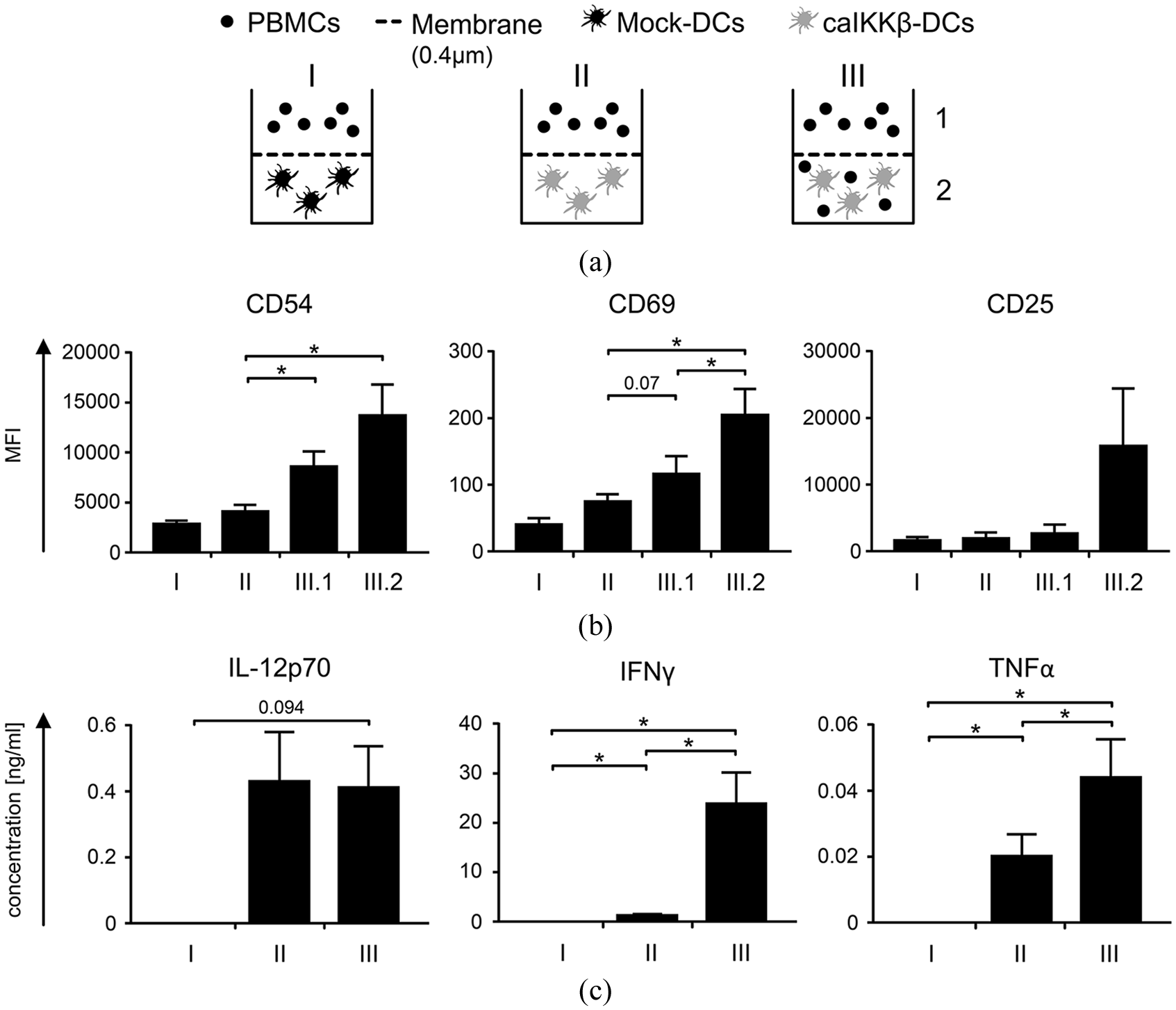
Cell–cell interaction is necessary for best NK-cell activation by caIKKβ-DCs.
Secretion of IFNγ was only sufficiently and significantly detectable when DCs and PBMCs were allowed to interact directly (Figure 3(c)). TNFα was strongly secreted when caIKKβ-DCs and PBMCs were co-cultured, intermediately when caIKKβ-DCs and PBMCs were separated and not at all in the mock condition (Figure 3(c)). These data show that caIKKβ-DCs and NK cells require direct cell–cell interaction for improved NK-cell activation. Activated NK cells seem to induce the activation of further NK cells, independently of direct cell contact.
caIKKβ-DCs can simultaneously activate both CD8+ T cells and NK cells
The classical function of DCs in therapeutic tumor vaccination is the activation of tumor-specific T cells that attack the tumor. Hence, it is essential that the DCs’ ability to activate CD8+ T cells is not diminished. On the other hand, it may be possible that the T cells that are stimulated by the DCs compete with the NK cells for the DC-mediated activation. To investigate this, we analyzed whether NK cells and T cells were in competition with one another or if they could both be activated simultaneously in a caIKKβ-DC/PBMC co-culture. Therefore, caIKKβ-RNA-electroporated and mock-electroporated DCs were loaded with a CD8+ T-cell epitope from the melanoma antigen MelanA, or were left untreated as a control. These DCs were co-cultured with autologous PBMCs at a cell ratio of 1:10. After 1

Stimulation of peripheral blood mononuclear cells (PBMCs) with caIKKβ-DCs leads to activation of both NK cells and CD8+ T cells.
caIKKβ-electroporated mature DCs induce NK cells that can lyse K562 target cells
One of the most desirable properties of DCs for their use in tumor vaccination is their ability to activate effector cells to initiate tumor killing. We could previously show that CD8+ T cells stimulated with caIKKβ-DCs were activated with a superior lytic capacity towards tumor cells compared with DCs matured with the standard protocol. 16 As NK cells could also eliminate tumor cells, a standard cytotoxicity assay was performed to determine whether caIKKβ-DCs could also stimulate NK cells to lyse tumor cells. Therefore, caIKKβ- or mock-transfected DCs were co-cultured with autologous PMBCs at a cell ratio of 1:2 and 1:10 (Figure 5(a)) or with autologous purified NK cells at a cell ratio of 5:1 and 1:1 (Figure 5(b)) for 1 week. The resulting cell population was then used in a cytotoxicity assay against K562 cells with a target-to-effector ratios of 1:20, 1:6, 1:2, and 3:2.

NK cells stimulated with caIKKβ-DCs can kill K562 cells.
Mock-electroporated DCs could not sufficiently activate NK cells as they were not able to lyse the target cells (Figure 5A, B). In contrast, the caIKKβ-DCs were able to stimulate PBMCs and also purified NK cells, resulting in NK cells that efficiently lysed the K562 cell line (Figure 5). Stimulated PMBCs were able to lyse K562 cells at a target-to-effector ratio of up to 1:2, reaching significance at a target-to-effector ratio of 1:20, when DCs and PMBCs had been co-cultured at a cell ratio of 1:10 (Figure 5(a)). caIKKβ-DC/PBMC co-cultures at a cell ratio of 1:2 also led to a cell population with an enhanced ability to lyse K562 cells, however, without reaching significance. Regarding purified NK cells, both caIKKβ/NK cell ratios of 5:1 and 1:1 were able to equip these NK cells with the ability to significantly lyse K562 cells at a target-to-effector ratio of 1:20. Purified NK cells that were stimulated with caIKKβ DCs were able to lyse K562 cells at a target-to-effector ratio of up to 1:2, when co-incubated at a ratio of 5:1 and even up to 1:0.6 when co-incubated at a ratio of 1:1.
Discussion
The DCs currently used for tumor vaccination were mainly optimized for induction of potent tumor-specific T cells, but the clinical efficacy observed after treatment with DCs as monotherapy suggested that an improvement of this approach is required. Therefore, next to new combinatorial approaches, it is of great importance to generate DCs with immunostimulatory functions beyond CTL induction.
Our group has established a method to enhance the activation of monocyte-derived DCs matured with the standard cytokine cocktail through subsequent transfection with a caIKKβ in order to additionally activate the NF-κB pathway. This strategy generated DCs with several advantageous features: (i) the activation of NF-κB led to an increased activation status of DCs by upregulation of several activation markers, while their ability to migrate towards lymphatic tissue remained intact; (ii) they spontaneously and continuously secreted IL-12p70; and, thus, (iii) activate CD8+ T cells that displayed a memory-like phenotype characterized by an upregulation of CD27 with a superior lytic capacity. 16 DCs matured with only the standard cocktail required CD4+ T cell help to secrete IL-12p70 and to induce CD8+ T cells with similar features. 40
In the study described here, we show that caIKKβ-DCs strongly activate NK cells in contrast to DCs generated with the standard protocol. Following contact with caIKKβ-DCs, activated NK cells were able to secrete high amounts of IFNγ and also some TNFα (Figure 2), which can promote further activation of DCs, 9 and naïve T cells 6 for induction of robust cytotoxic T-cell responses. Indeed, a clearly increased expansion of tumor antigen-specific CD8+ T cells by the caIKKβ-DC was found when compared with conventional DCs in presence of NK-cells (Figure 4). Nevertheless, in theory NK cells and T cells might compete for the DCs, thus resulting in a lower NK activation when the DCs had to stimulate both types of effector cells simultaneously. However, the fact that loading caIKKβ-DCs with an antigen resulted in the generation of specific CD8+ T cells and that this did not influence NK-cell activation (Figure 4) indicated that no competition between T-cell and NK-cell activation occurred in the utilized model system.
In addition, the DC-activated NK cells by themselves were able to effectively lyse the classical HLA-negative NK-cell target K562 (Figure 5). Hence, the simultaneous activation of CTL and NK cells would allow an attack on the tumor via tumor antigens presented in HLA class I and efficiently preempt the immune escape mechanism of HLA class I loss. The mechanisms by which the NK cells exert this killing remains to be further investigated; so far, we excluded degranulation as well as production of IFNγ and TNFα via CD107a and intracellular staining (data not shown) suggesting that cell-surface interaction might to play a role here.
The observation that even immature moDCs are in principle able to activate NK cells was made many years ago, but the classical maturation cocktail did not increase this ability. 41 Therefore, other groups have focused on creating improved protocols using alternative maturation mixtures, mostly containing different TLR agonists,17,27,28 inducing DCs to more efficiently activate effector cells. Anguille et al. used so-called IL-15-DCs by replacing IL-4 with IL-15 during the differentiation of DCs and then using TNFα, IFNγ, PGE2, and R-848 (a TLR-7/8 agonist) for maturation. 27 The maturation cocktail used by Massa et al. contained IFNγ and MLPA, 17 which is a ligand for TLR-4, whereas Mailliard et al. used a maturation mixture consisting of IFNα, IFNγ, TNFα, IL-1β, and a TLR-3 agonist (p-I:C), creating so-called α-type-1-polarized DCs (αDC1). 28
The caIKKβ-DCs, like the IL-15-DCs, αDC1, and DCs matured with MLPA and IFNγ were all able to effectively activate NK cells as shown in the upregulation of CD69 (Figure 1),17,42,43 CD25 (Figure 1),17,43 CD54 (Figure 1), and further activation markers. 43 caIKKβ DCs were able to activate both NK cells in co-cultures with PBMCs and also with purified NK cells showing that bystander cells were not necessary for NK-cell activation. NK cells activated through caIKKβ-DCs or MPLA and IFNγ matured DCs were both able to secrete IFNγ (Figure 2). 17 Both these DCs and also IL-15-DCs were able to induce NK cells to effectively kill certain tumor cell lines (Figure 5).17,43 Cytotoxicity of NK cells activated by αDC1s was not analyzed. 42 Regarding IFNγ production, IL-15-DCs alone were already able to secrete IFNγ themselves, whereas in IL-15-DC/NK co-culture the secretion of IFNγ did not increase significantly. 43 αDC1 were able to induce IFNγ production by NK cells, but only when αDC1 were co-cultured with PBMCs (or together with CD40L stimulation). In αDC1/NK cell co-cultures, neither IFNγ secretion was detectable, nor was an upregulation of CD69 seen, showing that co-factors (such as CD40L) are needed for NK-cell activation with αDC1. 42 In this context, it is noteworthy that CD40L is a bona fide activator of the NF-κB pathway.
The standard maturation cocktail contains PGE2 as it has been shown that it is important for the DCs’ ability to migrate to the lymph nodes (LNs).44,45 However, PGE2 interferes with the IL-12p70 secretion by DCs.46,47 Through electroporation of caIKKβ-RNA in DCs matured with the standard maturation cocktail, this problem could be overcome, as these DCs still could migrate towards the LN, but had the ability to secrete high amounts of IL-12p70. 16 For IL-15-DCs PGE2 was contained in their maturation cocktail, indeed creating DCs that could migrate towards the LN. However, these DCs were not able to secrete IL-12p70 when left alone, only gaining this ability when co-cultured with CD40L-transfected 3T3 mouse fibroblasts, representing the CD40–CD40L interaction between DCs and helper T cells. 27 Even though PGE2 was not included in the maturation cocktail to create αDC1s, these DCs were still able to migrate towards the corresponding chemokine, although not quite as well as DCs matured with the standard protocol. 28 Despite strong CCR7 expression on DCs matured with MPLA and IFNγ, these DCs did not show efficient migratory capacity towards CCL21, indicating a low potency to migrate towards the LN. 48 Both αDC1 and DCs matured with MPLA and IFNγ were able to secrete IL-12p70.17,28
The ability to secrete IL-12p70 is one of the most favorable features for vaccine DCs. IL-12p70 plays a crucial role in the development of a CD8+ T-cell memory, 49 and it is also important for a Th1 response. 50 Massa et al. showed that NK cells are highly dependent on IL-12p70 for the production of IFNγ, whereas IL-12p70 does not play a central role in the cytotoxicity of NK cells. 17 In line with others,2,20 we observed that the soluble factors secreted by the caIKKβ-DC, including IL-12p70, did not induce NK cells to secrete IFNγ, but that direct cell–cell interaction was required. An interesting observation was that once NK cells had become activated via direct interaction with caIKKβ-DCs, further NK cells that could not directly interact with these DCs were also slightly activated, as indicated by upregulation of CD54 and slightly CD69, but not CD25 (Figure 3). It is possible that IFNγ in concert with other cytokines produced by activated NK cells, led to the stimulation of further NK cells (as reviewed by Boehm et al. 51 ). This may indicate a positive feedback mechanism for NK cell recruitment but this process as well as the induced activation program within those NK cells requires further investigations.
In conclusion, caIKKβ-DCs meet many features for an optimal vaccination: they can migrate towards lymphatic tissue, secrete IL-12p70 for more than 2 days, activate CTL with a memory-like phenotype and NK cells. The possibility to activate the NF-κB pathway by mRNA electroporation is another advantage as this is a safe method approved and tested for clinical use.15,31 Therefore, we believe that caIKKβ-DCs are a powerful tool for anticancer vaccination and we are about to start testing these DCs in a phase I clinical trial.
Supplemental Material
Raw_data_used_for_this_manuscript_15.08.19 – Supplemental material for NF-κB activation triggers NK-cell stimulation by monocyte-derived dendritic cells
Supplemental material, Raw_data_used_for_this_manuscript_15.08.19 for NF-κB activation triggers NK-cell stimulation by monocyte-derived dendritic cells by Naomi C. Bosch, Reinhard E. Voll, Caroline J. Voskens, Stefanie Gross, Barbara Seliger, Gerold Schuler, Niels Schaft and Jan Dörrie in Therapeutic Advances in Medical Oncology
Supplemental Material
Supplemental_Figures_15.08.19 – Supplemental material for NF-κB activation triggers NK-cell stimulation by monocyte-derived dendritic cells
Supplemental material, Supplemental_Figures_15.08.19 for NF-κB activation triggers NK-cell stimulation by monocyte-derived dendritic cells by Naomi C. Bosch, Reinhard E. Voll, Caroline J. Voskens, Stefanie Gross, Barbara Seliger, Gerold Schuler, Niels Schaft and Jan Dörrie in Therapeutic Advances in Medical Oncology
Footnotes
Acknowledgements
We want to thank Dennis Harrer for fruitful discussions and Carmen Lorenz and Annett Hamann for technical assistance. We also thank Ton Schumacher for his help and advice concerning the production of peptide-HLA-tetramers and for providing the HLA-expression construct. We also express our gratitude to the voluntary blood donors and the medical staff for the acquisition of the blood. The present work was performed in fulfillment of the requirements for obtaining the degree of Dr. med. at the Martin-Luther University Halle-Wittenberg by Naomi C. Bosch.
Funding
The author(s) disclosed receipt of the following financial support for the research, authorship, and/or publication of this article: Preparatory work for this project was supported by grants from the German Cancer Aid (Deutsche Krebshilfe e.V; grant number 110265 to Jan Dörrie, Beatrice Schuler-Thurner, and Niels Schaft; grant number 111105 to Barbara Seliger and Chiara Massa; grant number 113311 to Barbara Seliger).
Conflict of interest statement
The authors declare the following potential conflict of interest: REV, GS, NS, and JD are named as inventors on a patent on caIKK-RNA-electroporated DCs (WO/2012/055551).
Supplemental material
Supplemental material for this article is available online.
References
Supplementary Material
Please find the following supplemental material available below.
For Open Access articles published under a Creative Commons License, all supplemental material carries the same license as the article it is associated with.
For non-Open Access articles published, all supplemental material carries a non-exclusive license, and permission requests for re-use of supplemental material or any part of supplemental material shall be sent directly to the copyright owner as specified in the copyright notice associated with the article.